IHCAB20

Items 1-50 of 7715

Set Ascending Direction
  • Applications
    Reactivities
    Conjugation
    Size
    Delivery Time
  • DDX58 Mouse Monoclonal Antibody [Clone ID: OTI6C1]

    HEK293T cells were transfected with the pCMV6-ENTRY control (Cat# PS100001, Left lane) or pCMV6-ENTRY DDX58 (Cat# RC217615, Right lane) cDNA for 48 hrs and lysed. Equivalent amounts of cell lysates (5 ug per lane) were separated by SDS-PAGE and immunoblotted with anti-DDX58(Cat# TA506141). Positive lysates LY415370 (100ug) and LC415370 (20ug) can be purchased separately from OriGene.

    HEK293T cells were transfected with the pCMV6-ENTRY control (Cat# PS100001, Left lane) or pCMV6-ENTRY DDX58 (Cat# RC217615, Right lane) cDNA for 48 hrs and lysed. Equivalent amounts of cell lysates (5 ug per lane) were separated by SDS-PAGE and immunoblotted with anti-DDX58(Cat# TA506141). Positive lysates LY415370 (100ug) and LC415370 (20ug) can be purchased separately from OriGene.
    DDX58 Mouse Monoclonal Antibody [Clone ID: OTI6C1] 1 Citation
    DDX58 mouse monoclonal antibody, clone OTI6C1 (formerly 6C1).
    ELISA (indirect), IF, IHC, WB
    Human
    Unconjugated
    100 ul
    $447.00
    In Stock*
  • Ribonuclease Inhibitor (RNH1) Mouse Monoclonal Antibody [Clone ID: OTI1B7]

    Figure A, Western blot analysis of overexpressed lysates (5ug per lane) from HEK293T cells transfected with empty plasmid (PS100001, NC), human RNH1 plasmid (RC208360, hRNH1), mouse RNH1 plasmid (MR207287, mRNH1) using anti-RNH1 antibody TA501981 (1:10000@1mg/ml). Figure B, Western blot analysis of the same samples as Figure A with anti-DDK antibody (TA180144, 1:10000@1mg/ml).

    Figure A, Western blot analysis of overexpressed lysates (5ug per lane) from HEK293T cells transfected with empty plasmid (PS100001, NC), human RNH1  plasmid (RC208360, hRNH1), mouse RNH1 plasmid (MR207287, mRNH1) using anti-RNH1 antibody TA501981 (1:10000@1mg/ml). Figure B, Western blot analysis of the same samples as Figure A with anti-DDK antibody (TA180144, 1:10000@1mg/ml).
    Ribonuclease Inhibitor (RNH1) Mouse Monoclonal Antibody [Clone ID: OTI1B7]
    RNH1 (Ribonuclease Inhibitor) mouse monoclonal antibody, clone OTI1B7 (formerly 1B7).
    FC, IF, IHC, Simple Western, WB
    Human
    Unconjugated
    100 ul
    $447.00
    In Stock*
  • P Glycoprotein (ABCB1) Mouse Monoclonal Antibody [Clone ID: OTI5B3]

    SF9 cells lysate (5 ug, left lane) and SF9 cells lysate expressing human recombinant protein fragment (5 ug, right lane) corresponding to amino acids 995-1280 of human ABCB1 (NP_000918) were separated by SDS-PAGE and immunoblotted with anti-ABCB1.

    SF9 cells lysate (5 ug, left lane) and SF9 cells lysate expressing human recombinant protein fragment (5 ug, right lane) corresponding to amino acids 995-1280 of human ABCB1 (NP_000918) were separated by SDS-PAGE and immunoblotted with anti-ABCB1.
    P Glycoprotein (ABCB1) Mouse Monoclonal Antibody [Clone ID: OTI5B3]
    ABCB1 (P Glycoprotein) mouse monoclonal antibody, clone OTI5B3 (formerly 5B3).
    IHC, WB
    Human, Mouse, Rat
    Unconjugated
    30 ul
    $200.00
    In Stock*
  • VEGFB Mouse Monoclonal Antibody [Clone ID: OTI2C4]

    HEK293T cells were transfected with the pCMV6-ENTRY control (Left lane) or pCMV6-ENTRY VEGFB (RC202426, Right lane) cDNA for 48 hrs and lysed. Equivalent amounts of cell lysates (5 ug per lane) were separated by SDS-PAGE and immunoblotted with anti-VEGFB. Positive lysates LY418732 (100ug) and LC418732 (20ug) can be purchased separately from OriGene.

    HEK293T cells were transfected with the pCMV6-ENTRY control (Left lane) or pCMV6-ENTRY VEGFB (RC202426, Right lane) cDNA for 48 hrs and lysed. Equivalent amounts of cell lysates (5 ug per lane) were separated by SDS-PAGE and immunoblotted with anti-VEGFB. Positive lysates LY418732 (100ug) and LC418732 (20ug) can be purchased separately from OriGene.
    VEGFB Mouse Monoclonal Antibody [Clone ID: OTI2C4]
    VEGFB mouse monoclonal antibody, clone OTI2C4 (formerly 2C4).
    IHC, WB
    Human, Mouse, Rat
    Unconjugated
    100 ul
    $447.00
    In Stock*
  • Apg3 (ATG3) Mouse Monoclonal Antibody [Clone ID: OTI2C12]

    Immunohistochemical staining of paraffin-embedded Human lymphoma tissue using anti-ATG3 mouse monoclonal antibody. Heat-induced epitope retrieval by EDTA solution buffer pH 8.0 at 120°C for 3 min.

    Immunohistochemical staining of paraffin-embedded Human lymphoma tissue using anti-ATG3 mouse monoclonal antibody. Heat-induced epitope retrieval by EDTA solution buffer pH 8.0 at 120°C for 3 min.
    Apg3 (ATG3) Mouse Monoclonal Antibody [Clone ID: OTI2C12]
    ATG3 mouse monoclonal antibody, clone OTI2C12 (formerly 2C12).
    FC, IF, IHC, WB
    Human, Mouse, Rat
    Unconjugated
    30 ul
    $200.00
    2 Days*
  • Myoneurin (MYNN) Mouse Monoclonal Antibody [Clone ID: OTI1C11]

    HEK293T cells were transfected with the pCMV6-ENTRY control (Left lane) or pCMV6-ENTRY MYNN (RC207080, Right lane) cDNA for 48 hrs and lysed. Equivalent amounts of cell lysates (5 ug per lane) were separated by SDS-PAGE and immunoblotted with anti-MYNN. Positive lysates LY412964 (100ug) and LC412964 (20ug) can be purchased separately from OriGene.

    HEK293T cells were transfected with the pCMV6-ENTRY control (Left lane) or pCMV6-ENTRY MYNN (RC207080, Right lane) cDNA for 48 hrs and lysed. Equivalent amounts of cell lysates (5 ug per lane) were separated by SDS-PAGE and immunoblotted with anti-MYNN. Positive lysates LY412964 (100ug) and LC412964 (20ug) can be purchased separately from OriGene.
    Myoneurin (MYNN) Mouse Monoclonal Antibody [Clone ID: OTI1C11]
    MYNN mouse monoclonal antibody, clone OTI1C11 (formerly 1C11).
    IHC, WB
    Human, Mouse, Rat
    Unconjugated
    100 ul
    $447.00
    In Stock*
  • LGR5 Mouse Monoclonal Antibody [Clone ID: OTI2A2]

    Figure from citation: Positive correlation between LSD1, c-Myc, β-catenin and LGR5 expression in human colorectal tumor tissues. Expression levels of LSD1, LGR5, β-catenin and c-Myc in consecutive sections from normal colon, adenoma and CRC tumor tissues. Dilution: 1:100 View Citation. Heat-induced epitope retrieval by EDTA solution buffer pH 8.0 at 120°C for 3 min.

    Figure from citation: Positive correlation between LSD1, c-Myc, β-catenin and LGR5 expression in human colorectal tumor tissues. Expression levels of LSD1, LGR5, β-catenin and c-Myc in consecutive sections from normal colon, adenoma and CRC tumor tissues. Dilution: 1:100 View Citation. Heat-induced epitope retrieval by EDTA solution buffer pH 8.0 at 120°C for 3 min.
    LGR5 Mouse Monoclonal Antibody [Clone ID: OTI2A2] 49 Citations
    LGR5 mouse monoclonal antibody, clone OTI2A2.
    FC, IF, IHC, IP, WB
    Human, Mouse, Rat
    Unconjugated
    100 ul
    $447.00
    In Stock*
  • KRAS Mouse Monoclonal Antibody [Clone ID: OTI2C1]

    Simple Western™ analysis of endogenous protein KRAS from HELA lysates (0.5 mg/mL) using KRAS Mouse Monoclonal Antibody #TA801672. The virtual lane view (left) shows the target (as indicated) at 1:50 dilution of primary antibody. The corresponding electropherogram view (right) plots chemiluminescence by molecular weight along the capillary at a 1:50 dilution of primary antibody. This experiment was performed under reducing conditions on the Jess™ Simple Western instrument from ProteinSimple, a Bio-Techne brand, using the 12–230 kDa Separation Module.

    Simple Western™ analysis of endogenous protein KRAS from HELA lysates (0.5 mg/mL) using KRAS Mouse Monoclonal Antibody #TA801672. The virtual lane view (left) shows the target (as indicated) at 1:50 dilution of primary antibody. The corresponding electropherogram view (right) plots chemiluminescence by molecular weight along the capillary at a 1:50  dilution of primary antibody. This experiment was performed under reducing conditions on the Jess™ Simple Western instrument from ProteinSimple, a Bio-Techne brand, using the 12–230 kDa Separation Module.
    KRAS Mouse Monoclonal Antibody [Clone ID: OTI2C1] 6 Citations
    KRAS mouse monoclonal antibody, clone OTI2C1 (formerly 2C1).
    IHC, Simple Western, WB
    Human, Mouse, Rat
    Unconjugated
    100 ul
    $447.00
    In Stock*
  • MAGEA4 Mouse Monoclonal Antibody [Clone ID: OTI1F9]

    Immunohistochemical staining of paraffin-embedded testis using anti-MAGE-A4 C/N TA505362 clone OTI1F9 mouse monoclonal antibody. HIER TEE buffer pH9 (B21-100) at 110C for 10 min, anti-MAGE-A4 diluted to 1:50. Detection was done with Polink1 Broad Mouse and Rabbit C/N D11-18 with DAB Kit. Image 40x magnification.

    Immunohistochemical staining of paraffin-embedded testis using anti-MAGE-A4 C/N TA505362 clone OTI1F9 mouse monoclonal antibody. HIER TEE buffer pH9 (B21-100)  at 110C for 10 min,  anti-MAGE-A4 diluted to 1:50. Detection was done with Polink1 Broad Mouse and Rabbit  C/N D11-18 with DAB Kit. Image 40x magnification.
    MAGEA4 Mouse Monoclonal Antibody [Clone ID: OTI1F9] 3 Citations
    MAGEA4 mouse monoclonal antibody, clone OTI1F9 (formerly 1F9).
    FC, IHC, WB
    Human
    Unconjugated
    100 ul
    $447.00
    In Stock*
  • CD1C Mouse Monoclonal Antibody [Clone ID: OTI2F4]

    HEK293T cells were transfected with the pCMV6-ENTRY control (Cat# PS100001, Left lane) or pCMV6-ENTRY CD1C (Cat# RC218490, Right lane) cDNA for 48 hrs and lysed. Equivalent amounts of cell lysates (5 ug per lane) were separated by SDS-PAGE and immunoblotted with anti-CD1C(Cat# TA505411). Positive lysates LY419762 (100ug) and LC419762 (20ug) can be purchased separately from OriGene.

    HEK293T cells were transfected with the pCMV6-ENTRY control (Cat# PS100001, Left lane) or pCMV6-ENTRY CD1C (Cat# RC218490, Right lane) cDNA for 48 hrs and lysed. Equivalent amounts of cell lysates (5 ug per lane) were separated by SDS-PAGE and immunoblotted with anti-CD1C(Cat# TA505411). Positive lysates LY419762 (100ug) and LC419762 (20ug) can be purchased separately from OriGene.
    CD1C Mouse Monoclonal Antibody [Clone ID: OTI2F4] 4 Citations
    CD1C mouse monoclonal antibody, clone OTI2F4 (formerly 2F4).
    IF, IHC, WB
    Human
    Unconjugated
    100 ul
    $447.00
    In Stock*
  • CD163 Mouse Monoclonal Antibody [Clone ID: OTI2G12]

    Figure A, Western blot analysis of overexpressed lysates(25ug per lane) from HEK293T cells transfected with empty plasmid (PS100001, lane 1) , human CD163 plasmid (RC208557, lane 2), mouse CD163 plasmid (MR216798, lane 3) , rat CD163 plasmid (RR206586, lane 4) using anti-CD163 antibody TA506380 (1:500). Figure B, Western blot analysis of the same samples as figure A with anti-DDK antibody (TA180144, 1:1000)

    Figure A, Western blot analysis of overexpressed lysates(25ug per lane)  from  HEK293T cells transfected with empty plasmid (PS100001, lane 1) , human CD163 plasmid (RC208557, lane 2), mouse CD163 plasmid (MR216798, lane 3) , rat CD163 plasmid (RR206586, lane 4) using anti-CD163 antibody TA506380 (1:500). Figure B, Western blot analysis of  the same samples as figure A with anti-DDK antibody (TA180144, 1:1000)
    CD163 Mouse Monoclonal Antibody [Clone ID: OTI2G12] 8 Citations
    CD163 mouse monoclonal antibody, clone OTI2G12 (formerly 2G12).
    IF, IHC, WB
    Human
    Unconjugated
    100 ul
    $447.00
    In Stock*
  • PADI4 Mouse Monoclonal Antibody [Clone ID: OTI4H5]

    Immunohistochemical staining of paraffin-embedded Human breast tissue within the normal limits using anti-PADI4 mouse monoclonal antibody. Heat-induced epitope retrieval by EDTA solution buffer pH 8.0 at 120°C for 3 min.

    Immunohistochemical staining of paraffin-embedded Human breast tissue within the normal limits using anti-PADI4 mouse monoclonal antibody. Heat-induced epitope retrieval by EDTA solution buffer pH 8.0 at 120°C for 3 min.
    PADI4 Mouse Monoclonal Antibody [Clone ID: OTI4H5] 8 Citations
    PADI4 mouse monoclonal antibody, clone OTI4H5 (formerly 4H5).
    FC, IF, IHC, WB
    Human, Mouse, Rat
    Unconjugated
    100 ul
    $447.00
    In Stock*
  • CD26 (DPP4) Mouse Monoclonal Antibody [Clone ID: OTI11D7]

    Figure A, Western blot analysis of overexpressed lysates(25ug per lane) from HEK293T cells transfected with empty plasmid (PS100001, lane 1) , human DPP4 plasmid (RC209466, lane 2), mouse DPP4 plasmid (MR222698, lane 3) using anti-DPP4 antibody TA500733 (1:500). Figure B, Western blot analysis of the same samples as figure A with anti-DDK antibody (TA180144, 1:1000)

    Figure A, Western blot analysis of overexpressed lysates(25ug per lane)  from  HEK293T cells transfected with empty plasmid (PS100001, lane 1) , human DPP4 plasmid (RC209466, lane 2), mouse DPP4 plasmid (MR222698, lane 3) using anti-DPP4 antibody TA500733 (1:500). Figure B, Western blot analysis of  the same samples as figure A with anti-DDK antibody (TA180144, 1:1000)
    CD26 (DPP4) Mouse Monoclonal Antibody [Clone ID: OTI11D7] 12 Citations
    Anti-DPP4 (CD26) mouse monoclonal antibody, clone OTI11D7 (formerly 11D7).
    FC, IF, IHC, WB
    Human
    Unconjugated
    100 ul
    $447.00
    In Stock*
  • beta IV Tubulin (TUBB4A) Mouse Monoclonal Antibody [Clone ID: OTI5C1]

    Simple Western™ analysis of endogenous protein TUBB4A from HELA lysates (0.2 mg/mL) using TUBB4A Mouse Monoclonal Antibody #TA503129. The virtual lane view (left) shows the target (as indicated) at 1:50 dilution of primary antibody. The corresponding electropherogram view (right) plots chemiluminescence by molecular weight along the capillary at a 1:50 dilution of primary antibody. This experiment was performed under reducing conditions on the Jess™ Simple Western instrument from ProteinSimple, a Bio-Techne brand, using the 12–230 kDa Separation Module.

    Simple Western™ analysis of endogenous protein TUBB4A from HELA lysates (0.2 mg/mL) using TUBB4A Mouse Monoclonal Antibody #TA503129. The virtual lane view (left) shows the target (as indicated) at 1:50 dilution of primary antibody. The corresponding electropherogram view (right) plots chemiluminescence by molecular weight along the capillary at a 1:50  dilution of primary antibody. This experiment was performed under reducing conditions on the Jess™ Simple Western instrument from ProteinSimple, a Bio-Techne brand, using the 12–230 kDa Separation Module.
    beta IV Tubulin (TUBB4A) Mouse Monoclonal Antibody [Clone ID: OTI5C1] 29 Citations
    Mouse monoclonal anti-TUBB4(beta IV Tubulin) antibody, clone 5C1, Loading, clone OTI5C1 (formerly 5C1).
    FC, IF, IHC, Simple Western, WB
    Dog, Human, Monkey, Mouse, Rat
    Unconjugated
    100 ul
    $447.00
    In Stock*
  • NNMT Mouse Monoclonal Antibody [Clone ID: OTI3D8]

    Simple Western™ analysis of endogenous protein NNMT from HELA lysates (0.5 mg/mL) using NNMT Mouse Monoclonal Antibody #TA502624. The virtual lane view (left) shows the target (as indicated) at 1:50 dilution of primary antibody. The corresponding electropherogram view (right) plots chemiluminescence by molecular weight along the capillary at a 1:50 dilution of primary antibody. This experiment was performed under reducing conditions on the Jess™ Simple Western instrument from ProteinSimple, a Bio-Techne brand, using the 12–230 kDa Separation Module.

    Simple Western™ analysis of endogenous protein NNMT from HELA lysates (0.5 mg/mL) using NNMT Mouse Monoclonal Antibody #TA502624. The virtual lane view (left) shows the target (as indicated) at 1:50 dilution of primary antibody. The corresponding electropherogram view (right) plots chemiluminescence by molecular weight along the capillary at a 1:50 dilution of primary antibody. This experiment was performed under reducing conditions on the Jess™ Simple Western instrument from ProteinSimple, a Bio-Techne brand, using the 12–230 kDa Separation Module.
    NNMT Mouse Monoclonal Antibody [Clone ID: OTI3D8] 1 Citation
    NNMT mouse monoclonal antibody, clone OTI3D8 (formerly 3D8).
    FC, IF, IHC, Simple Western, WB
    Human, Mouse, Rat
    Unconjugated
    100 ul
    $447.00
    In Stock*
  • SAMHD1 Mouse Monoclonal Antibody

    Simple Western™ analysis of endogenous protein SAMHD1 from THP-1 lysates (0.1 mg/mL) using SAMHD1 Mouse Monoclonal Antibody #TA502157. The virtual lane view (left) shows the target (as indicated) at 1:50 dilution of primary antibody. The corresponding electropherogram view (right) plots chemiluminescence by molecular weight along the capillary at a 1:50 dilution of primary antibody. This experiment was performed under reducing conditions on the Jess™ Simple Western instrument from ProteinSimple, a Bio-Techne brand, using the 12–230 kDa Separation Module.

    Simple Western™ analysis of endogenous protein SAMHD1 from THP-1 lysates (0.1 mg/mL) using SAMHD1 Mouse Monoclonal Antibody #TA502157. The virtual lane view (left) shows the target (as indicated) at 1:50 dilution of primary antibody. The corresponding electropherogram view (right) plots chemiluminescence by molecular weight along the capillary at a 1:50  dilution of primary antibody. This experiment was performed under reducing conditions on the Jess™ Simple Western instrument from ProteinSimple, a Bio-Techne brand, using the 12–230 kDa Separation Module.
    SAMHD1 Mouse Monoclonal Antibody 4 Citations
    SAMHD1 mouse monoclonal antibody, clone OTI1A1 (formerly 1A1).
    FC, IF, IHC, Simple Western, WB
    Human, Monkey, Mouse, Rat
    Unconjugated
    100 ul
    $447.00
    In Stock*
  • Protein Kinase A regulatory subunit I alpha (PRKAR1A) Mouse Monoclonal Antibody [Clone ID: OTI6C7]

    Simple Western™ analysis of endogenous protein PRKAR1A from HEK293T lysates (0.2 mg/mL) using PRKAR1A Mouse Monoclonal Antibody #TA500641. The virtual lane view (left) shows the target (as indicated) at 1:50 dilution of primary antibody. The corresponding electropherogram view (right) plots chemiluminescence by molecular weight along the capillary at a 1:50 dilution of primary antibody. This experiment was performed under reducing conditions on the Jess™ Simple Western instrument from ProteinSimple, a Bio-Techne brand, using the 12–230 kDa Separation Module.

    Simple Western™ analysis of endogenous protein PRKAR1A from HEK293T lysates (0.2 mg/mL) using PRKAR1A Mouse Monoclonal Antibody #TA500641. The virtual lane view (left) shows the target (as indicated) at 1:50 dilution of primary antibody. The corresponding electropherogram view (right) plots chemiluminescence by molecular weight along the capillary at a 1:50  dilution of primary antibody. This experiment was performed under reducing conditions on the Jess™ Simple Western instrument from ProteinSimple, a Bio-Techne brand, using the 12–230 kDa Separation Module.
    Protein Kinase A regulatory subunit I alpha (PRKAR1A) Mouse Monoclonal Antibody [Clone ID: OTI6C7] 7 Citations
    Anti-PRKAR1A (PKA regulatory subunit I alpha) mouse monoclonal antibody, clone OTI6C7 (formerly 6C7).
    IF, IHC, Simple Western, WB
    Human, Mouse, Rat
    Unconjugated
    100 ul
    $447.00
    In Stock*
  • beta Actin (ACTB) Mouse Monoclonal Antibody [Clone ID: OTI1]

    Immunohistochemical staining of paraffin-embedded normal tonsil using ACTB antibody TA811000 clone OTI1 mouse monoclonal antibody. Protocol used HIER TEE pH9.0 (cat# B21-100)and anti-ACTB at 1:1000 dilution. Detection was done with Polink1 Broad Mouse and Rabbit C/N D11-18 with DAB Kit. Image 40x magnification.

    Immunohistochemical staining of paraffin-embedded normal tonsil using ACTB antibody TA811000 clone OTI1 mouse  monoclonal antibody.  Protocol used HIER TEE pH9.0 (cat# B21-100)and anti-ACTB  at 1:1000 dilution. Detection was done with Polink1 Broad Mouse and Rabbit  C/N D11-18 with DAB Kit. Image 40x magnification.
    beta Actin (ACTB) Mouse Monoclonal Antibody [Clone ID: OTI1] 93 Citations
    beta Actin Mouse Monoclonal Antibody, Clone OTI1, Loading Control.
    IHC, Simple Western, WB
    Dog, Human, Monkey, Mouse, Rat
    Unconjugated
    100 ul
    $447.00
    In Stock*
  • IFIT1 Mouse Monoclonal Antibody [Clone ID: OTI3G8]

    Figure A, Western blot analysis of overexpressed lysates(25ug per lane) from HEK293T cells transfected with empty plasmid (PS100001, lane 1) , human IFIT1 plasmid (RC203320, lane 2), mouse IFIT1 plasmid (MR207393, lane 3) using anti-IFIT1 antibody TA500948 (1:500). Figure B, Western blot analysis of the same samples as figure A with anti-DDK antibody (TA180144, 1:1000)

    Figure A, Western blot analysis of overexpressed lysates(25ug per lane)  from  HEK293T cells transfected with empty plasmid (PS100001, lane 1) , human IFIT1 plasmid (RC203320, lane 2), mouse IFIT1 plasmid (MR207393, lane 3) using anti-IFIT1 antibody TA500948 (1:500). Figure B, Western blot analysis of  the same samples as figure A with anti-DDK antibody (TA180144, 1:1000)
    IFIT1 Mouse Monoclonal Antibody [Clone ID: OTI3G8] 10 Citations
    IFIT1 mouse monoclonal antibody, clone OTI3G8 (formerly 3G8).
    FC, IF, IHC, WB
    Human, Mouse, Rat
    Unconjugated
    100 ul
    $447.00
    In Stock*
  • LMAN1 Mouse Monoclonal Antibody [Clone ID: OTI1A8]

    Simple Western™ analysis of endogenous protein LMAN1 from HEK293T lysates (0.2 mg/mL) using LMAN1 Mouse Monoclonal Antibody #TA502137. The virtual lane view (left) shows the target (as indicated) at 1:50 dilution of primary antibody. The corresponding electropherogram view (right) plots chemiluminescence by molecular weight along the capillary at a 1:50 dilution of primary antibody. This experiment was performed under reducing conditions on the Jess™ Simple Western instrument from ProteinSimple, a Bio-Techne brand, using the 12–230 kDa Separation Module.

    Simple Western™ analysis of endogenous protein LMAN1 from HEK293T lysates (0.2 mg/mL) using LMAN1 Mouse Monoclonal Antibody #TA502137. The virtual lane view (left) shows the target (as indicated) at 1:50 dilution of primary antibody. The corresponding electropherogram view (right) plots chemiluminescence by molecular weight along the capillary at a 1:50  dilution of primary antibody. This experiment was performed under reducing conditions on the Jess™ Simple Western instrument from ProteinSimple, a Bio-Techne brand, using the 12–230 kDa Separation Module.
    LMAN1 Mouse Monoclonal Antibody [Clone ID: OTI1A8]
    LMAN1 mouse monoclonal antibody, clone OTI1A8 (formerly 1A8).
    FC, IF, IHC, Simple Western, WB
    Dog, Human, Monkey, Mouse, Rat
    Unconjugated
    100 ul
    $447.00
    In Stock*
  • ALOX15 Mouse Monoclonal Antibody [Clone ID: OTI3G8]

    Western blot analysis of overexpressed lysates(15ug per lane) from HEK293T cells transfected with empty plasmid (PS100001, lane 1) , human ALOX15 plasmid (RC206621, lane 2), mouse ALOX15 plasmid (MR226258, lane 3) using anti-ALOX15 antibody TA504250 (1:500).

    Western blot analysis of overexpressed lysates(15ug per lane)  from  HEK293T cells transfected with empty plasmid (PS100001, lane 1) , human ALOX15 plasmid (RC206621, lane 2), mouse ALOX15 plasmid (MR226258, lane 3)  using anti-ALOX15  antibody TA504250 (1:500).
    ALOX15 Mouse Monoclonal Antibody [Clone ID: OTI3G8] 2 Citations
    ALOX15 mouse monoclonal antibody, clone OTI3G8 (formerly 3G8).
    IF, IHC, WB
    Human, Mouse, Rat
    Unconjugated
    100 ul
    $447.00
    In Stock*
  • SAMHD1 Mouse Monoclonal Antibody [Clone ID: OTI3F5]

    Figure A, Western blot analysis of overexpressed lysates(25ug per lane) from HEK293T cells transfected with empty plasmid (PS100001, lane 1) , human SAMHD1 plasmid (RC206013, lane 2) using anti-SAMHD1 antibody TA502024 (1:500). Figure B, Western blot analysis of the same samples as figure A with anti-DDK antibody (TA180144, 1:1000)

    Figure A, Western blot analysis of  overexpressed lysates(25ug per lane)  from  HEK293T cells transfected with empty plasmid (PS100001, lane 1) , human SAMHD1 plasmid (RC206013, lane 2) using anti-SAMHD1 antibody TA502024 (1:500). Figure B, Western blot analysis of  the same samples as figure A with anti-DDK antibody (TA180144, 1:1000)
    SAMHD1 Mouse Monoclonal Antibody [Clone ID: OTI3F5] 13 Citations
    SAMHD1 mouse monoclonal antibody, clone OTI3F5 (formerly 3F5).
    FC, IF, IHC, WB
    Human, Monkey, Mouse, Rat
    Unconjugated
    100 ul
    $447.00
    In Stock*
  • ROMO1 Mouse Monoclonal Antibody [Clone ID: OTI2C12]

    Figure A, Western blot analysis of overexpressed lysates(25ug per lane) from HEK293T cells transfected with empty plasmid (PS100001, lane 1) , human ROMO1 plasmid (RC210655, lane 2), mouse ROMO1 plasmid (MR215950, lane 3) using anti-ROMO1 antibody TA505580 (1:500). Figure B, Western blot analysis of the same samples as figure A with anti-DDK antibody (TA180144, 1:1000)

    Figure A, Western blot analysis of overexpressed lysates(25ug per lane)  from  HEK293T cells transfected with empty plasmid (PS100001, lane 1) , human ROMO1 plasmid (RC210655, lane 2), mouse ROMO1 plasmid (MR215950, lane 3) using anti-ROMO1 antibody TA505580 (1:500). Figure B, Western blot analysis of  the same samples as figure A with anti-DDK antibody (TA180144, 1:1000)
    ROMO1 Mouse Monoclonal Antibody [Clone ID: OTI2C12] 14 Citations
    ROMO1 mouse monoclonal antibody, clone OTI2C12 (formerly 2C12).
    IF, IHC, WB
    Dog, Human, Monkey, Mouse, Rat
    Unconjugated
    100 ul
    $447.00
    In Stock*
  • TLE 1 (TLE1) Mouse Monoclonal Antibody [Clone ID: OTI1F5]

    Figure A, Western blot analysis of overexpressed lysates(15ug per lane) from HEK293T cells transfected with empty plasmid (PS100001, lane 1) , human TLE1 plasmid (RC204037, lane 2), mouse TLE1 plasmid (MR210559, lane 3) using anti-TLE1 antibody TA800294 (1:500). Figure B, Western blot analysis of the same samples as figure A with anti-DDK antibody (TA180144, 1:1000)

    Figure A, Western blot analysis of overexpressed lysates(15ug per lane)  from  HEK293T cells transfected with empty plasmid (PS100001, lane 1) , human TLE1 plasmid (RC204037, lane 2), mouse TLE1 plasmid (MR210559, lane 3)  using anti-TLE1  antibody TA800294 (1:500). Figure B, Western blot analysis of  the same samples as figure A with anti-DDK antibody (TA180144, 1:1000)
    TLE 1 (TLE1) Mouse Monoclonal Antibody [Clone ID: OTI1F5]
    TLE1 mouse monoclonal antibody, clone OTI1F5 (formerly 1F5).
    FC, IF, IHC, WB
    Human, Mouse, Rat
    Unconjugated
    100 ul
    $447.00
    In Stock*
  • PFKP Mouse Monoclonal Antibody [Clone ID: OTI1D6]

    Simple Western™ analysis of endogenous protein PFKP from MCF-7 lysates (0.1 mg/mL) using PFKP Mouse Monoclonal Antibody #TA503982. The virtual lane view (left) shows the target (as indicated) at 1:50 dilution of primary antibody. The corresponding electropherogram view (right) plots chemiluminescence by molecular weight along the capillary at a 1:50 dilution of primary antibody. This experiment was performed under reducing conditions on the Jess™ Simple Western instrument from ProteinSimple, a Bio-Techne brand, using the 12–230 kDa Separation Module.

    Simple Western™ analysis of endogenous protein PFKP from MCF-7 lysates (0.1 mg/mL) using PFKP Mouse Monoclonal Antibody #TA503982. The virtual lane view (left) shows the target (as indicated) at 1:50 dilution of primary antibody. The corresponding electropherogram view (right) plots chemiluminescence by molecular weight along the capillary at a 1:50  dilution of primary antibody. This experiment was performed under reducing conditions on the Jess™ Simple Western instrument from ProteinSimple, a Bio-Techne brand, using the 12–230 kDa Separation Module.
    PFKP Mouse Monoclonal Antibody [Clone ID: OTI1D6]
    PFKP mouse monoclonal antibody, clone OTI1D6 (formerly 1D6).
    FC, IF, IHC, Simple Western, WB
    Human
    Unconjugated
    100 ul
    $447.00
    In Stock*
  • Cytokeratin 8 (KRT8) Mouse Monoclonal Antibody [Clone ID: OTI1B12]

    HEK293T cells were transfected with the pCMV6-ENTRY control (Cat# PS100001, Left lane) or pCMV6-ENTRY KRT8 (Cat# RC209570, Right lane) cDNA for 48 hrs and lysed. Equivalent amounts of cell lysates (5 ug per lane) were separated by SDS-PAGE and immunoblotted with anti-KRT8 (Cat# TA500021)(1:500).

    HEK293T cells were transfected with the pCMV6-ENTRY control (Cat# PS100001, Left lane) or pCMV6-ENTRY KRT8 (Cat# RC209570, Right lane) cDNA for 48 hrs and lysed. Equivalent amounts of cell lysates (5 ug per lane) were separated by SDS-PAGE and immunoblotted with anti-KRT8 (Cat# TA500021)(1:500).
    Cytokeratin 8 (KRT8) Mouse Monoclonal Antibody [Clone ID: OTI1B12] 3 Citations
    CK8 (Keratin 8) mouse monoclonal antibody, clone OTI1B12 (formerly 1B12).
    IF, IHC, Simple Western, WB
    Dog, Human, Monkey, Mouse, Rat
    Unconjugated
    100 ul
    $447.00
    In Stock*
  • BCAT1 Mouse Monoclonal Antibody [Clone ID: OTI3F5]

    Simple Western™ analysis of endogenous protein BCAT1 from HepG2 lysates (0.5 mg/mL) using BCAT1 Mouse Monoclonal Antibody #TA504360. The virtual lane view (left) shows the target (as indicated) at 1:20 dilution of primary antibody. The corresponding electropherogram view (right) plots chemiluminescence by molecular weight along the capillary at a 1:20 dilutios of primary antibody. This experiment was performed under reducing conditions on the Jess™ Simple Western instrument from ProteinSimple, a Bio-Techne brand, using the 12–230 kDa Separation Module.

    Simple Western™ analysis of endogenous protein BCAT1 from HepG2 lysates (0.5 mg/mL) using BCAT1 Mouse Monoclonal Antibody #TA504360. The virtual lane view (left) shows the target (as indicated) at 1:20 dilution of primary antibody. The corresponding electropherogram view (right) plots chemiluminescence by molecular weight along the capillary at a 1:20  dilutios of primary antibody. This experiment was performed under reducing conditions on the Jess™ Simple Western instrument from ProteinSimple, a Bio-Techne brand, using the 12–230 kDa Separation Module.
    BCAT1 Mouse Monoclonal Antibody [Clone ID: OTI3F5] 14 Citations
    BCAT1 mouse monoclonal antibody, clone OTI3F5 (formerly 3F5).
    IHC, Simple Western, WB
    Dog, Human, Mouse
    Unconjugated
    100 ul
    $447.00
    In Stock*
  • STING (TMEM173) Mouse Monoclonal Antibody [Clone ID: OTI4H1]

    Immunohistochemical staining of paraffin-embedded Human prostate tissue within the normal limits using anti-TMEM173 mouse monoclonal antibody. Heat-induced epitope retrieval by EDTA solution buffer pH 8.0 at 120°C for 3 min.

    Immunohistochemical staining of paraffin-embedded Human prostate tissue within the normal limits using anti-TMEM173 mouse monoclonal antibody. Heat-induced epitope retrieval by EDTA solution buffer pH 8.0 at 120°C for 3 min.
    STING (TMEM173) Mouse Monoclonal Antibody [Clone ID: OTI4H1] 1 Citation
    TMEM173 mouse monoclonal antibody, clone OTI4H1 (formerly 4H1).
    IHC, Simple Western, WB
    Human
    Unconjugated
    100 ul
    $447.00
    In Stock*
  • Von Hippel Lindau (VHL) Mouse Monoclonal Antibody [Clone ID: OTI1E1]

    Immunohistochemical staining of paraffin-embedded Adenocarcinoma of Human colon tissue using anti-VHL mouse monoclonal antibody. Heat-induced epitope retrieval by EDTA solution buffer pH 8.0 at 120°C for 3 min.

    Immunohistochemical staining of paraffin-embedded Adenocarcinoma of Human colon tissue using anti-VHL mouse monoclonal antibody. Heat-induced epitope retrieval by EDTA solution buffer pH 8.0 at 120°C for 3 min.
    Von Hippel Lindau (VHL) Mouse Monoclonal Antibody [Clone ID: OTI1E1] 2 Citations
    VHL (Von Hippel Lindau) mouse monoclonal antibody, clone OTI1E1 (formerly 1E1).
    IHC, WB
    Human, Mouse, Rat
    Unconjugated
    100 ul
    $447.00
    In Stock*
  • GAD67 (GAD1) Mouse Monoclonal Antibody [Clone ID: OTI3G9]

    HEK293T cells were transfected with the pCMV6-ENTRY control (Cat# PS100001, Left lane) or pCMV6-ENTRY GAD1 (Cat# RC207226, Right lane) cDNA for 48 hrs and lysed. Equivalent amounts of cell lysates (5 ug per lane) were separated by SDS-PAGE and immunoblotted with anti-GAD1 (Cat# TA500329). Positive lysates LY400290 (100ug) and LC400290 (20ug) can be purchased separately from OriGene.

    HEK293T cells were transfected with the pCMV6-ENTRY control (Cat# PS100001, Left lane) or pCMV6-ENTRY GAD1 (Cat# RC207226, Right lane) cDNA for 48 hrs and lysed. Equivalent amounts of cell lysates (5 ug per lane) were separated by SDS-PAGE and immunoblotted with anti-GAD1 (Cat# TA500329). Positive lysates LY400290 (100ug) and LC400290 (20ug) can be purchased separately from OriGene.
    GAD67 (GAD1) Mouse Monoclonal Antibody [Clone ID: OTI3G9] 2 Citations
    GAD1 (GAD67) mouse monoclonal antibody, clone OTI3G9 (formerly 3G9).
    IF, IHC, IP, WB
    Human, Monkey, Mouse, Rat
    Unconjugated
    100 ul
    $447.00
    In Stock*
  • IL6 Mouse Monoclonal Antibody [Clone ID: OTI3G9]

    Figure A, Western blot analysis of overexpressed lysates (5ug per lane) from HEK293T cells transfected with empty plasmid (PS100001, NC), human IL6 plasmid (RC202078, hIL6), mouse IL6 plasmid (MR227281, mIL6), rat IL6 plasmid (RR201203, rIL6) using anti-IL6 antibody TA500067 (1:20000@1mg/ml). Figure B, Western blot analysis of the same samples as figure A with anti-DDK antibody (TA180144, 1:10000@1mg/ml)

    Figure A, Western blot analysis of overexpressed lysates (5ug per lane) from HEK293T cells transfected with empty plasmid (PS100001, NC), human IL6  plasmid (RC202078, hIL6), mouse IL6 plasmid (MR227281, mIL6), rat IL6 plasmid (RR201203, rIL6) using anti-IL6 antibody TA500067 (1:20000@1mg/ml). Figure B, Western blot analysis of  the same samples as figure A with anti-DDK antibody (TA180144, 1:10000@1mg/ml)
    IL6 Mouse Monoclonal Antibody [Clone ID: OTI3G9] 19 Citations
    IL6 mouse monoclonal antibody, clone OTI3G9 (formerly 3G9).
    IF, IHC, WB
    Human
    Unconjugated
    100 ul
    $447.00
    In Stock*
  • SLUG (SNAI2) Mouse Monoclonal Antibody [Clone ID: OTI1A6]

    HEK293T cells transfected with either RC202365 overexpress plasmid (Red) or empty vector control plasmid (Blue) were immunostained by anti-SNAI2 antibody (TA800167), and then analyzed by flow cytometry.

    HEK293T cells transfected with either RC202365 overexpress plasmid (Red) or empty vector control plasmid (Blue) were immunostained by anti-SNAI2 antibody (TA800167), and then analyzed by flow cytometry.
    SLUG (SNAI2) Mouse Monoclonal Antibody [Clone ID: OTI1A6] 5 Citations
    SNAI2 (SLUG) mouse monoclonal antibody, clone OTI1A6 (formerly 1A6).
    FC, IHC, WB
    Human, Mouse, Rat
    Unconjugated
    100 ul
    $447.00
    In Stock*
  • TRPM4 Mouse Monoclonal Antibody [Clone ID: OTI10H5]

    Simple Western™ analysis of endogenous protein TRPM4 from Jurkat lysates (0.2 mg/mL) using TRPM4 Mouse Monoclonal Antibody #TA500381. The virtual lane view (left) shows the target (as indicated) at 1:50 dilution of primary antibody. The corresponding electropherogram view (right) plots chemiluminescence by molecular weight along the capillary at a 1:50 dilution of primary antibody. This experiment was performed under reducing conditions on the Jess™ Simple Western instrument from ProteinSimple, a Bio-Techne brand, using the 12–230 kDa Separation Module.

    Simple Western™ analysis of endogenous protein TRPM4 from Jurkat lysates (0.2 mg/mL) using TRPM4 Mouse Monoclonal Antibody #TA500381. The virtual lane view (left) shows the target (as indicated) at 1:50 dilution of primary antibody. The corresponding electropherogram view (right) plots chemiluminescence by molecular weight along the capillary at a 1:50  dilution of primary antibody. This experiment was performed under reducing conditions on the Jess™ Simple Western instrument from ProteinSimple, a Bio-Techne brand, using the 12–230 kDa Separation Module.
    TRPM4 Mouse Monoclonal Antibody [Clone ID: OTI10H5] 12 Citations
    TRPM4 mouse monoclonal antibody, clone OTI10H5 (formerly 10H5).
    IHC, Simple Western, WB
    Human
    Unconjugated
    100 ul
    $447.00
    In Stock*
  • RGS5 Mouse Monoclonal Antibody [Clone ID: OTI1C1]

    Figure A, Western blot analysis of overexpressed lysates(25ug per lane) from HEK293T cells transfected with empty plasmid (PS100001, lane 1) , human RGS5 plasmid (RC206857, lane 2), mouse RGS5 plasmid (MR201631, lane 3) using anti-RGS5 antibody TA503075 (1:500). Figure B, Western blot analysis of the same samples as figure A with anti-DDK antibody (TA180144, 1:1000)

    Figure A, Western blot analysis of overexpressed lysates(25ug per lane)  from  HEK293T cells transfected with empty plasmid (PS100001, lane 1) , human RGS5 plasmid (RC206857, lane 2), mouse RGS5 plasmid (MR201631, lane 3) using anti-RGS5 antibody TA503075 (1:500). Figure B, Western blot analysis of  the same samples as figure A with anti-DDK antibody (TA180144, 1:1000)
    RGS5 Mouse Monoclonal Antibody [Clone ID: OTI1C1] 5 Citations
    RGS5 mouse monoclonal antibody, clone OTI1C1 (formerly 1C1).
    FC, IF, IHC, WB
    Human
    Unconjugated
    100 ul
    $447.00
    In Stock*
  • AKT1 Mouse Monoclonal Antibody [Clone ID: OTI4D6]

    Simple Western™ analysis of endogenous protein AKT1 from HepG2 lysates (0.2 mg/mL) using AKT1 Mouse Monoclonal Antibody #TA504230. The virtual lane view (left) shows the target (as indicated) at 1:50 dilution of primary antibody. The corresponding electropherogram view (right) plots chemiluminescence by molecular weight along the capillary at a 1:50 dilution of primary antibody. This experiment was performed under reducing conditions on the Jess™ Simple Western instrument from ProteinSimple, a Bio-Techne brand, using the 12–230 kDa Separation Module.

    Simple Western™ analysis of endogenous protein AKT1 from HepG2 lysates (0.2 mg/mL) using AKT1 Mouse Monoclonal Antibody #TA504230. The virtual lane view (left) shows the target (as indicated) at 1:50 dilution of primary antibody. The corresponding electropherogram view (right) plots chemiluminescence by molecular weight along the capillary at a 1:50  dilution of primary antibody. This experiment was performed under reducing conditions on the Jess™ Simple Western instrument from ProteinSimple, a Bio-Techne brand, using the 12–230 kDa Separation Module.
    AKT1 Mouse Monoclonal Antibody [Clone ID: OTI4D6] 8 Citations
    AKT1 mouse monoclonal antibody, clone OTI4D6 (formerly 4D6).
    FC, IF, IHC, Simple Western, WB
    Dog, Human, Monkey, Mouse, Rat
    Unconjugated
    100 ul
    $447.00
    In Stock*
  • TIA1 Mouse Monoclonal Antibody [Clone ID: OTI1D7]

    Simple Western™ analysis of endogenous protein TIA1 from HELA lysates (0.1 mg/mL) using TIA1 Mouse Monoclonal Antibody #TA800657. The virtual lane view (left) shows the target (as indicated) at 1:50 dilution of primary antibody. The corresponding electropherogram view (right) plots chemiluminescence by molecular weight along the capillary at a 1:50 dilution of primary antibody. This experiment was performed under reducing conditions on the Jess™ Simple Western instrument from ProteinSimple, a Bio-Techne brand, using the 12–230 kDa Separation Module.

    Simple Western™ analysis of endogenous protein TIA1 from HELA lysates (0.1 mg/mL) using TIA1 Mouse Monoclonal Antibody #TA800657. The virtual lane view (left) shows the target (as indicated) at 1:50 dilution of primary antibody. The corresponding electropherogram view (right) plots chemiluminescence by molecular weight along the capillary at a 1:50  dilution of primary antibody. This experiment was performed under reducing conditions on the Jess™ Simple Western instrument from ProteinSimple, a Bio-Techne brand, using the 12–230 kDa Separation Module.
    TIA1 Mouse Monoclonal Antibody [Clone ID: OTI1D7]
    TIA1 mouse monoclonal antibody, clone OTI1D7 (formerly 1D7).
    IHC, Simple Western, WB
    Human, Mouse, Rat
    Unconjugated
    100 ul
    $447.00
    In Stock*
  • BTN3A2 Mouse Monoclonal Antibody [Clone ID: OTI1A6]

    HEK293T cells were transfected with the pCMV6-ENTRY control (Cat# PS100001, Left lane) or pCMV6-ENTRY BTN3A2 (Cat# RC201183, Right lane) cDNA for 48 hrs and lysed. Equivalent amounts of cell lysates (5 ug per lane) were separated by SDS-PAGE and immunoblotted with anti-BTN3A2(Cat# TA500730). Positive lysates LY402082 (100ug) and LC402082 (20ug) can be purchased separately from OriGene.

    HEK293T cells were transfected with the pCMV6-ENTRY control (Cat# PS100001, Left lane) or pCMV6-ENTRY BTN3A2 (Cat# RC201183, Right lane) cDNA for 48 hrs and lysed. Equivalent amounts of cell lysates (5 ug per lane) were separated by SDS-PAGE and immunoblotted with anti-BTN3A2(Cat# TA500730). Positive lysates LY402082 (100ug) and LC402082 (20ug) can be purchased separately from OriGene.
    BTN3A2 Mouse Monoclonal Antibody [Clone ID: OTI1A6] 2 Citations
    Anti-BTN3A2 mouse monoclonal antibody, clone OTI1A6 (formerly 1A6).
    IF, IHC, WB
    Human
    Unconjugated
    100 ul
    $447.00
    In Stock*
  • DRP1 (DNM1L) Mouse Monoclonal Antibody [Clone ID: OTI4F6]

    Simple Western™ analysis of endogenous protein DNM1L from HELA lysates (0.2 mg/mL) using DNM1L Mouse Monoclonal Antibody #TA506153. The virtual lane view (left) shows the target (as indicated) at 1:50 dilution of primary antibody. The corresponding electropherogram view (right) plots chemiluminescence by molecular weight along the capillary at a 1:50 dilution of primary antibody. This experiment was performed under reducing conditions on the Jess™ Simple Western instrument from ProteinSimple, a Bio-Techne brand, using the 12–230 kDa Separation Module.

    Simple Western™ analysis of endogenous protein DNM1L from HELA lysates (0.2 mg/mL) using DNM1L Mouse Monoclonal Antibody #TA506153. The virtual lane view (left) shows the target (as indicated) at 1:50 dilution of primary antibody. The corresponding electropherogram view (right) plots chemiluminescence by molecular weight along the capillary at a 1:50  dilution of primary antibody. This experiment was performed under reducing conditions on the Jess™ Simple Western instrument from ProteinSimple, a Bio-Techne brand, using the 12–230 kDa Separation Module.
    DRP1 (DNM1L) Mouse Monoclonal Antibody [Clone ID: OTI4F6] 4 Citations
    DNM1L mouse monoclonal antibody, clone OTI4F6 (formerly 4F6).
    IHC, Simple Western, WB
    Dog, Human, Monkey, Mouse, Rat
    Unconjugated
    100 ul
    $447.00
    In Stock*
  • CD19 Mouse Monoclonal Antibody [Clone ID: OTI3B10]

    HEK293T cells were transfected with the pCMV6-ENTRY control (Cat# PS100001, Left lane) or pCMV6-ENTRY CD19 (Cat# RC202922, Right lane) cDNA for 48 hrs and lysed. Equivalent amounts of cell lysates (5 ug per lane) were separated by SDS-PAGE and immunoblotted with anti-CD19(Cat# TA506236). Positive lysates LY400678 (100ug) and LC400678 (20ug) can be purchased separately from OriGene.

    HEK293T cells were transfected with the pCMV6-ENTRY control (Cat# PS100001, Left lane) or pCMV6-ENTRY CD19 (Cat# RC202922, Right lane) cDNA for 48 hrs and lysed. Equivalent amounts of cell lysates (5 ug per lane) were separated by SDS-PAGE and immunoblotted with anti-CD19(Cat# TA506236). Positive lysates LY400678 (100ug) and LC400678 (20ug) can be purchased separately from OriGene.
    CD19 Mouse Monoclonal Antibody [Clone ID: OTI3B10] 4 Citations
    CD19 mouse monoclonal antibody, clone OTI3B10.
    FC, IF, IHC, WB
    Human
    Unconjugated
    100 ul
    $447.00
    In Stock*
  • Kindlin 2 (FERMT2) Mouse Monoclonal Antibody [Clone ID: OTI9E4]

    Simple Western™ analysis of endogenous protein FERMT2 from HepG2 lysates (0.2 mg/mL) using FERMT2 Mouse Monoclonal Antibody #TA500504. The virtual lane view (left) shows the target (as indicated) at 1:50 dilution of primary antibody. The corresponding electropherogram view (right) plots chemiluminescence by molecular weight along the capillary at a 1:50 dilution of primary antibody. This experiment was performed under reducing conditions on the Jess™ Simple Western instrument from ProteinSimple, a Bio-Techne brand, using the 12–230 kDa Separation Module.

    Simple Western™ analysis of endogenous protein FERMT2 from HepG2 lysates (0.2 mg/mL) using FERMT2 Mouse Monoclonal Antibody #TA500504. The virtual lane view (left) shows the target (as indicated) at 1:50 dilution of primary antibody. The corresponding electropherogram view (right) plots chemiluminescence by molecular weight along the capillary at a 1:50  dilution of primary antibody. This experiment was performed under reducing conditions on the Jess™ Simple Western instrument from ProteinSimple, a Bio-Techne brand, using the 12–230 kDa Separation Module.
    Kindlin 2 (FERMT2) Mouse Monoclonal Antibody [Clone ID: OTI9E4]
    Anti-FERMT2 mouse monoclonal antibody, clone OTI9E4 (formerly 9E4).
    IF, IHC, IP, Simple Western, WB
    Dog, Human, Monkey, Mouse, Rat
    Unconjugated
    100 ul
    $447.00
    In Stock*
  • CD32A (FCGR2A) Mouse Monoclonal Antibody [Clone ID: OTI9C6]

    HEK293T cells were transfected with the pCMV6-ENTRY control (Cat# PS100001, Left lane) or pCMV6-ENTRY FCGR2A (Cat# RC205786, Right lane) cDNA for 48 hrs and lysed. Equivalent amounts of cell lysates (5 ug per lane) were separated by SDS-PAGE and immunoblotted with anti-FCGR2A(Cat# TA500645). Positive lysates LY402871 (100ug) and LC402871 (20ug) can be purchased separately from OriGene.

    HEK293T cells were transfected with the pCMV6-ENTRY control (Cat# PS100001, Left lane) or pCMV6-ENTRY FCGR2A (Cat# RC205786, Right lane) cDNA for 48 hrs and lysed. Equivalent amounts of cell lysates (5 ug per lane) were separated by SDS-PAGE and immunoblotted with anti-FCGR2A(Cat# TA500645). Positive lysates LY402871 (100ug) and LC402871 (20ug) can be purchased separately from OriGene.
    CD32A (FCGR2A) Mouse Monoclonal Antibody [Clone ID: OTI9C6] 1 Citation
    FCGR2A (CD32) mouse monoclonal antibody, clone OTI9C6 (formerly 9C6).
    IF, IHC, IP, WB
    Human
    Unconjugated
    100 ul
    $447.00
    In Stock*
  • ID3 Mouse Monoclonal Antibody [Clone ID: OTI8B3]

    Figure A, Western blot analysis of overexpressed lysates (5ug per lane) from HEK293T cells transfected with empty plasmid (PS100001, NC), human ID3 plasmid (RC200583, hID3), mouse ID3 plasmid (MR227292, mID3) using anti-ID3 antibody TA500713 (1:500@1mg/ml). Figure B, Western blot analysis of the same samples as figure A with anti-DDK antibody (TA180144, 1:10000@1mg/ml)

    Figure A, Western blot analysis of overexpressed lysates (5ug per lane) from HEK293T cells transfected with empty plasmid (PS100001, NC), human ID3  plasmid (RC200583, hID3), mouse ID3 plasmid (MR227292, mID3) using anti-ID3 antibody TA500713 (1:500@1mg/ml). Figure B, Western blot analysis of  the same samples as figure A with anti-DDK antibody (TA180144, 1:10000@1mg/ml)
    ID3 Mouse Monoclonal Antibody [Clone ID: OTI8B3]
    ID3 mouse monoclonal antibody, clone OTI8B3 (formerly 8B3).
    FC, IF, IHC, WB
    Human, Mouse, Rat
    Unconjugated
    100 ul
    $447.00
    In Stock*
  • RBPMS Mouse Monoclonal Antibody [Clone ID: OTI3B7]

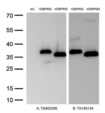
    Figure A, Western blot analysis of overexpressed lysates (15ug per lane) from HEK293T cells transfected with empty plasmid (PS100001, NC), human RBPMS plasmid (RC200248, hRBPMS), mouse RBPMS plasmid (MR201952, mRBPMS) using anti-RBPMS antibody TA800206 (1:10000@1mg/ml). Figure B, Western blot analysis of the same human RBPMS and mouse RBPMS samples as figure A with anti-DDK antibody (TA180144, 1:10000@1mg/ml)

    Figure A, Western blot analysis of overexpressed lysates (15ug per lane) from HEK293T cells transfected with empty plasmid (PS100001, NC), human RBPMS plasmid (RC200248, hRBPMS), mouse RBPMS plasmid (MR201952, mRBPMS) using anti-RBPMS antibody TA800206 (1:10000@1mg/ml). Figure B, Western blot analysis of the same human RBPMS and mouse RBPMS samples as figure A with anti-DDK antibody (TA180144, 1:10000@1mg/ml)
    RBPMS Mouse Monoclonal Antibody [Clone ID: OTI3B7]
    RBPMS mouse monoclonal antibody, clone OTI3B7 (formerly 3B7).
    FC, IHC, WB
    Human, Mouse, Rat
    Unconjugated
    100 ul
    $447.00
    In Stock*
  • PRRX1 Mouse Monoclonal Antibody [Clone ID: OTI1E10]

    Figure A, Western blot analysis of overexpressed lysates(25ug per lane) from HEK293T cells transfected with empty plasmid (PS100001, lane 1) , human PRRX1 plasmid (RC210393, lane 2), mouse PRRX1 v2 plasmid (MR202408, lane 3) , mouse PRRX1 v1 plasmid (MR226699, lane 4) using anti-PRRX1 antibody TA803116 (1:500). Figure B, Western blot analysis of the same samples as figure A with anti-DDK antibody (TA180144, 1:1000)

    Figure A, Western blot analysis of overexpressed lysates(25ug per lane)  from  HEK293T cells transfected with empty plasmid (PS100001, lane 1) , human PRRX1 plasmid (RC210393, lane 2), mouse PRRX1 v2 plasmid (MR202408, lane 3) , mouse PRRX1 v1 plasmid (MR226699, lane 4)  using anti-PRRX1 antibody TA803116 (1:500). Figure B, Western blot analysis of  the same samples as figure A with anti-DDK antibody (TA180144, 1:1000)
    PRRX1 Mouse Monoclonal Antibody [Clone ID: OTI1E10] 11 Citations
    PRRX1 mouse monoclonal antibody, clone OTI1E10 (formerly 1E10).
    IHC, WB
    Human, Mouse, Rat
    Unconjugated
    100 ul
    $447.00
    In Stock*
  • FOLR2 Mouse Monoclonal Antibody [Clone ID: OTI4G6] 1 Citation
    FOLR2 mouse monoclonal antibody, clone OTI4G6 (formerly 4G6).
    IHC, WB
    Human
    Unconjugated
    100 ul
    $447.00
    In Stock*
  • PTCH1 Mouse Monoclonal Antibody [Clone ID: OTI5C7]

    Figure A, Western blot analysis of overexpressed lysates(25ug per lane) from HEK293T cells transfected with empty plasmid (PS100001, NC) , human PTCH1 plasmid (RC216999, hPTCH1), mouse PTCH1 plasmid (MR227010, mPTCH1) using anti-PTCH1 antibody TA500123(1:500). Figure B, Western blot analysis of the same samples as figure A with anti-DDK antibody (TA180144, 1:1000)

    Figure A, Western blot analysis of overexpressed lysates(25ug per lane)  from  HEK293T cells transfected with empty plasmid (PS100001, NC) , human PTCH1 plasmid (RC216999, hPTCH1), mouse PTCH1 plasmid (MR227010, mPTCH1) using anti-PTCH1  antibody TA500123(1:500). Figure B, Western blot analysis of  the same samples as figure A with anti-DDK antibody (TA180144, 1:1000)
    PTCH1 Mouse Monoclonal Antibody [Clone ID: OTI5C7] 1 Citation
    Patched1 (Patched) mouse monoclonal antibody, clone OTI5C7 (formerly 5C7).
    IF, IHC, WB
    Human, Mouse, Rat
    Unconjugated
    100 ul
    $447.00
    In Stock*
  • LIM Kinase 1 (LIMK1) Mouse Monoclonal Antibody [Clone ID: OTI6B4]

    Simple Western™ analysis of endogenous protein LIMK1 from HELA lysates (0.2 mg/mL) using LIMK1 Mouse Monoclonal Antibody #TA503012. The virtual lane view (left) shows the target (as indicated) at 1:50 dilution of primary antibody. The corresponding electropherogram view (right) plots chemiluminescence by molecular weight along the capillary at a 1:50 dilution of primary antibody. This experiment was performed under reducing conditions on the Jess™ Simple Western instrument from ProteinSimple, a Bio-Techne brand, using the 12–230 kDa Separation Module.

    Simple Western™ analysis of endogenous protein LIMK1 from HELA lysates (0.2 mg/mL) using LIMK1 Mouse Monoclonal Antibody #TA503012. The virtual lane view (left) shows the target (as indicated) at 1:50 dilution of primary antibody. The corresponding electropherogram view (right) plots chemiluminescence by molecular weight along the capillary at a 1:50  dilution of primary antibody. This experiment was performed under reducing conditions on the Jess™ Simple Western instrument from ProteinSimple, a Bio-Techne brand, using the 12–230 kDa Separation Module.
    LIM Kinase 1 (LIMK1) Mouse Monoclonal Antibody [Clone ID: OTI6B4]
    LIMK1 (LIM Kinase 1 ) mouse monoclonal antibody, clone OTI6B4 (formerly 6B4).
    FC, IF, IHC, Simple Western, WB
    Dog, Human, Monkey, Mouse, Rat
    Unconjugated
    100 ul
    $447.00
    In Stock*
  • Alpha B Crystallin (CRYAB) Mouse Monoclonal Antibody [Clone ID: OTI6D11]

    HEK293T cells were transfected with the pCMV6-ENTRY control (Cat# PS100001, Left lane) or pCMV6-ENTRY CRYAB (Cat# RC202718, Right lane) cDNA for 48 hrs and lysed. Equivalent amounts of cell lysates (5 ug per lane) were separated by SDS-PAGE and immunoblotted with anti-CRYAB (Cat# TA500583). Positive lysates LY419682 (100ug) and LC419682 (20ug) can be purchased separately from OriGene.

    HEK293T cells were transfected with the pCMV6-ENTRY control (Cat# PS100001, Left lane) or pCMV6-ENTRY CRYAB (Cat# RC202718, Right lane) cDNA for 48 hrs and lysed. Equivalent amounts of cell lysates (5 ug per lane) were separated by SDS-PAGE and immunoblotted with anti-CRYAB (Cat# TA500583). Positive lysates LY419682 (100ug) and LC419682 (20ug) can be purchased separately from OriGene.
    Alpha B Crystallin (CRYAB) Mouse Monoclonal Antibody [Clone ID: OTI6D11] 2 Citations
    CRYAB (Alpha B Crystallin) mouse monoclonal antibody, clone OTI6D11 (formerly 6D11).
    FC, IF, IHC, IP, Simple Western, WB
    Human, Monkey, Mouse, Rat
    Unconjugated
    100 ul
    $447.00
    In Stock*
  • Natriuretic Peptide Receptor C (NPR3) Mouse Monoclonal Antibody [Clone ID: OTI4C3]

    Figure A, Western blot analysis of overexpressed lysates(25ug per lane) from HEK293T cells transfected with empty plasmid (PS100001, lane 1) , human NPR3 plasmid (RC219453, lane 2), mouse NPR3 v2 plasmid (MR208587, lane 3), mouse NPR3 v1 plasmid (MR227616, lane 4), rat NPR3 plasmid (RR203170, lane 5) using anti-NPR3 antibody TA501021 (1:500). Figure B, Western blot analysis of the same samples as figure A with anti-DDK antibody (TA180144, 1:1000)

    Figure A, Western blot analysis of overexpressed lysates(25ug per lane)  from  HEK293T cells transfected with empty plasmid (PS100001, lane 1) , human NPR3 plasmid (RC219453, lane 2), mouse NPR3 v2 plasmid (MR208587, lane 3), mouse NPR3 v1 plasmid (MR227616, lane 4), rat NPR3 plasmid (RR203170, lane 5) using anti-NPR3 antibody TA501021 (1:500). Figure B, Western blot analysis of  the same samples as figure A with anti-DDK antibody (TA180144, 1:1000)
    Natriuretic Peptide Receptor C (NPR3) Mouse Monoclonal Antibody [Clone ID: OTI4C3]
    Anti-NPR3 (Natriuretic Peptide Receptor C) mouse monoclonal antibody, clone OTI4C3 (formerly 4C3).
    FC, IF, IHC, WB
    Human
    Unconjugated
    100 ul
    $447.00
    In Stock*
  • PANK2 Mouse Monoclonal Antibody [Clone ID: OTI3H9]

    Immunohistochemical staining of paraffin-embedded Human liver tissue within the normal limits using anti-PANK2 mouse monoclonal antibody. Heat-induced epitope retrieval by EDTA solution buffer pH 8.0 at 120°C for 3 min.

    Immunohistochemical staining of paraffin-embedded Human liver tissue within the normal limits using anti-PANK2 mouse monoclonal antibody. Heat-induced epitope retrieval by EDTA solution buffer pH 8.0 at 120°C for 3 min.
    PANK2 Mouse Monoclonal Antibody [Clone ID: OTI3H9] 6 Citations
    Anti-PANK2 mouse monoclonal antibody, clone OTI3H9 (formerly 3H9).
    FC, IF, IHC, WB
    Dog, Human, Monkey, Mouse, Rat
    Unconjugated
    100 ul
    $447.00
    In Stock*