Dendritic Cells
Dendritic cells (DCs) are bone-marrow-derived immune cells found in blood, lymphoid organs, and various tissue of the body.
First discovered by Ralph Steinman and Zanvil A. Cohn in the 1970s, named for their tree-like branches or "dendrites", these cells patrol our bodies, ingest pathogens, process them, and present fragments (antigens) to T-cells, thus initiating a targeted immune response [2]. They play an essential role in the induction of adaptive immune response against infectious agents and in generating tolerance against self-antigens [3].
Unlike other immune cells, dendritic cells are not just a single entity, but a complex assortment of subtypes with varying functions. These subtypes include conventional dendritic cells (cDC1 and cDC2), plasmacytoid dendritic cells (pDCs), monocyte-derived dendritic cells (moDCs), and Langerhans cells (LCs).
Dendritic Cell Antibody Panel
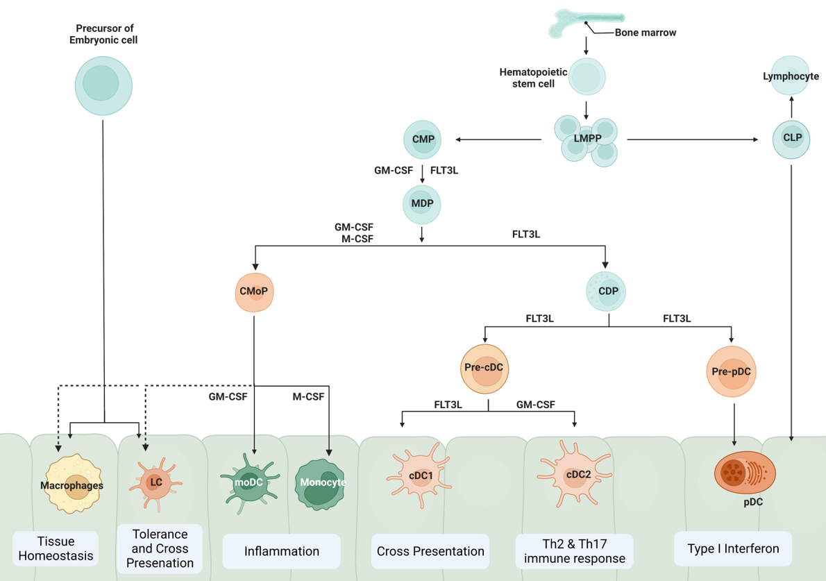
Several models are proposed to describe the development and differentiation of dendritic cells (DCs). These models are based on experimental evidence and observations and aim to explain different DC subsets' sequential stages and lineage relationships. According to the classical model, it proposes a common myeloid progenitor (CMP) as the precursor of both monocytes/macrophages and DCs. It suggests that DCs arise from a common DC progenitor (CDP), which gives rise to pre-DCs that subsequently differentiate into conventional DCs (cDCs) and plasmacytoid DCs (pDCs).
Fig 1. Dendritic cell differentiation from bone marrow [1]
Conventional dendritic cells (cDCs), also known as myeloid dendritic cells or classical dendritic cells, are a type of antigen-presenting cell (APC), cDCs originate from hematopoietic stem cells, specifically from the common myeloid progenitor in the bone marrow. The maturation pathway involves several stages, with differentiating cells passing through a monocyte-like stage before fully maturing into dendritic cells. In humans and mice, conventional dendritic cells are divided into two main subtypes: cDC1 and cDC2, each with distinct functions and markers [2].
cDC1: These cells are very effective at presenting antigens to CD8+ T cells, a type of cytotoxic T cell that can kill infected or cancerous cells. They play a key role in responses against viruses and cancer. They are known to secrete interleukin-12, as well as type I and type III interferons, and are believed to promote Th1 helper T cell and natural killer responses. In mice, they express the cell surface protein XCR1, while in humans, they can be identified by the presence of CD141 (or BDCA-3).
cDC2: These cells are specialized in presenting antigens to CD4+ T cells, a type of helper T cell that helps coordinate the immune response. They are particularly involved in responses to extracellular bacteria and fungi. In both mice and humans, they express the cell surface protein CD11c and the integrin CD172a (or SIRP-alpha)
Plasmacytoid dendritic cells (pDCs), are round plasma-shaped cells specialized for the production of large amounts of type I and type III interferon in response to viral infection. They are less abundant compared to conventional DCs. In humans, pDCs can be identified by the expression of several specific cell surface proteins, including CD123 (the IL-3 receptor alpha chain), BDCA-2, and BDCA-4.
Other subtypes of DCs:
- Langerhans Cells (LCs): These cells reside in the skin and mucosal tissues and can migrate to lymph nodes upon antigen capture.
- Monocyte-Derived Dendritic Cells (MoDCs): These are not a distinct lineage but rather a functional state that monocytes can differentiate into, especially during inflammation.
- Inflammatory Dendritic Cells: These are cells derived from monocytes during inflammation.
| Dendritic Cell Subtype | Cell Surface Marker | Secreted Molecules |
|---|---|---|
| Conventional Dendritic Cells | CD1b | IDO |
| CD11c | IL-1Beta | |
| CD14 | IL-6 | |
| CD40 | IL-8 | |
| CD49 | IL-12 | |
| CD141 | IL-15 | |
| CD172a | IL-23 | |
| CD197 | BATF3 | |
| CD205 | KLF4 | |
| CD207 | ||
| CD206 | ||
| CD273 | ||
| CD282 | ||
| CD284 | ||
| CD369 | ||
| CD370 | ||
| CD11b | ||
| CD13 | ||
| CD33 | ||
| CD80 | ||
| CD83 | ||
| CD66 | ||
| Plasmacytoid Dendritic Cell | CD123 | IFN alpha |
| CD283 | IFN beta | |
| CD303 | E2-2 | |
| CD304 | IRF8 | |
| CD370 | IRF7 | |
| CD45RA | TCF4 | |
| Langerhan Dendritic Cell | CD1C | |
| EpCAM | ||
| CD207 | ||
| CD172 alpha | ||
| Monocyte Derived Dendritic Cell | CD14 | IRF4 |
| CD1A | KLF4 | |
| CD1C | ZBTB46 | |
| CD209 | ||
| FCER1 | ||
| ITGAM | ||
| MRC1 | ||
| SIRPA |
Table 1: Human dendritic cell markers [3][4].
References
- Khan, Farhan Ullah, et al. "Dendritic cells and their immunotherapeutic potential for treating type 1 diabetes." International journal of molecular sciences 23.9 (2022): 4885.
- Puhr S, Lee J, Zvezdova E, Zhou YJ, Liu K. Dendritic cell development-History, advances, and open questions. Semin Immunol. 2015 Dec;27(6):388-96. doi: 10.1016/j.smim.2016.03.012. Epub 2016 Mar 31. PMID: 27040276; PMCID: PMC5032838.
- Schlitzer A, Zhang W, Song M, Ma X. Recent advances in understanding dendritic cell development, classification, and phenotype. F1000Res. 2018 Sep 26;7:F1000 Faculty Rev-1558. doi: 10.12688/f1000research.14793.1. PMID: 30345015; PMCID: PMC6173131.
- Rhodes JW, Tong O, Harman AN, Turville SG. Human Dendritic Cell Subsets, Ontogeny, and Impact on HIV Infection. Front Immunol. 2019 May 16;10:1088. doi: 10.3389/fimmu.2019.01088. PMID: 31156637; PMCID: PMC6532592.

