
Total Exosome Isolation Kit · Lipid Nanoprobe Technology
MagExo™ EV Isolation Kit
Capture the full spectrum of extracellular vesicles to power EV-based multi-omics studies.
A fast, antibody-free extracellular vesicles (EV) isolation protocol built on lipid nanoprobe technology. By targeting the natural lipid bilayer of EVs, MagExo enables direct, high-efficiency capture without harsh processing — delivering high-quality EVs for in-depth molecular profiling within 30 minutes.
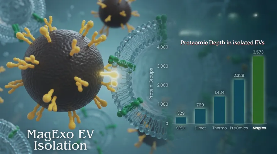
Total EV capture efficiency
One-step, antibody-free workflow
Lipoprotein contaminant depletion
EV-derived DNA, RNA, and protein compatible with every multi-omics workflow — NGS, immunoassays, and LC-MS/MS
Order
Available Product Kits
Choose the MagExo EV isolation kit optimized for your downstream application — multi-omics or LC-MS/MS proteomics.
| Product | SKU | Size | Applications | Price | Add to Cart |
|---|---|---|---|---|---|
| MagExo™ EV Isolation Kit for Multi-Omics | 10 reactions |
DNARNANGSImmunoassay qPCR/dPCR
|
$550 | ||
| 25 reactions | $1,150 | ||||
| MagExo™ EV Isolation Kit for LC/MS | 10 reactions |
LC-MS/MS Proteomics
|
$550 | ||
| 25 reactions | $1,150 |
Validated Performance
Supporting Data
From EV marker confirmation to liquid biopsy, MagExo delivers reproducible total exosome isolation with clean, multi-omics-ready cargo across diverse sample types.
Clinical utility of EVs for Cancer studies
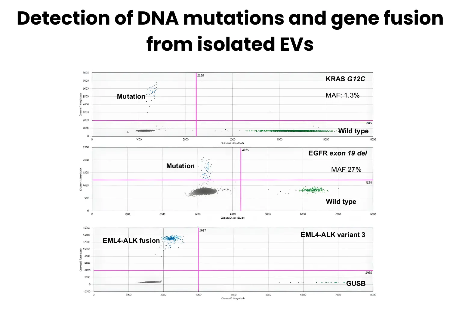
DNA point mutation (KRAS and EGFR) and gene fusion (ALK-ELM4) were detected in EVs isolated from plasma of NSCLC patients, demonstrating mutation and gene fusion analysis from EV-derived DNA as a minimally invasive liquid biopsy source.
Deep proteomic coverage from 100 µL Plasma
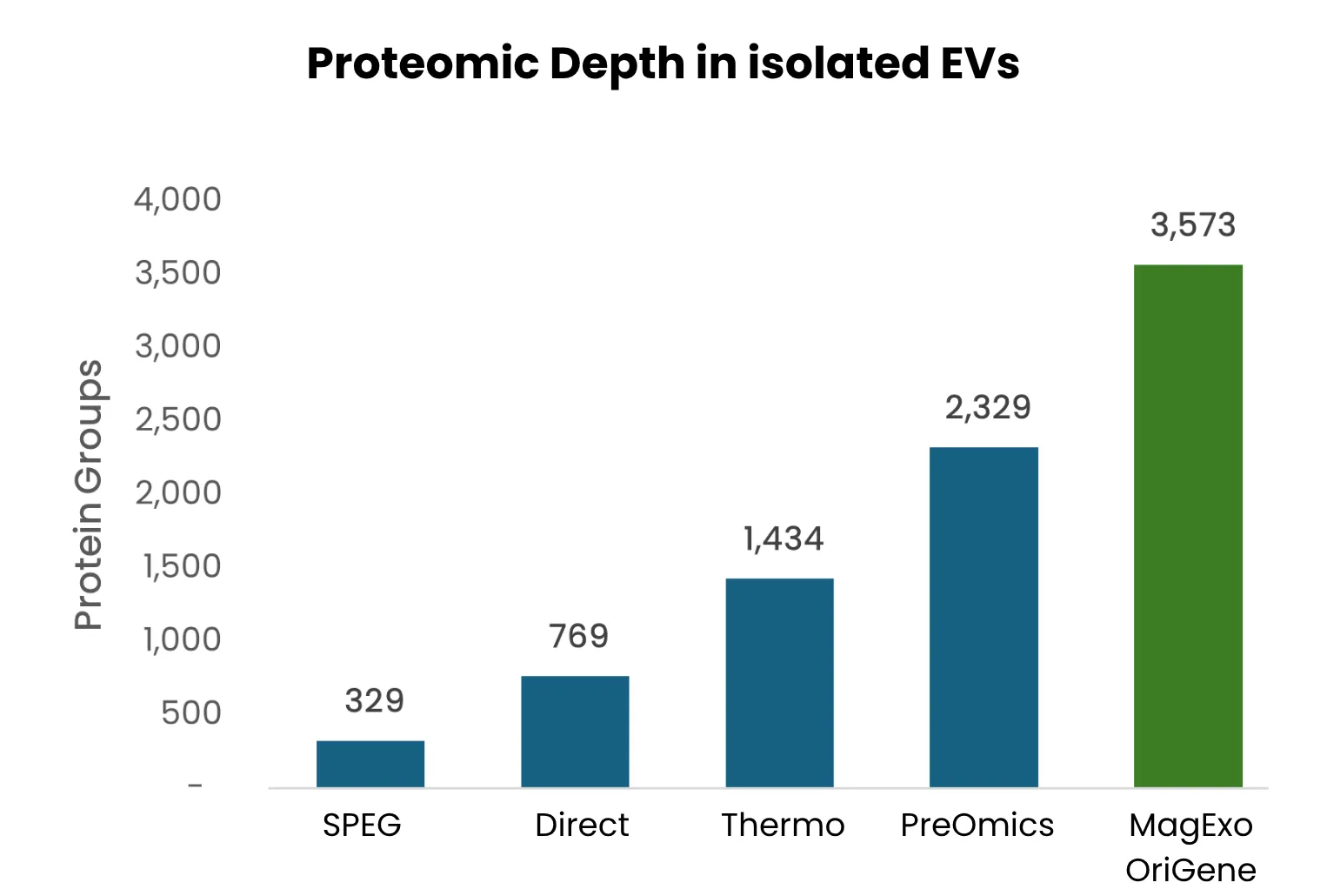
De-identified pooled human plasma was analyzed across multiple mass-spec workflows. EV enrichment identified ~3,573 protein groups — substantially more than other methods.
Proteomics-ready EVs from low-input plasma
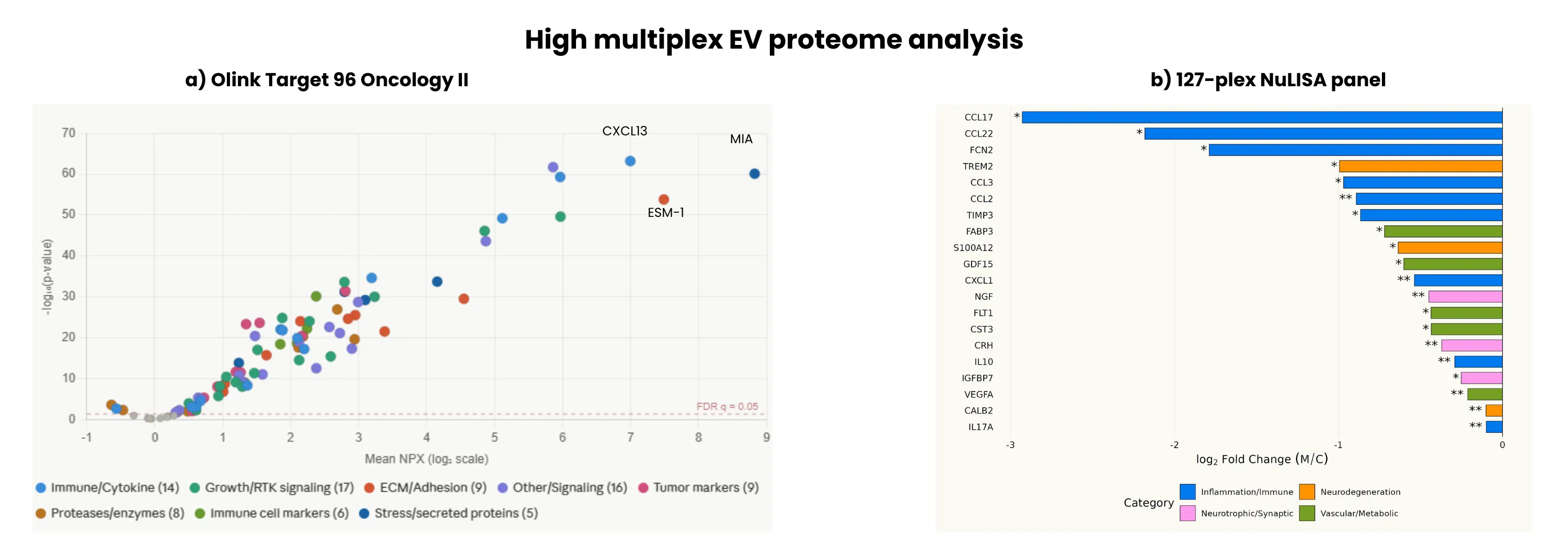
EVs isolated from as little as 100 µL of plasma enable sensitive detection of disease-relevant protein features via Olink Target 96 Oncology II and 127-plex NULISA panels.
Robust recovery of EV protein markers
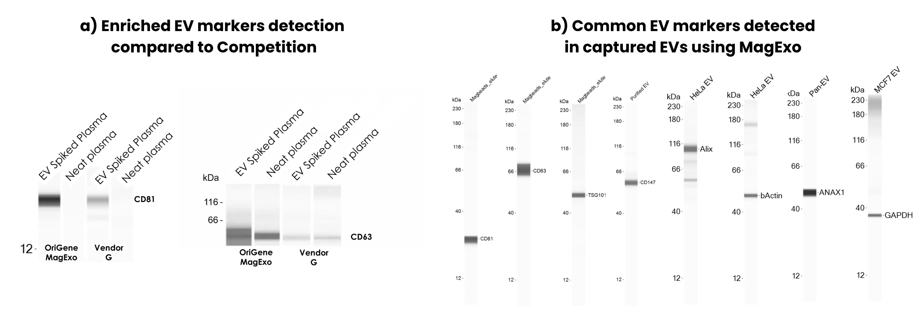
Captured EVs were lysed in RIPA buffer and analyzed by Simple Western. (a) MagExo demonstrated higher EV yield than comparable products. (b) Established exosome markers (CD9, CD81, TSG101) were readily detected, confirming EV identity and preservation of biological integrity.
Lipoprotein-depleted EV preparation
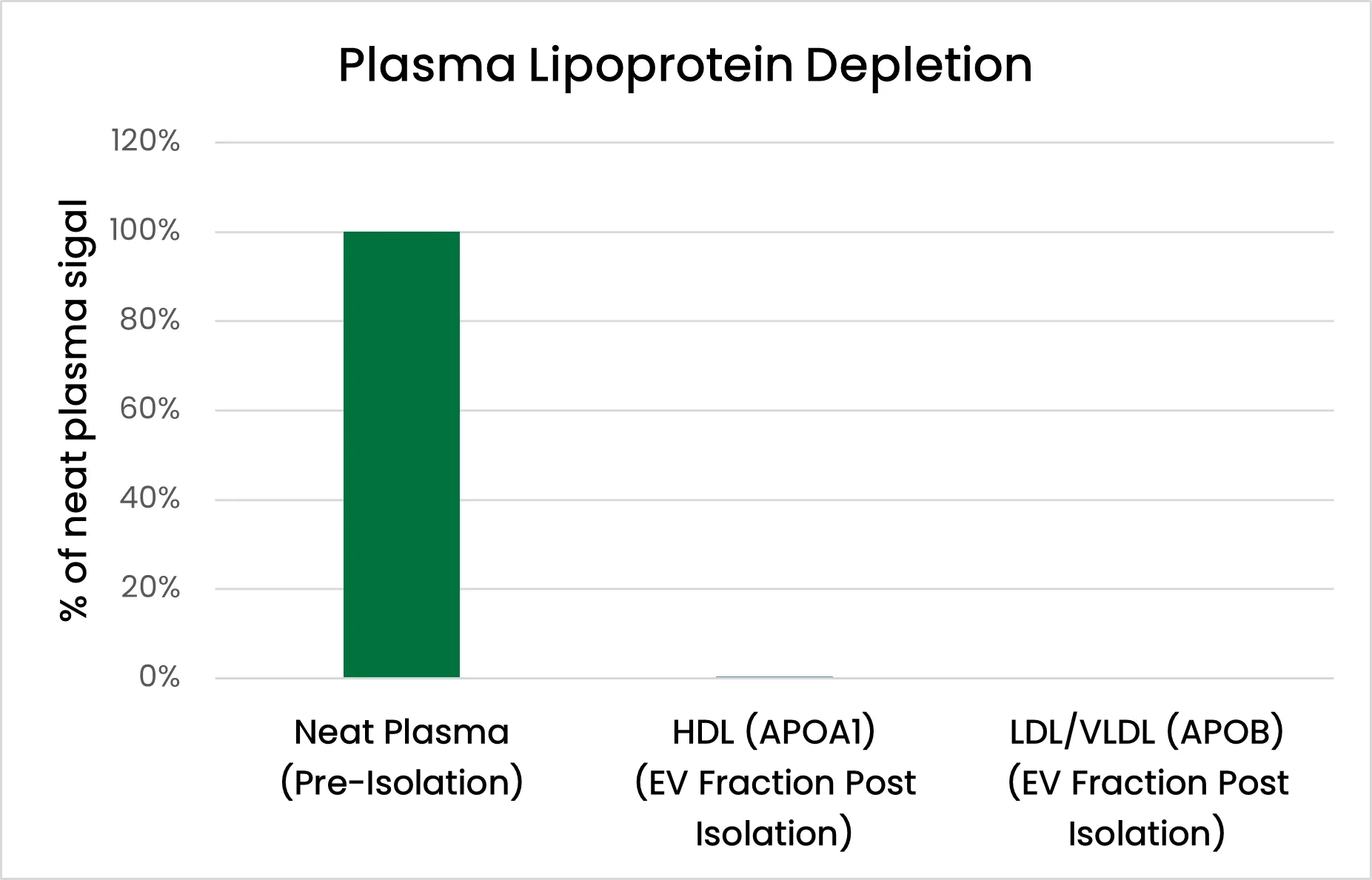
LC/MS analysis of neat plasma versus MagExo-enriched plasma EVs shows ~99% removal of HDL (ApoA1) and LDL/VLDL (ApoB), indicating efficient depletion of lipoprotein contaminants.
High-efficiency recovery of EV cargo nucleic acids
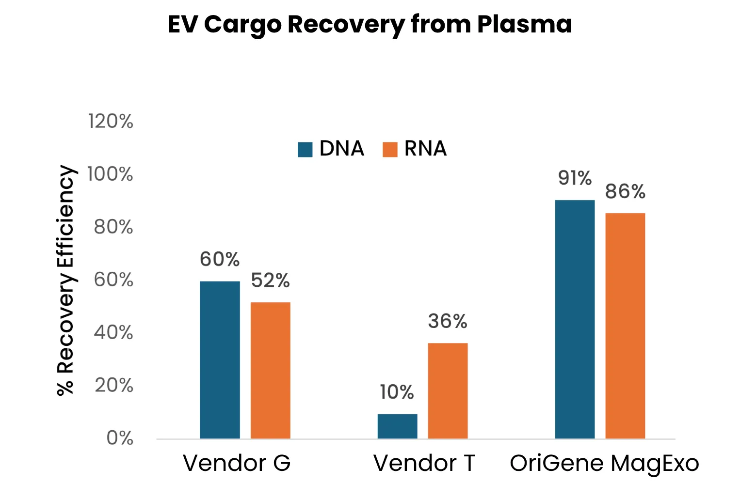
Consistent recovery of >80% of DNA and RNA cargo from cell-derived EVs spiked into diverse sample types, outperforming comparable kits. Quantified using the Qubit DNA/RNA HS assay.
Maximum Preservation of Cargo
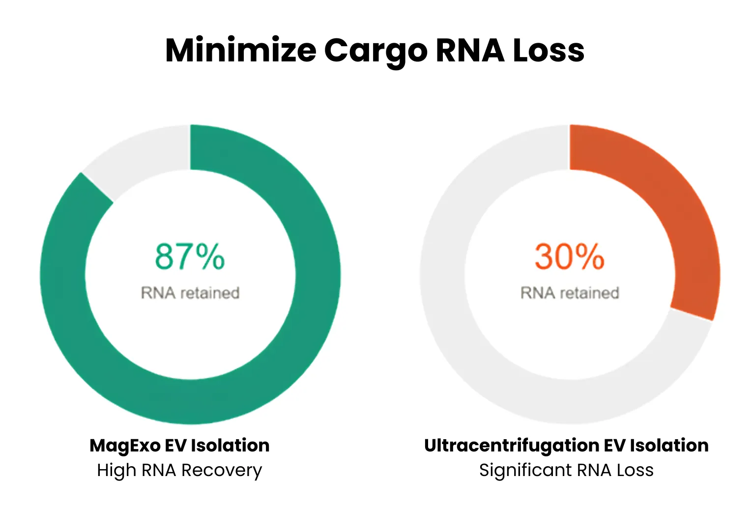
The MagExo isolation method preserves RNA content while reducing protein contaminants. Compared to ultracentrifugation, EV RNA isolated with MagExo shows improved retention — indicating reduced loss of RNA during isolation.
Enrichment and integrity of EV nucleic acid cargo
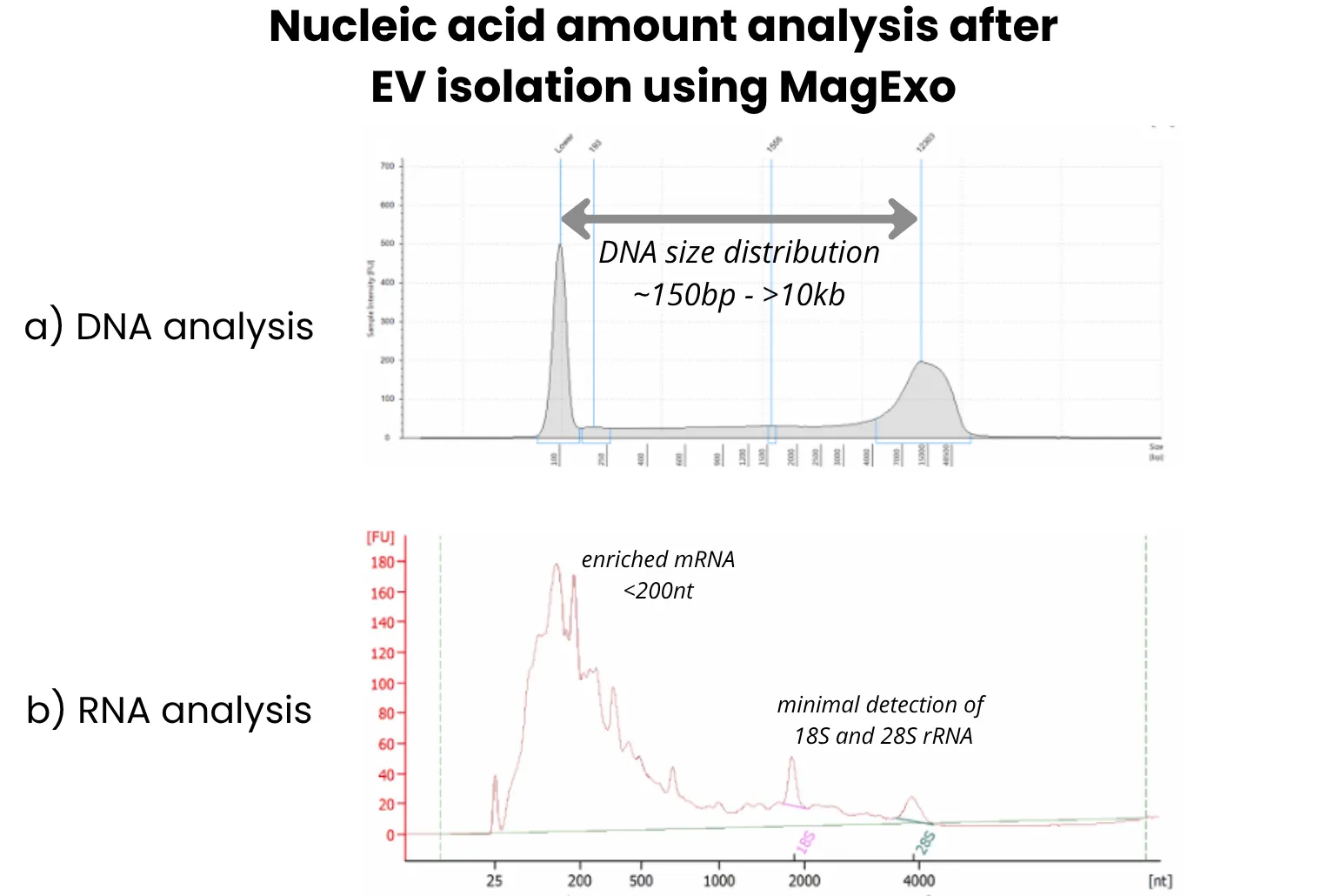
MagExo-captured EVs yield DNA and RNA of high integrity. (a) EV DNA shows broad size distribution from ~150–200 bp to >10 kb. (b) RNA profiling shows enrichment of small RNAs (<200 nt), particularly miRNAs, with minimal rRNA.
The Technology
Lipid Nanoprobe–Based EV Isolation
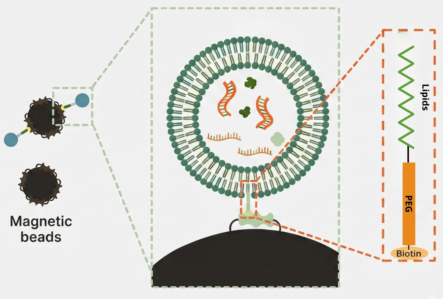
Capture magnetic beads are coated with lipid-based probes that interact with EV lipid bilayers, enabling broad-spectrum capture of EVs rapidly. (Read Publication, Nature Biomedical Engineering, 2017)
- ✓Total EV isolation — >90% EV capture efficiency across sample types.
- ✓High-quality cargo — Captured EVs harbor high-quality nucleic acids (DNA and RNA) and unique proteins.
- ✓Simple workflow — one magnetic step completed in 5-30 minutes.
Exosome Isolation Protocol
Outline of the Procedure
A streamlined, five-step exosome isolation protocol from sample to multi-omics-ready EVs.
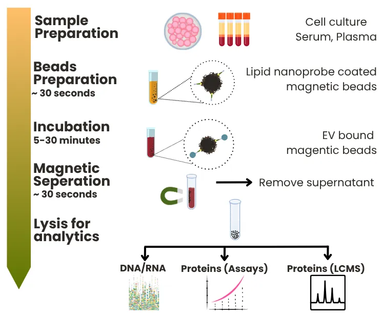
Sample Prep
Cell culture, serum, or plasma.
Beads Prep
~30 secLipid nanoprobe–coated magnetic beads.
Mix & Incubate
5–30 minEVs bind the magnetic beads.
Magnetic Separation
~30 secRemove supernatant.
Isolated EVs
Ready for DNA/RNA, assays & LC-MS.
Method Comparison
How MagExo compares with other EV purification methods
Across the metrics that matter — speed, cost, scalability, purity, and specificity — lipid nanoprobe-based MagExo leads the field.
| Method | Speed | Cost | Scalability | Purity | Specificity |
|---|---|---|---|---|---|
| Lipid nanoprobe-Based MagExo™ | |||||
| Ultracentrifugation | |||||
| Precipitation | |||||
| Size Exclusion (SEC) | |||||
| Tangential Flow Filtration | |||||
| Positive Charged Resin | |||||
| Immuno-Magnetic Beads |
Comparison based on internal survey data. Final results depend on experimental design and method implementation. ●●● strong · ●● moderate · ● limited.
Background
What are extracellular vesicles?
Extracellular vesicles (EVs) are nanoscale, membrane-bound particles released by nearly all cell types that play a central role in intercellular communication. Present in biological fluids such as blood, urine, saliva, cerebrospinal fluid, and cell culture media, EVs carry functional cargo — proteins, lipids, DNA, mRNA, and microRNA — that can modulate recipient cell behavior.
Based on size and biogenesis, EVs are commonly classified into exosomes, microvesicles, and apoptotic bodies. EVs are increasingly leveraged in biomarker discovery, liquid biopsy development, and translational research across oncology, immunology, neuroscience, and regenerative medicine.
Answers
Frequently Asked Questions
The MagExo™ EV Isolation Kit is a fast, antibody-free extracellular vesicles (EV) isolation kit built on lipid nanoprobe technology. It enables direct, high-efficiency capture of total EVs without harsh processing — delivering high-quality EVs for in-depth molecular profiling within 30 minutes.
Capture magnetic beads are coated with lipid-based probes that interact with EV lipid bilayers via a lipid-PEG-biotin anchor. This enables broad-spectrum, antibody-free capture of total EVs in a single magnetic step.
The MagExo protocol is completed in under 30 minutes from sample to isolated EVs. It is a one-step, antibody-free magnetic bead-based workflow with no ultracentrifugation required.
MagExo is compatible with plasma, serum, urine, cerebrospinal fluid (CSF), and cell culture media. It works across diverse sample types for total EV isolation.
MagExo-isolated EVs are compatible with NGS, immunoassay proteomics, and LC-MS/MS workflows. Common exosome markers (CD9, CD81, CD63, TSG101) are readily detected in MagExo isolates, confirming EV identity and cargo integrity.
