
Validated antibodies for EV characterization
Confirm vesicle identity, intactness, and purity — and probe disease-related cargo — with validated antibody targets organized by the questions every EV experiment has to answer. All validated in EV samples using OriGene enrichment products.
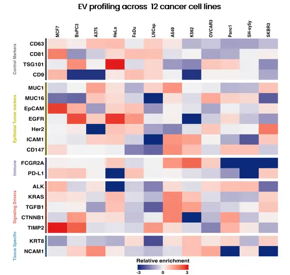
EV marker profiling across 12 cancer cell lines — relative enrichment
Four marker classes, one rigorous EV profile
Robust EV studies confirm what's present, that vesicles are intact, that the prep is clean, and — for translational work — which disease markers are carried. This panel maps every antibody to one of those four questions, in line with MISEV characterization principles.
Membrane markers
Positive tetraspanin & surface markers that confirm extracellular vesicles are present.
Luminal & cytosolic
Inner-cargo markers that distinguish intact vesicles from free, co-purified protein.
Negative / contaminant
Markers that should be absent — demonstrating freedom from cellular debris & lipoproteins.
Disease-related
Oncology & CNS markers for EV biomarker discovery and liquid-biopsy research.
EV marker antibodies by marker class
Each card maps validated antibody targets to their functional role in EV characterization. Every SKU links directly to its product page for datasheets, citations, and ordering.
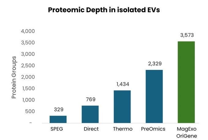
Magnetic Bead · Total EV
MagExo™ EV Isolation Kit
Total EV isolation directly from serum and plasma, engineered for downstream EV multi-omics. Delivers clean, intact vesicles ready for proteomics, RNA/miRNA, and LC/MS workflows.
Polymer Precipitation
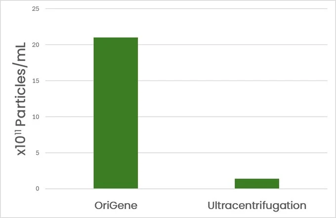
EV Isolation Reagent
A powerful alternative to ultracentrifugation and chromatography. Recovers more EVs than ultracentrifugation from cell culture media with a simple mix → incubate → resuspend protocol.
Magnetic Immunocapture
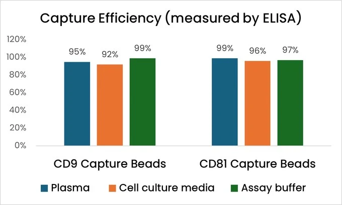
EV/Exosome Immunocapture Kits
High-affinity antibodies on magnetic beads enrich specific EV subpopulations with >90% capture efficiency and low protein carryover — cleaner preps for sensitive downstream assays.
Membrane markers
Positive tetraspanin & surface markers that confirm EVs are present. Validated by Western blot against EV isolates from multiple cell lines.
- CD9 TA813476 Classic exosome marker
- CD63 TA803574 Late endosomal tetraspanin
- CD81 TA343281 TSPAN28 · exosome enriched
- CD147 TA501212 BSG · membrane glycoprotein
- MFGE8 TA809279 Lactadherin · MFG-E8
Luminal & cytosolic markers
Inner-cargo markers that confirm intact vesicles and rule out co-purified free protein. Validated in EVs isolated using MagExo™, Immunocapture, and precipitation methods.
- TSG101 TA385476 ESCRT-I component
- ALIX TA507312 PDCD6IP · ESCRT accessory
- SDCBP TA504796 Syntenin-1 · exosome biogenesis
- HSP70 TA500772 Heat shock protein 70
- HSP90 TA500494 Heat shock protein 90
- GAPDH TA890003 Cytosolic loading control
- ANXA1 TA500967 Annexin A1
- ANXA10 TA500882 Annexin A10
Negative / contaminant markers
Proteins that should be absent from a clean EV preparation — demonstrating freedom from cellular debris and co-isolated lipoproteins.
- Calnexin TA336279 CANX · ER contamination marker
- ApoE TA805357 Apolipoprotein E · lipoprotein marker
Disease-related cargo
Oncology & CNS markers for EV cargo profiling, biomarker discovery, and liquid-biopsy research. Validated for EV inputs at clinically relevant antibody dilutions.
- EpCAM UM500097 TACSTD1 · epithelial marker
- EGFR TA506224 Receptor tyrosine kinase
- HER2 TA503524 ERBB2 · breast/GI oncology
- KRAS TA801672 Driver oncogene · Simple Western
- MUC1 TA800769 Mucin-1 · cancer glycoprotein
- MUC16 TA801281 CA125 · ovarian biomarker
- NCAM1 UM800086 CD56 · neuronal marker
- FOLH1 TA504509 PSMA · prostate biomarker
- PFN1 TA501243 Profilin-1 · actin dynamics
- RNH1 TA601981 Ribonuclease inhibitor 1
- SIRT1 TA809831 Sirtuin-1 · epigenetic regulator
- TIA1 TA800658 RNA-binding protein
Membrane markers
Positive tetraspanin & surface markers that confirm EVs are present. Validated by Western blot against EV isolates from multiple cell lines.
- CD9 TA813476 Classic exosome marker
- CD63 TA803574 Late endosomal tetraspanin
- CD81 TA343281 TSPAN28 · exosome enriched
- CD147 TA501212 BSG · membrane glycoprotein
- MFGE8 TA809279 Lactadherin · MFG-E8
Luminal & cytosolic markers
Inner-cargo markers that confirm intact vesicles and rule out co-purified free protein. Validated in EVs isolated using MagExo™, Immunocapture, and precipitation methods.
- TSG101 TA385476 ESCRT-I component
- ALIX TA507312 PDCD6IP · ESCRT accessory
- SDCBP TA504796 Syntenin-1 · exosome biogenesis
- HSP70 TA500772 Heat shock protein 70
- HSP90 TA500494 Heat shock protein 90
- GAPDH TA890003 Cytosolic loading control
- ANXA1 TA500967 Annexin A1
- ANXA10 TA500882 Annexin A10
Negative / contaminant markers
Proteins that should be absent from a clean EV preparation — demonstrating freedom from cellular debris and co-isolated lipoproteins.
- Calnexin TA336279 CANX · ER contamination marker
- ApoE TA805357 Apolipoprotein E · lipoprotein marker
Disease-related cargo
Oncology & CNS markers for EV cargo profiling, biomarker discovery, and liquid-biopsy research. Validated for EV inputs at clinically relevant antibody dilutions.
- EpCAM UM500097 TACSTD1 · epithelial marker
- EGFR TA506224 Receptor tyrosine kinase
- HER2 TA503524 ERBB2 · breast/GI oncology
- KRAS TA801672 Driver oncogene · Simple Western
- MUC1 TA800769 Mucin-1 · cancer glycoprotein
- MUC16 TA801281 CA125 · ovarian biomarker
- NCAM1 UM800086 CD56 · neuronal marker
- FOLH1 TA504509 PSMA · prostate biomarker
- PFN1 TA501243 Profilin-1 · actin dynamics
- RNH1 TA501981 Ribonuclease inhibitor 1
- SIRT1 TA809831 Sirtuin-1 · epigenetic regulator
- TIA1 TA800658 RNA-binding protein
Validation you can stake an experiment on
Each marker is validated on extracellular vesicles isolated using OriGene's own enrichment products — including MagExo™ magnetic isolation, CD9/CD81 Immunocapture Kits, and Precipitation Reagent — from multiple cell lines and biofluid types. Biomarker targets are additionally confirmed on the automated Simple Western platform, proving these antibodies work end-to-end within a single OriGene EV workflow.
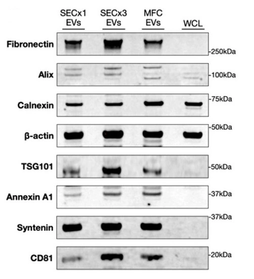
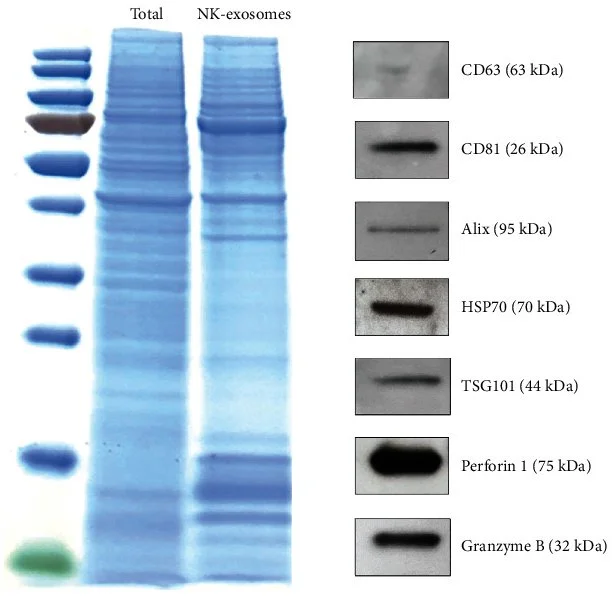
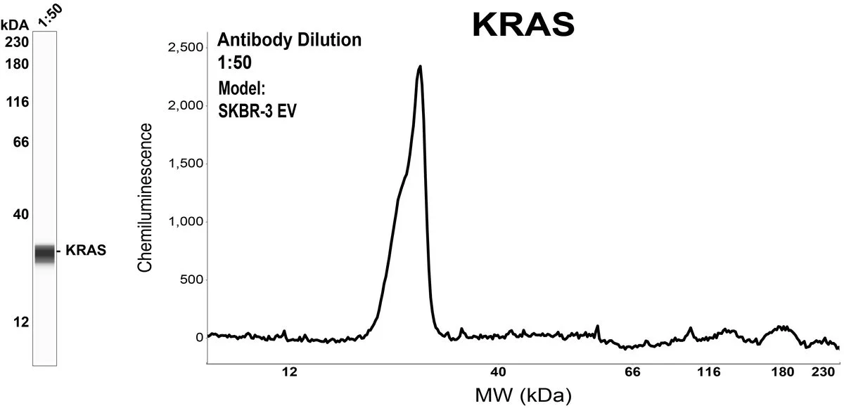
Figure 1. EV markers by Western blot in EVs isolated by size-exclusion chromatography (SECx1, SECx3) and MagExo™ magnetic capture (MFC) versus whole-cell lysate (WCL). Syntenin-1, CD81, TSG101, Alix, and Annexin A1 enrich in EV fractions; Calnexin (ER) and Fibronectin are negative controls. Antibody: Syntenin-1 / SDCBP (TA504796).
Figure 2. Canonical EV markers — CD81, CD63, Alix, HSP70, TSG101 — detected by Western blot in NK-cell–derived exosomes enriched by immunocapture, relative to total cell lysate. Antibody: CD81 (TA343281).
Figure 3. KRAS oncoprotein detected by automated Simple Western (capillary immunoassay) in EVs from SKBR-3 cells enriched by MagExo™ magnetic isolation — a single clean peak at expected MW, 1:50 dilution. Antibody: KRAS (TA801672).
All EV samples isolated using OriGene enrichment products (MagExo™, CD9/CD81 Immunocapture Kits, or Precipitation Reagent). Representative data from internal OriGene EV characterization studies.
Target-specific EV immunoprecipitation
While total-EV isolation captures the full vesicle spectrum, many biomarker discovery and clinical assay workflows require isolation of specific EV subpopulations. OriGene's validated antibodies can be conjugated to magnetic beads for immunoprecipitation of EVs carrying your marker of choice — enabling cleaner signal and higher biological specificity than bulk methods.
How magnetic bead IP capture works
- 1
Select your target antibody
Choose from validated EV Identity, Intactness, or Biomarker antibodies — or use your own. Targets like EpCAM, EGFR, HER2, or NCAM1 enable tumor- or neuron-derived EV enrichment.
- 2
Conjugate to OriGene magnetic beads
Antibody is coupled to protein A/G or streptavidin-coated magnetic beads using OriGene's bead conjugation service or standard protocols.
- 3
Incubate with EV-containing biofluid
Pre-cleared serum, plasma, CSF, urine, or conditioned medium is incubated with conjugated beads. Target EVs bind via the surface marker; unbound vesicles are washed away.
- 4
Elute and characterize
Captured EVs proceed directly to downstream characterization — Western blot with the panel above, ELISA, Simple Western, miRNA profiling, or proteomics.
Example capture targets
These validated antibodies can be used as capture reagents for subpopulation-specific EV isolation:
Need a capture antibody not listed here? OriGene's catalog covers thousands of human protein targets — contact our team to identify the best reagent for your subpopulation of interest.
Ready to start your EV project?
Our technical team can help you select the right isolation method, confirm sample compatibility, scope an assay development workflow, or arrange a bulk or custom order.
