
EV Solutions
from isolation to
clinical insight
High-quality tools for EV isolation, detection, and characterization.
OriGene offers a comprehensive range of extracellular vesicle research solutions, providing scientists with high-quality tools for EV isolation, detection, and characterization. Supporting applications from cancer research to biomarker discovery and translational studies, our portfolio helps researchers advance scientific understanding and generate meaningful clinical insights.
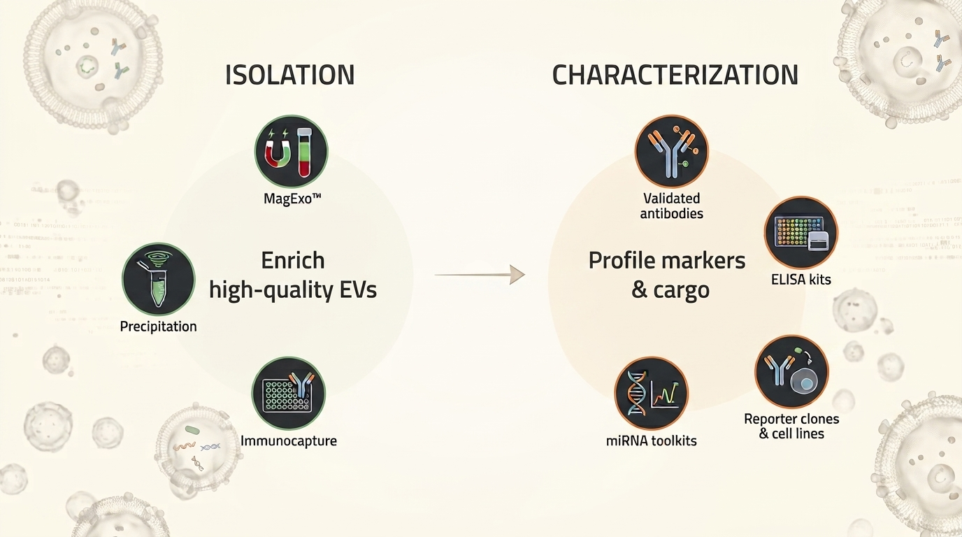
The portfolio at a glance
Two stages. One integrated workflow.
Enrich high-quality EVs
Validated across multiple sample types — choose your method based on sample input and downstream application.
- ✓ MagExo™ — total EV from serum/plasma for multi-omics & clinical utilities
- ✓ Precipitation — fast total EV recovery from cell culture media
- ✓ Immunocapture — marker-specific EV subpopulation enrichment
Profile markers & cargo
Four validated tool families to confirm EV identity, quantify subpopulations, and study functional cargo.
- ✓ Validated antibodies — surface markers & disease targets
- ✓ ELISA kits — quantify EV subpopulations from biofluids
- ✓ miRNA toolkits — expression profiling & target validation
- ✓ Reporter clones & cell lines — visualize EV biogenesis (coming soon!)
Profile markers & cargo
Four validated tool families to confirm EV identity, quantify subpopulations, and study functional cargo.
- ✓ Validated antibodies — surface markers & disease targets
- ✓ ELISA kits — quantify EV subpopulations from biofluids
- ✓ miRNA toolkits — expression profiling & target validation
- ✓ Reporter clones & cell lines — visualize EV biogenesis (coming soon!)
Stage 01 · Isolation
Recover EVs your way — no ultracentrifuge required
From total-EV recovery to targeted subpopulation enrichment, pick the method that fits your sample and your downstream assay.
Magnetic Bead · Total EV
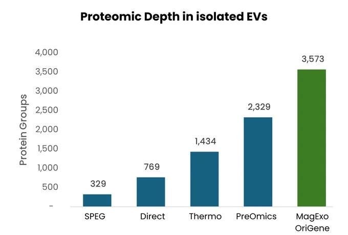
MagExo™ EV Isolation Kit
Total EV isolation directly from serum and plasma, engineered for downstream EV multi-omics. Delivers clean, intact vesicles ready for proteomics, RNA/miRNA, and LC/MS workflows.
Polymer Precipitation
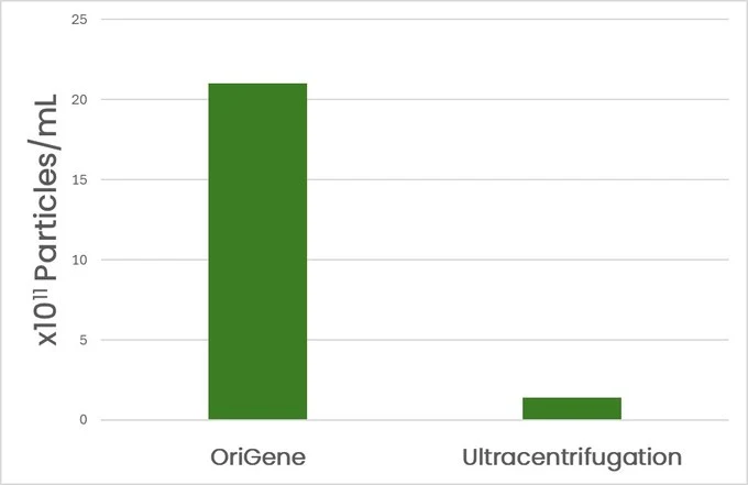
EV Isolation Reagent
A powerful alternative to ultracentrifugation and chromatography. Recovers more EVs than ultracentrifugation from cell culture media with a simple mix → incubate → resuspend protocol.
Magnetic Immunocapture
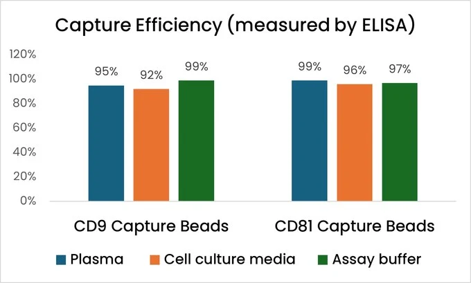
EV/Exosome Immunocapture Kits
High-affinity antibodies on magnetic beads enrich specific EV subpopulations with >90% capture efficiency and low protein carryover — cleaner preps for sensitive downstream assays.
Magnetic Bead · Total EV
MagExo™ EV Isolation Kit
Total EV isolation directly from serum and plasma, engineered for downstream EV multi-omics. Delivers clean, intact vesicles ready for proteomics, RNA/miRNA, and LC/MS workflows.
Polymer Precipitation
EV Isolation Reagent
A powerful alternative to ultracentrifugation and chromatography. Recovers more EVs than ultracentrifugation from cell culture media with a simple mix → incubate → resuspend protocol.
Magnetic Immunocapture
EV/Exosome Immunocapture Kits
High-affinity antibodies on magnetic beads enrich specific EV subpopulations with >90% capture efficiency and low protein carryover — cleaner preps for sensitive downstream assays.
Stage 02 · Characterization
Profile markers & cargo
After enrichment, confirm EV identity, measure subpopulation abundance, and study functional cargo with four validated tool families — all designed to work with OriGene isolation products.
Validated EV Antibodies
Antibodies validated for EV surface markers (CD9, CD63, CD81) and disease-associated cargo proteins — enabling identity confirmation and target-specific profiling across oncology, neurology, and immunology.
EV ELISA Kits
Quantify CD9+ and CD81+ EV subpopulations directly from native biofluids — no prior isolation required.
miRNA Toolkits
A complete miRNA analysis toolkit for EV cargo studies — sensitive qPCR detection assays for expression profiling and 3'UTR reporter vectors for functional miRNA target validation.
Reporter Clones & Cell Lines
Fluorescently tagged EV marker constructs and stable reporter cell lines for direct visualization of EV biogenesis, secretion, and cellular uptake — for mechanistic studies in vitro and in vivo.
Validated EV Antibodies
Antibodies validated for EV surface markers (CD9, CD63, CD81) and disease-associated cargo proteins — enabling identity confirmation and target-specific profiling across oncology, neurology, and immunology.
EV ELISA Kits
Quantify CD9+ and CD81+ EV subpopulations directly from native biofluids — no prior isolation required. Paired recombinant protein standards support custom ELISA development and inter-assay standardization.
miRNA Toolkits
A complete miRNA analysis toolkit for EV cargo studies — sensitive qPCR detection assays for expression profiling and 3'UTR reporter vectors for functional miRNA target validation.
Reporter Clones & Cell Lines
Fluorescently tagged EV marker constructs and stable reporter cell lines for direct visualization of EV biogenesis, secretion, and cellular uptake — for mechanistic studies in vitro and in vivo.
Method selector
Match your sample to the right isolation method
Choose based on your starting material, application, and downstream workflow.
| Criteria | MagExo™ ★ | EV Isolation Reagent | Immunocapture Kits |
|---|---|---|---|
| Best for | Clinical biofluids · assay development | High-yield total EVs (cell culture) | Targeted EV subpopulations |
| Validated sample types | Human serum, plasma | Cell culture media | Biofluids & cell culture media |
| EV population recovered | Total EVs, omics-grade | Total EVs | Marker-specific (CD9+, CD81+) |
| Equipment needed | Magnetic stand | None — bench protocol | Magnetic stand |
| Downstream applications | Proteomics · RNA · LC-MS · assay dev | WB, ELISA, molecular profiling | Biomarker assays, clean preps |
| Clinical suitability | Clinical biomarker & IVD research | Research use | Research use |
| Explore MagExo™ → | Learn more → | Learn more → |
Ready to start your EV project?
Our technical team can help you select the right isolation method, confirm sample compatibility, scope an assay development workflow, or arrange a bulk or custom order.
Ready to start your EV project?
Our technical team can help you select the right isolation method, confirm sample compatibility, scope an assay development workflow, or arrange a bulk or custom order.
Tell us about your project
Ready to start your EV project?
Our technical team can help you select the right isolation method, confirm sample compatibility, scope an assay development workflow, or arrange a bulk or custom order.
Our technical team typically responds within 1–2 business days.
Ready to start your EV project?
Our technical team can help you select the right isolation method, confirm sample compatibility, scope an assay development workflow, or arrange a bulk or custom order.
