Neuroscience

Items 1-50 of 2986

Set Ascending Direction
  • Applications
    Reactivities
    Conjugation
    Size
    Delivery Time
  • Transglutaminase 2 (TGM2) Mouse Monoclonal Antibody (Biotin conjugated) [Clone ID: OTI4H5]

    HEK293T cells were transfected with the pCMV6-ENTRY control (Cat# PS100001, Left lane) or pCMV6-ENTRY TGM2 (Cat# RC217353, Right lane) cDNA for 48 hrs and lysed. Equivalent amounts of cell lysates (5 ug per lane) were separated by SDS-PAGE and immunoblotted with anti-TGM2 (Cat# TA809245). Positive lysates LY401458 (100ug) and LC401458 (20ug) can be purchased separately from OriGene.

    HEK293T cells were transfected with the pCMV6-ENTRY control (Cat# PS100001, Left lane) or pCMV6-ENTRY TGM2 (Cat# RC217353, Right lane) cDNA for 48 hrs and lysed. Equivalent amounts of cell lysates (5 ug per lane) were separated by SDS-PAGE and immunoblotted with anti-TGM2 (Cat# TA809245). Positive lysates LY401458 (100ug) and LC401458 (20ug) can be purchased separately from OriGene.
    Transglutaminase 2 (TGM2) Mouse Monoclonal Antibody (Biotin conjugated) [Clone ID: OTI4H5]
    TGM2 mouse monoclonal antibody,clone OTI4H5, Biotinylated.
    IHC, WB
    Human
    Biotin
    100 ul
    $509.00
    2 Weeks*
  • Clathrin light chain (CLTA) Mouse Monoclonal Antibody (Biotin conjugated) [Clone ID: OTI2F7]

    HEK293T cells were transfected with the pCMV6-ENTRY control (Left lane) or pCMV6-ENTRY CLTA (RC203505, Right lane) cDNA for 48 hrs and lysed. Equivalent amounts of cell lysates (5 ug per lane) were separated by SDS-PAGE and immunoblotted with anti-CLTA (1:2000). Positive lysates LY400696 (100ug) and LC400696 (20ug) can be purchased separately from OriGene.

    HEK293T cells were transfected with the pCMV6-ENTRY control (Left lane) or pCMV6-ENTRY CLTA (RC203505, Right lane) cDNA for 48 hrs and lysed. Equivalent amounts of cell lysates (5 ug per lane) were separated by SDS-PAGE and immunoblotted with anti-CLTA (1:2000). Positive lysates LY400696 (100ug) and LC400696 (20ug) can be purchased separately from OriGene.
    Clathrin light chain (CLTA) Mouse Monoclonal Antibody (Biotin conjugated) [Clone ID: OTI2F7]
    CLTA mouse monoclonal antibody,clone OTI2F7, Biotinylated.
    IHC, WB
    Human, Mouse, Rat
    Biotin
    100 ul
    $509.00
    2 Weeks*
  • BAX Rabbit Polyclonal Antibody

    HEK293T cells were transfected with the pCMV6-ENTRY control (Left lane) or pCMV6-ENTRY BAX (RC204369, Right lane) cDNA for 48 hrs and lysed. Equivalent amounts of cell lysates (5 ug per lane) were separated by SDS-PAGE and immunoblotted with Rabbit polycl. Positive lysates LY408504 (100ug) and LC408504 (20ug) can be purchased separately from OriGene.

    HEK293T cells were transfected with the pCMV6-ENTRY control (Left lane) or pCMV6-ENTRY BAX (RC204369, Right lane) cDNA for 48 hrs and lysed. Equivalent amounts of cell lysates (5 ug per lane) were separated by SDS-PAGE and immunoblotted with Rabbit polycl. Positive lysates LY408504 (100ug) and LC408504 (20ug) can be purchased separately from OriGene.
    BAX Rabbit Polyclonal Antibody 3 Citations
    Rabbit polyclonal BAX Antibody.
    IHC, WB
    Human, Mouse, Rat
    Unconjugated
    30 ul
    $188.00
    In Stock*
  • GAPDH Mouse Monoclonal Antibody [Clone ID: OTI2D9]

    Simple Western™ analysis of endogenous protein GAPDH from HEK293T lysates (0.2 mg/mL) using GAPDH Mouse Monoclonal Antibody #TA802519. The virtual lane view (left) shows the target (as indicated) at 1:50 dilution of primary antibody. The corresponding electropherogram view (right) plots chemiluminescence by molecular weight along the capillary at a 1:50 dilution of primary antibody. This experiment was performed under reducing conditions on the Jess™ Simple Western instrument from ProteinSimple, a Bio-Techne brand, using the 12–230 kDa Separation Module.

    Simple Western™ analysis of endogenous protein GAPDH from HEK293T lysates (0.2 mg/mL) using GAPDH Mouse Monoclonal Antibody #TA802519. The virtual lane view (left) shows the target (as indicated) at 1:50 dilution of primary antibody. The corresponding electropherogram view (right) plots chemiluminescence by molecular weight along the capillary at a 1:50  dilution of primary antibody. This experiment was performed under reducing conditions on the Jess™ Simple Western instrument from ProteinSimple, a Bio-Techne brand, using the 12–230 kDa Separation Module.
    GAPDH Mouse Monoclonal Antibody [Clone ID: OTI2D9] 154 Citations
    Mouse monoclonal anti-GAPDH antibody, clone OTI2D9 (formerly 2D9), loading control.
    Simple Western, WB
    Dog, Human, Monkey, Mouse, Rat
    Unconjugated
    100 ul
    $447.00
    In Stock*
  • PGP9.5 (UCHL1) Mouse Monoclonal Antibody [Clone ID: UMAB244]

    Immunohistochemical staining of paraffin-embedded Human colon tissue within the normal limits using anti-UCHL1 (PGP9.5) mouse monoclonal antibody. (Heat-induced epitope retrieval by 1mM EDTA in 10mM Tris buffer (pH8.5) at 120°C for 3min, UM800136) (1:600)

    Immunohistochemical staining of paraffin-embedded Human colon tissue within the normal limits using anti-UCHL1 (PGP9.5) mouse monoclonal antibody. (Heat-induced epitope retrieval by 1mM EDTA in 10mM Tris buffer (pH8.5) at 120°C for 3min, UM800136) (1:600)
    PGP9.5 (UCHL1) Mouse Monoclonal Antibody [Clone ID: UMAB244]
    UCHL1 (PGP9.
    10k-ChIP, IHC, WB
    Human, Mouse, Rat
    Unconjugated
    100 ul
    $610.00
    4 Weeks*
  • IL1 beta (IL1B) Mouse Monoclonal Antibody [Clone ID: OTI3E1]

    Figure A, Western blot analysis of overexpressed lysates(25ug per lane) from HEK293T cells transfected with empty plasmid (PS100001, NC) , human IL1B plasmid (RC202079, hIL1B), mouse IL1B plasmid (MR226719, mIL1B), rat IL1B plasmid (RR211023, rIL1B) using anti-IL1B antibody TA506443 (1:500). Figure B, Western blot analysis of the same samples as figure A with anti-DDK antibody (TA180144, 1:1000)

    Figure A, Western blot analysis of overexpressed lysates(25ug per lane)  from  HEK293T cells transfected with empty plasmid (PS100001, NC) , human IL1B plasmid (RC202079, hIL1B), mouse IL1B  plasmid (MR226719, mIL1B), rat IL1B plasmid (RR211023, rIL1B) using anti-IL1B antibody TA506443 (1:500). Figure B, Western blot analysis of  the same samples as figure A with anti-DDK antibody (TA180144, 1:1000)
    IL1 beta (IL1B) Mouse Monoclonal Antibody [Clone ID: OTI3E1] 8 Citations
    IL1B mouse monoclonal antibody, clone OTI3E1 (formerly 3E1).
    IF, IHC, WB
    Human
    Unconjugated
    100 ul
    $447.00
    In Stock*
  • p53 (TP53) Mouse Monoclonal Antibody [Clone ID: UMAB62]

    Immunohistochemical staining of paraffin-embedded Adenocarcinoma of Human breast tissue using anti-TP53 mouse monoclonal antibody. (UM500053; heat-induced epitope retrieval by 10mM citric buffer, pH6.0, 120°C for 3min)

    Immunohistochemical staining of paraffin-embedded Adenocarcinoma of Human breast tissue using anti-TP53 mouse monoclonal antibody. (UM500053; heat-induced epitope retrieval by 10mM citric buffer, pH6.0, 120°C for 3min)
    p53 (TP53) Mouse Monoclonal Antibody [Clone ID: UMAB62] 2 Citations
    Purified P53 (TP53) mouse monoclonal antibody, clone UMAB62.
    10k-ChIP, IF, IHC, WB
    Human
    Unconjugated
    100 ul
    $610.00
    In Stock*
  • GAPDH Mouse Monoclonal Antibody [Clone ID: OTI2D9]

    Simple Western™ analysis of endogenous protein GAPDH from HEK293T lysates (0.2 mg/mL) using GAPDH Mouse Monoclonal Antibody #TA802519. The virtual lane view (left) shows the target (as indicated) at 1:50 dilution of primary antibody. The corresponding electropherogram view (right) plots chemiluminescence by molecular weight along the capillary at a 1:50 dilution of primary antibody. This experiment was performed under reducing conditions on the Jess™ Simple Western instrument from ProteinSimple, a Bio-Techne brand, using the 12–230 kDa Separation Module.

    Simple Western™ analysis of endogenous protein GAPDH from HEK293T lysates (0.2 mg/mL) using GAPDH Mouse Monoclonal Antibody #TA802519. The virtual lane view (left) shows the target (as indicated) at 1:50 dilution of primary antibody. The corresponding electropherogram view (right) plots chemiluminescence by molecular weight along the capillary at a 1:50  dilution of primary antibody. This experiment was performed under reducing conditions on the Jess™ Simple Western instrument from ProteinSimple, a Bio-Techne brand, using the 12–230 kDa Separation Module.
    GAPDH Mouse Monoclonal Antibody [Clone ID: OTI2D9] 154 Citations
    Mouse monoclonal anti-GAPDH antibody, clone OTI2D9 (formerly 2D9), loading control.
    Simple Western, WB
    Dog, Human, Monkey, Mouse, Rat
    Unconjugated
    30 ul
    $200.00
    In Stock*
  • GAPDH Mouse Monoclonal Antibody (HRP conjugated) [Clone ID: OTI2D9]

    Simple Western™ analysis of endogenous protein GAPDH from HEK293T lysates (0.2 mg/mL) using GAPDH Mouse Monoclonal Antibody #TA802519. The virtual lane view (left) shows the target (as indicated) at 1:50 dilution of primary antibody. The corresponding electropherogram view (right) plots chemiluminescence by molecular weight along the capillary at a 1:50 dilution of primary antibody. This experiment was performed under reducing conditions on the Jess™ Simple Western instrument from ProteinSimple, a Bio-Techne brand, using the 12–230 kDa Separation Module.

    Simple Western™ analysis of endogenous protein GAPDH from HEK293T lysates (0.2 mg/mL) using GAPDH Mouse Monoclonal Antibody #TA802519. The virtual lane view (left) shows the target (as indicated) at 1:50 dilution of primary antibody. The corresponding electropherogram view (right) plots chemiluminescence by molecular weight along the capillary at a 1:50  dilution of primary antibody. This experiment was performed under reducing conditions on the Jess™ Simple Western instrument from ProteinSimple, a Bio-Techne brand, using the 12–230 kDa Separation Module.
    GAPDH Mouse Monoclonal Antibody (HRP conjugated) [Clone ID: OTI2D9] 154 Citations
    Mouse monoclonal anti-GAPDH antibody HRP conjugated, clone OTI2D9, Loading control.
    Simple Western, WB
    Dog, Human, Monkey, Mouse, Rat
    HRP
    100 ul
    $509.00
    In Stock*
  • CD10 (MME) Mouse Monoclonal Antibody [Clone ID: UMAB235]

    Immunohistochemical staining of paraffin-embedded Human breast tissue within the normal limits using anti-MME (CD10) mouse monoclonal antibody. (Heat-induced epitope retrieval by 1mM EDTA in 10mM Tris buffer (pH9.0) at 120°C for 3min, UM800127) (1:600)

    Immunohistochemical staining of paraffin-embedded Human breast tissue within the normal limits using anti-MME (CD10) mouse monoclonal antibody. (Heat-induced epitope retrieval by 1mM EDTA in 10mM Tris buffer (pH9.0) at 120°C for 3min, UM800127) (1:600)
    CD10 (MME) Mouse Monoclonal Antibody [Clone ID: UMAB235] 3 Citations
    MME/CD10 mouse monoclonal antibody, clone UMAB235.
    FC, IHC, WB
    Human, Mouse, Rat
    Unconjugated
    100 ul
    $610.00
    In Stock*
  • Prolactin (PRL) Mouse Monoclonal Antibody [Clone ID: OTI6B1]

    Figure A, Western blot analysis of overexpressed lysates (25ug per lane) from HEK293T cells transfected with empty plasmid (PS100001, NC), human Prolactin plasmid (RC205627, hProlactin), mouse Prolactin plasmid (MR227036, mProlactin) using anti-Prolactin antibody TA500719 (1:20000@1mg/ml). Figure B, Western blot analysis of the same samples as figure A with anti-DDK antibody (TA180144, 1:10000@1mg/ml)

    Figure A, Western blot analysis of overexpressed lysates (25ug per lane) from HEK293T cells transfected with empty plasmid (PS100001, NC), human Prolactin  plasmid (RC205627, hProlactin), mouse Prolactin plasmid (MR227036, mProlactin) using anti-Prolactin antibody TA500719 (1:20000@1mg/ml). Figure B, Western blot analysis of  the same samples as figure A with anti-DDK antibody (TA180144, 1:10000@1mg/ml)
    Prolactin (PRL) Mouse Monoclonal Antibody [Clone ID: OTI6B1] 1 Citation
    PRL (Prolactin) mouse monoclonal antibody, clone OTI6B1 (formerly 6B1).
    FC, IF, IHC, WB
    Human
    Unconjugated
    100 ul
    $447.00
    In Stock*
  • p53 (TP53) Mouse Monoclonal Antibody [Clone ID: OTI5E2]

    Figure A, Western blot analysis of overexpressed lysates (15ug per lane) from HEK293T cells transfected with empty plasmid (PS100001, NC), human TP53 plasmid (RC200003, hTP53), mouse TP53 plasmid (MR206086, mTP53), rat TP53 plasmid (RR201296, rTP53) using anti-TP53 antibody TA502870 (1:10000@1mg/ml). Figure B, Western blot analysis of the same samples as figure A with anti-DDK antibody (TA180144, 1:10000@1mg/ml)

    Figure A, Western blot analysis of overexpressed lysates (15ug per lane) from HEK293T cells transfected with empty plasmid (PS100001, NC), human TP53 plasmid (RC200003, hTP53), mouse TP53 plasmid (MR206086, mTP53), rat TP53 plasmid (RR201296, rTP53) using anti-TP53 antibody TA502870 (1:10000@1mg/ml). Figure B, Western blot analysis of the same samples as figure A with anti-DDK antibody (TA180144, 1:10000@1mg/ml)
    p53 (TP53) Mouse Monoclonal Antibody [Clone ID: OTI5E2] 5 Citations
    TP53 mouse monoclonal antibody, clone OTI5E2 (formerly 5E2).
    FC, IF, IHC, WB
    Human, Monkey, Mouse, Rat
    Unconjugated
    100 ul
    $447.00
    In Stock*
  • PGP9.5 (UCHL1) Mouse Monoclonal Antibody [Clone ID: UMAB244]

    Immunohistochemical staining of paraffin-embedded Human colon tissue within the normal limits using anti-UCHL1 (PGP9.5) mouse monoclonal antibody. (Heat-induced epitope retrieval by 1mM EDTA in 10mM Tris buffer (pH8.5) at 120°C for 3min, UM800136) (1:600)

    Immunohistochemical staining of paraffin-embedded Human colon tissue within the normal limits using anti-UCHL1 (PGP9.5) mouse monoclonal antibody. (Heat-induced epitope retrieval by 1mM EDTA in 10mM Tris buffer (pH8.5) at 120°C for 3min, UM800136) (1:600)
    PGP9.5 (UCHL1) Mouse Monoclonal Antibody [Clone ID: UMAB244]
    Carrier-free (BSA/glycerol-free) UCHL1 (PGP9.
    10k-ChIP, IHC, WB
    Human, Mouse, Rat
    Unconjugated
    100 ug
    $680.00
    In Stock*
  • VMAT2 (SLC18A2) Mouse Monoclonal Antibody [Clone ID: OTI9E11]

    Figure A, Western blot analysis of overexpressed lysates (5ug per lane) from HEK293T cells transfected with empty plasmid (PS100001, NC), human VMAT2 plasmid (RC221342, hVMAT2) using anti-VMAT2 antibody TA500506 (1:20000@1mg/ml). Figure B, Western blot analysis of the same samples as figure A with anti-DDK antibody (TA180144, 1:10000@1mg/ml)

    Figure A, Western blot analysis of overexpressed lysates (5ug per lane) from HEK293T cells transfected with empty plasmid (PS100001, NC), human VMAT2 plasmid (RC221342, hVMAT2) using anti-VMAT2 antibody TA500506 (1:20000@1mg/ml). Figure B, Western blot analysis of the same samples as figure A with anti-DDK antibody (TA180144, 1:10000@1mg/ml)
    VMAT2 (SLC18A2) Mouse Monoclonal Antibody [Clone ID: OTI9E11] 6 Citations
    Anti-SLC18A2 (VMAT2) mouse monoclonal antibody, clone OTI9E11 (formerly 9E11).
    FC, IF, IHC, WB
    Human
    Unconjugated
    100 ul
    $447.00
    In Stock*
  • ERK2 (MAPK1) Mouse Monoclonal Antibody [Clone ID: OTI6F8]

    HEK293T cells were transfected with the pCMV6-ENTRY control (Cat# PS100001, Left lane) or pCMV6-ENTRY MAPK1 (Cat# RC204703, Right lane) cDNA for 48 hrs and lysed. Equivalent amounts of cell lysates (5 ug per lane) were separated by SDS-PAGE and immunoblotted with anti-MAPK1 (Cat# TA500468). Positive lysates LY408481 (100ug) and LC408481 (20ug) can be purchased separately from OriGene.

    HEK293T cells were transfected with the pCMV6-ENTRY control (Cat# PS100001, Left lane) or pCMV6-ENTRY MAPK1 (Cat# RC204703, Right lane) cDNA for 48 hrs and lysed. Equivalent amounts of cell lysates (5 ug per lane) were separated by SDS-PAGE and immunoblotted with anti-MAPK1 (Cat# TA500468). Positive lysates LY408481 (100ug) and LC408481 (20ug) can be purchased separately from OriGene.
    ERK2 (MAPK1) Mouse Monoclonal Antibody [Clone ID: OTI6F8] 5 Citations
    Anti-MAPK1 (ERK2) mouse monoclonal antibody, clone OTI6F8 (formerly 6F8).
    FC, IHC, IP, WB
    Dog, Human, Monkey, Mouse, Rat
    Unconjugated
    100 ul
    $447.00
    In Stock*
  • CD10 (MME) Mouse Monoclonal Antibody [Clone ID: UMAB235]

    Immunohistochemical staining of paraffin-embedded Human breast tissue within the normal limits using anti-MME (CD10) mouse monoclonal antibody. (Heat-induced epitope retrieval by 1mM EDTA in 10mM Tris buffer (pH9.0) at 120°C for 3min, UM800127) (1:600)

    Immunohistochemical staining of paraffin-embedded Human breast tissue within the normal limits using anti-MME (CD10) mouse monoclonal antibody. (Heat-induced epitope retrieval by 1mM EDTA in 10mM Tris buffer (pH9.0) at 120°C for 3min, UM800127) (1:600)
    CD10 (MME) Mouse Monoclonal Antibody [Clone ID: UMAB235] 3 Citations
    Carrier-free (BSA/glycerol-free) MME/CD10 mouse monoclonal antibody, clone UMAB235.
    FC, IHC, WB
    Human, Mouse, Rat
    Unconjugated
    100 ug
    $680.00
    3 Days*
  • NDUFB10 Mouse Monoclonal Antibody [Clone ID: OTI4F1]

    HEK293T cells were transfected with the pCMV6-ENTRY control (Left lane) or pCMV6-ENTRY NDUFB10 (RC200526, Right lane) cDNA for 48 hrs and lysed. Equivalent amounts of cell lysates (5 ug per lane) were separated by SDS-PAGE and immunoblotted with anti-NDUFB10 (TA505207). Positive lysates LY417917 (100ug) and LC417917 (20ug) can be purchased separately from OriGene.

    HEK293T cells were transfected with the pCMV6-ENTRY control (Left lane) or pCMV6-ENTRY NDUFB10 (RC200526, Right lane) cDNA for 48 hrs and lysed. Equivalent amounts of cell lysates (5 ug per lane) were separated by SDS-PAGE and immunoblotted with anti-NDUFB10 (TA505207). Positive lysates LY417917 (100ug) and LC417917 (20ug) can be purchased separately from OriGene.
    NDUFB10 Mouse Monoclonal Antibody [Clone ID: OTI4F1]
    NDUFB10 mouse monoclonal antibody, clone OTI4F1 (formerly 4F1).
    IF, IHC, WB
    Dog, Human, Monkey, Mouse, Rat
    Unconjugated
    100 ul
    $447.00
    In Stock*
  • BID Mouse Monoclonal Antibody [Clone ID: UMAB139]

    Immunohistochemical staining of paraffin-embedded Adenocarcinoma of Human breast tissue using anti-BID mouse monoclonal antibody. (UM500101, heat-induced epitope retrieval by 1mM EDTA in 10mM Tris, pH8.0, 120°C for 3min)

    Immunohistochemical staining of paraffin-embedded Adenocarcinoma of Human breast tissue using anti-BID mouse monoclonal antibody. (UM500101, heat-induced epitope retrieval by 1mM EDTA in 10mM Tris, pH8.0, 120°C for 3min)
    BID Mouse Monoclonal Antibody [Clone ID: UMAB139] 1 Citation
    BID mouse monoclonal antibody,clone UMAB139.
    IHC, WB
    Human
    Unconjugated
    100 ul
    $610.00
    In Stock*
  • NDUFB10 Mouse Monoclonal Antibody [Clone ID: OTI1H6]

    Figure A, Western blot analysis of overexpressed lysates (15ug per lane) from HEK293T cells transfected with empty plasmid (PS100001, NC), human NDUFB10 plasmid (RC200526, hNDUFB10), mouse NDUFB10 plasmid (MR201544, mNDUFB10) using anti-NDUFB10 antibody TA505113 (1:10000@1mg/ml). Figure B, Western blot analysis of the same human NDUFB10 and mouse NDUFB10 samples as figure A with anti-DDK antibody (TA180144, 1:10000@1mg/ml)

    Figure A, Western blot analysis of overexpressed lysates (15ug per lane) from HEK293T cells transfected with empty plasmid (PS100001, NC), human NDUFB10 plasmid (RC200526, hNDUFB10), mouse NDUFB10 plasmid (MR201544, mNDUFB10) using anti-NDUFB10 antibody TA505113 (1:10000@1mg/ml). Figure B, Western blot analysis of the same human NDUFB10 and mouse NDUFB10 samples as figure A with anti-DDK antibody (TA180144, 1:10000@1mg/ml)
    NDUFB10 Mouse Monoclonal Antibody [Clone ID: OTI1H6]
    Carrier-free (BSA/glycerol-free) NDUFB10 mouse monoclonal antibody, clone OTI1H6 (formerly 1H6).
    FC, IHC, WB
    Human, Mouse, Rat
    Unconjugated
    100 ug
    $620.00
    3 Days*
  • Ubiquinol Cytochrome C Reductase Core Protein I (UQCRC1) Mouse Monoclonal Antibody [Clone ID: OTI1G6]

    Equivalent amounts of cell lysates (30 ug per lane) of wild-type HeLa cells(WT) and UQCRC1-Knockdown HeLa cells(KD) were separated by SDS-PAGE and immunoblotted with anti-UQCRC1 monoclonal antibody TA800089(1:2000).Then the blotted membrane was stripped and reprobed with anti-HSP90AA1 antibody as a loading control.

    Equivalent amounts of  cell lysates (30 ug per lane) of  wild-type HeLa cells(WT) and UQCRC1-Knockdown HeLa cells(KD)  were separated by SDS-PAGE and immunoblotted with anti-UQCRC1 monoclonal antibody TA800089(1:2000).Then the blotted membrane was stripped and reprobed with anti-HSP90AA1 antibody as a loading control.
    Ubiquinol Cytochrome C Reductase Core Protein I (UQCRC1) Mouse Monoclonal Antibody [Clone ID: OTI1G6] 1 Citation
    UQCRC1 mouse monoclonal antibody, clone OTI1G6 (formerly 1G6).
    FC, IHC, WB
    Dog, Human, Monkey, Mouse, Rat
    Unconjugated
    100 ul
    $447.00
    In Stock*
  • SDHB Mouse Monoclonal Antibody [Clone ID: OTI3F5]

    Simple Western™ analysis of endogenous protein SDHB from HEK293T lysates (0.2 mg/mL) using SDHB Mouse Monoclonal Antibody #TA808102. The virtual lane view (left) shows the target (as indicated) at 1:50 dilution of primary antibody. The corresponding electropherogram view (right) plots chemiluminescence by molecular weight along the capillary at a 1:50 dilution of primary antibody. This experiment was performed under reducing conditions on the Jess™ Simple Western instrument from ProteinSimple, a Bio-Techne brand, using the 12–230 kDa Separation Module.

    Simple Western™ analysis of endogenous protein SDHB from HEK293T lysates (0.2 mg/mL) using SDHB Mouse Monoclonal Antibody #TA808102. The virtual lane view (left) shows the target (as indicated) at 1:50 dilution of primary antibody. The corresponding electropherogram view (right) plots chemiluminescence by molecular weight along the capillary at a 1:50  dilution of primary antibody. This experiment was performed under reducing conditions on the Jess™ Simple Western instrument from ProteinSimple, a Bio-Techne brand, using the 12–230 kDa Separation Module.
    SDHB Mouse Monoclonal Antibody [Clone ID: OTI3F5] 1 Citation
    SDHB mouse monoclonal antibody, clone OTI3F5 (formerly 3F5).
    IHC, Simple Western, WB
    Human, Mouse, Rat
    Unconjugated
    100 ul
    $447.00
    In Stock*
  • POLR2E Mouse Monoclonal Antibody [Clone ID: OTI3B5]

    HEK293T cells were transfected with the pCMV6-ENTRY control (Cat# PS100001, Left lane) or pCMV6-ENTRY POLR2E (Cat# RC201266, Right lane) cDNA for 48 hrs and lysed. Equivalent amounts of cell lysates (5 ug per lane) were separated by SDS-PAGE and immunoblotted with anti-POLR2E (Cat# TA502548). Positive lysates LY419161 (100ug) and LC419161 (20ug) can be purchased separately from OriGene.

    HEK293T cells were transfected with the pCMV6-ENTRY control (Cat# PS100001, Left lane) or pCMV6-ENTRY POLR2E (Cat# RC201266, Right lane) cDNA for 48 hrs and lysed. Equivalent amounts of cell lysates (5 ug per lane) were separated by SDS-PAGE and immunoblotted with anti-POLR2E (Cat# TA502548). Positive lysates LY419161 (100ug) and LC419161 (20ug) can be purchased separately from OriGene.
    POLR2E Mouse Monoclonal Antibody [Clone ID: OTI3B5]
    POLR2E mouse monoclonal antibody, clone OTI3B5 (formerly 3B5).
    FC, IF, IHC, WB
    Dog, Human, Monkey, Mouse, Rat
    Unconjugated
    100 ul
    $447.00
    In Stock*
  • UBC6e (UBE2J1) Mouse Monoclonal Antibody [Clone ID: OTI3D1]

    HEK293T cells were transfected with the pCMV6-ENTRY control (Cat# PS100001, Left lane) or pCMV6-ENTRY UBE2J1 (Cat# RC202826, Right lane) cDNA for 48 hrs and lysed. Equivalent amounts of cell lysates (5 ug per lane) were separated by SDS-PAGE and immunoblotted with anti-UBE2J1 (Cat# TA504988). Positive lysates LY414230 (100ug) and LC414230 (20ug) can be purchased separately from OriGene.

    HEK293T cells were transfected with the pCMV6-ENTRY control (Cat# PS100001, Left lane) or pCMV6-ENTRY UBE2J1 (Cat# RC202826, Right lane) cDNA for 48 hrs and lysed. Equivalent amounts of cell lysates (5 ug per lane) were separated by SDS-PAGE and immunoblotted with anti-UBE2J1 (Cat# TA504988). Positive lysates LY414230 (100ug) and LC414230 (20ug) can be purchased separately from OriGene.
    UBC6e (UBE2J1) Mouse Monoclonal Antibody [Clone ID: OTI3D1] 1 Citation
    UBE2J1 mouse monoclonal antibody, clone OTI3D1 (formerly 3D1).
    FC, IHC, WB
    Dog, Human, Monkey, Mouse, Rat
    Unconjugated
    100 ul
    $447.00
    In Stock*
  • ERAB (HSD17B10) Mouse Monoclonal Antibody [Clone ID: OTI10B4]

    HEK293T cells were transfected with the pCMV6-ENTRY control (Cat# PS100001, Left lane) or pCMV6-ENTRY HSD17B10 (Cat# RC201734, Right lane) cDNA for 48 hrs and lysed. Equivalent amounts of cell lysates (5 ug per lane) were separated by SDS-PAGE and immunoblotted with anti-HSD17B10(Cat# TA500686). Positive lysates LY401426 (100ug) and LC401426 (20ug) can be purchased separately from OriGene.

    HEK293T cells were transfected with the pCMV6-ENTRY control (Cat# PS100001, Left lane) or pCMV6-ENTRY HSD17B10 (Cat# RC201734, Right lane) cDNA for 48 hrs and lysed. Equivalent amounts of cell lysates (5 ug per lane) were separated by SDS-PAGE and immunoblotted with anti-HSD17B10(Cat# TA500686). Positive lysates LY401426 (100ug) and LC401426 (20ug) can be purchased separately from OriGene.
    ERAB (HSD17B10) Mouse Monoclonal Antibody [Clone ID: OTI10B4]
    Anti-HSD17B10 mouse monoclonal antibody, clone OTI10B4 (formerly 10B4).
    FC, IF, IHC, WB
    Human, Mouse, Rat
    Unconjugated
    100 ul
    $447.00
    In Stock*
  • ERK2 (MAPK1) Mouse Monoclonal Antibody [Clone ID: OTI6E5]

    HEK293T cells were transfected with the pCMV6-ENTRY control (Left lane) or pCMV6-ENTRY MAPK1 (RC204703, Right lane) cDNA for 48 hrs and lysed. Equivalent amounts of cell lysates (5 ug per lane) were separated by SDS-PAGE and immunoblotted with anti-MAPK1. Positive lysates LY408481 (100ug) and LC408481 (20ug) can be purchased separately from OriGene.

    HEK293T cells were transfected with the pCMV6-ENTRY control (Left lane) or pCMV6-ENTRY MAPK1 (RC204703, Right lane) cDNA for 48 hrs and lysed. Equivalent amounts of cell lysates (5 ug per lane) were separated by SDS-PAGE and immunoblotted with anti-MAPK1. Positive lysates LY408481 (100ug) and LC408481 (20ug) can be purchased separately from OriGene.
    ERK2 (MAPK1) Mouse Monoclonal Antibody [Clone ID: OTI6E5]
    Anti-MAPK1 (ERK2) mouse monoclonal antibody, clone OTI6E5 (formerly 6E5).
    IHC, IP, WB
    Dog, Human, Monkey, Mouse, Rat
    Unconjugated
    100 ul
    $447.00
    In Stock*
  • NDUFS2 Mouse Monoclonal Antibody [Clone ID: OTI1D5]

    HEK293T cells were transfected with the pCMV6-ENTRY control (Cat# PS100001, Left lane) or pCMV6-ENTRY NDUFS2 (Cat# RC203485, Right lane) cDNA for 48 hrs and lysed. Equivalent amounts of cell lysates (5 ug per lane) were separated by SDS-PAGE and immunoblotted with anti-NDUFS2 (Cat# TA802340). Positive lysates LY417919 (100ug) and LC417919 (20ug) can be purchased separately from OriGene.

    HEK293T cells were transfected with the pCMV6-ENTRY control (Cat# PS100001, Left lane) or pCMV6-ENTRY NDUFS2 (Cat# RC203485, Right lane) cDNA for 48 hrs and lysed. Equivalent amounts of cell lysates (5 ug per lane) were separated by SDS-PAGE and immunoblotted with anti-NDUFS2 (Cat# TA802340). Positive lysates LY417919 (100ug) and LC417919 (20ug) can be purchased separately from OriGene.
    NDUFS2 Mouse Monoclonal Antibody [Clone ID: OTI1D5]
    NDUFS2 mouse monoclonal antibody,clone OTI1D5.
    IHC, WB
    Human
    Unconjugated
    100 ul
    $447.00
    In Stock*
  • Lipoprotein lipase (LPL) Mouse Monoclonal Antibody [Clone ID: OTI3A10]

    HEK293T cells were transfected with the pCMV6-ENTRY control (Cat# PS100001, Left lane) or pCMV6-ENTRY LPL (Cat# RC203766, Right lane) cDNA for 48 hrs and lysed. Equivalent amounts of cell lysates (5 ug per lane) were separated by SDS-PAGE and immunoblotted with anti-LPL (Cat# TA503793). Positive lysates LY400089 (100ug) and LC400089 (20ug) can be purchased separately from OriGene.

    HEK293T cells were transfected with the pCMV6-ENTRY control (Cat# PS100001, Left lane) or pCMV6-ENTRY LPL (Cat# RC203766, Right lane) cDNA for 48 hrs and lysed. Equivalent amounts of cell lysates (5 ug per lane) were separated by SDS-PAGE and immunoblotted with anti-LPL (Cat# TA503793). Positive lysates LY400089 (100ug) and LC400089 (20ug) can be purchased separately from OriGene.
    Lipoprotein lipase (LPL) Mouse Monoclonal Antibody [Clone ID: OTI3A10] 1 Citation
    LPL (Lipoprotein lipase) mouse monoclonal antibody, clone OTI3A10 (formerly 3A10).
    FC, IHC, WB
    Human, Mouse, Rat
    Unconjugated
    100 ul
    $447.00
    In Stock*
  • CD10 (MME) Mouse Monoclonal Antibody [Clone ID: OTI3D11]

    HEK293T cells were transfected with the pCMV6-ENTRY control (Cat# PS100001, Left lane) or pCMV6-ENTRY MME (Cat# RC223013, Right lane) cDNA for 48 hrs and lysed. Equivalent amounts of cell lysates (5 ug per lane) were separated by SDS-PAGE and immunoblotted with anti-MME (Cat# TA504507)(1:500).

    HEK293T cells were transfected with the pCMV6-ENTRY control (Cat# PS100001, Left lane) or pCMV6-ENTRY MME (Cat# RC223013, Right lane) cDNA for 48 hrs and lysed. Equivalent amounts of cell lysates (5 ug per lane) were separated by SDS-PAGE and immunoblotted with anti-MME (Cat# TA504507)(1:500).
    CD10 (MME) Mouse Monoclonal Antibody [Clone ID: OTI3D11] 1 Citation
    MME (CD10) mouse monoclonal antibody, clone OTI3D11 (formerly 3D11).
    FC, WB
    Human, Mouse, Rat
    Unconjugated
    100 ul
    $447.00
    In Stock*
  • ATP5F1B Mouse Monoclonal Antibody [Clone ID: OTI1F7]

    HEK293T cells were transfected with the pCMV6-ENTRY control (Left lane) or pCMV6-ENTRY ATP5B (RC201638, Right lane) cDNA for 48 hrs and lysed. Equivalent amounts of cell lysates (5 ug per lane) were separated by SDS-PAGE and immunoblotted with anti-ATP5B. Positive lysates LY400637 (100ug) and LC400637 (20ug) can be purchased separately from OriGene.

    HEK293T cells were transfected with the pCMV6-ENTRY control (Left lane) or pCMV6-ENTRY ATP5B (RC201638, Right lane) cDNA for 48 hrs and lysed. Equivalent amounts of cell lysates (5 ug per lane) were separated by SDS-PAGE and immunoblotted with anti-ATP5B. Positive lysates LY400637 (100ug) and LC400637 (20ug) can be purchased separately from OriGene.
    ATP5F1B Mouse Monoclonal Antibody [Clone ID: OTI1F7]
    Anti-ATP5B mouse monoclonal antibody, clone OTI1F7 (formerly 1F7).
    FC, IF, IHC, WB
    Dog, Human, Monkey
    Unconjugated
    100 ul
    $447.00
    In Stock*
  • CD10 (MME) Mouse Monoclonal Antibody [Clone ID: UMAB236]

    Immunohistochemical staining of paraffin-embedded Human breast tissue within the normal limits using anti-MME (CD10) mouse monoclonal antibody. (Heat-induced epitope retrieval by 1mM EDTA in 10mM Tris buffer (pH9.0) at 120°C for 3min, UM800128) (1:600)

    Immunohistochemical staining of paraffin-embedded Human breast tissue within the normal limits using anti-MME (CD10) mouse monoclonal antibody. (Heat-induced epitope retrieval by 1mM EDTA in 10mM Tris buffer (pH9.0) at 120°C for 3min, UM800128) (1:600)
    CD10 (MME) Mouse Monoclonal Antibody [Clone ID: UMAB236] 2 Citations
    Carrier-free (BSA/glycerol-free) MME/CD10 mouse monoclonal antibody, clone UMAB236.
    10k-ChIP, FC, IHC
    Human, Mouse, Rat
    Unconjugated
    100 ug
    $680.00
    3 Days*
  • Prolactin (PRL) Mouse Monoclonal Antibody [Clone ID: OTI6B1]

    Figure A, Western blot analysis of overexpressed lysates (25ug per lane) from HEK293T cells transfected with empty plasmid (PS100001, NC), human Prolactin plasmid (RC205627, hProlactin), mouse Prolactin plasmid (MR227036, mProlactin) using anti-Prolactin antibody TA500719 (1:20000@1mg/ml). Figure B, Western blot analysis of the same samples as figure A with anti-DDK antibody (TA180144, 1:10000@1mg/ml)

    Figure A, Western blot analysis of overexpressed lysates (25ug per lane) from HEK293T cells transfected with empty plasmid (PS100001, NC), human Prolactin  plasmid (RC205627, hProlactin), mouse Prolactin plasmid (MR227036, mProlactin) using anti-Prolactin antibody TA500719 (1:20000@1mg/ml). Figure B, Western blot analysis of  the same samples as figure A with anti-DDK antibody (TA180144, 1:10000@1mg/ml)
    Prolactin (PRL) Mouse Monoclonal Antibody [Clone ID: OTI6B1] 1 Citation
    Carrier-free (BSA/glycerol-free) PRL mouse monoclonal antibody, clone OTI6B1 (formerly 6B1).
    FC, IF, IHC, WB
    Human
    Unconjugated
    100 ug
    $620.00
    3 Days*
  • CDK5 Mouse Monoclonal Antibody [Clone ID: OTI2H6]

    HEK293T cells were transfected with the pCMV6-ENTRY control (Cat# PS100001, Left lane) or pCMV6-ENTRY CDK5 (Cat# RC200342, Right lane) cDNA for 48 hrs and lysed. Equivalent amounts of cell lysates (5 ug per lane) were separated by SDS-PAGE and immunoblotted with anti-CDK5 (Cat# TA500397). Positive lysates LY401536 (100ug) and LC401536 (20ug) can be purchased separately from OriGene.

    HEK293T cells were transfected with the pCMV6-ENTRY control (Cat# PS100001, Left lane) or pCMV6-ENTRY CDK5 (Cat# RC200342, Right lane) cDNA for 48 hrs and lysed. Equivalent amounts of cell lysates (5 ug per lane) were separated by SDS-PAGE and immunoblotted with anti-CDK5 (Cat# TA500397). Positive lysates LY401536 (100ug) and LC401536 (20ug) can be purchased separately from OriGene.
    CDK5 Mouse Monoclonal Antibody [Clone ID: OTI2H6] 3 Citations
    CDK5 mouse monoclonal antibody, clone OTI2H6 (formerly 2H6).
    FC, IF, WB
    Dog, Human, Mouse, Rat
    Unconjugated
    100 ul
    $447.00
    In Stock*
  • NDUFB9 Mouse Monoclonal Antibody [Clone ID: OTI13H11]

    HEK293T cells were transfected with the pCMV6-ENTRY control (Cat# PS100001, Left lane) or pCMV6-ENTRY NDUFB9 (Cat# RC200223, Right lane) cDNA for 48 hrs and lysed. Equivalent amounts of cell lysates (5 ug per lane) were separated by SDS-PAGE and immunoblotted with anti-NDUFB9 (Cat# TA502568). Positive lysates LY417578 (100ug) and LC417578 (20ug) can be purchased separately from OriGene.

    HEK293T cells were transfected with the pCMV6-ENTRY control (Cat# PS100001, Left lane) or pCMV6-ENTRY NDUFB9 (Cat# RC200223, Right lane) cDNA for 48 hrs and lysed. Equivalent amounts of cell lysates (5 ug per lane) were separated by SDS-PAGE and immunoblotted with anti-NDUFB9 (Cat# TA502568). Positive lysates LY417578 (100ug) and LC417578 (20ug) can be purchased separately from OriGene.
    NDUFB9 Mouse Monoclonal Antibody [Clone ID: OTI13H11]
    NDUFB9 mouse monoclonal antibody, clone OTI13H11 (formerly 13H11).
    FC, IHC, WB
    Human, Mouse, Rat
    Unconjugated
    100 ul
    $447.00
    In Stock*
  • EDG2 (LPAR1) Mouse Monoclonal Antibody [Clone ID: OTI1G6]

    Figure A, Western blot analysis of overexpressed lysates(25ug per lane) from HEK293T cells transfected with empty plasmid (PS100001, lane 1) , human LPAR1 plasmid (RC206065, lane 2), mouse LPAR1 plasmid (MR205601, lane 3), rat LPAR1 plasmid (RR210163, lane 4) using anti-LPAR1 antibody TA503737 (1:500). Figure B, Western blot analysis of the same samples as figure A with anti-DDK antibody (TA180144, 1:1000)

    Figure A, Western blot analysis of overexpressed lysates(25ug per lane)  from  HEK293T cells transfected with empty plasmid (PS100001, lane 1) , human LPAR1 plasmid (RC206065, lane 2), mouse LPAR1 plasmid (MR205601, lane 3), rat LPAR1 plasmid (RR210163, lane 4) using anti-LPAR1 antibody TA503737 (1:500). Figure B, Western blot analysis of  the same samples as figure A with anti-DDK antibody (TA180144, 1:1000)
    EDG2 (LPAR1) Mouse Monoclonal Antibody [Clone ID: OTI1G6] 1 Citation
    LPAR1 (EDG2) mouse monoclonal antibody, clone OTI1G6 (formerly 1G6).
    FC, IF, WB
    Human, Mouse, Rat
    Unconjugated
    100 ul
    $447.00
    In Stock*
  • GAPDH Mouse Monoclonal Antibody [Clone ID: OTI2F8]

    HEK293T cells were transfected with the pCMV6-ENTRY control (Cat# PS100001, Left lane) or pCMV6-ENTRY GAPDH (Cat# RC202309, Right lane) cDNA for 48 hrs and lysed. Equivalent amounts of cell lysates (5 ug per lane) were separated by SDS-PAGE and immunoblotted with anti-GAPDH (Cat# TA802563). Positive lysates LY419567 (100ug) and LC419567 (20ug) can be purchased separately from OriGene.

    HEK293T cells were transfected with the pCMV6-ENTRY control (Cat# PS100001, Left lane) or pCMV6-ENTRY GAPDH (Cat# RC202309, Right lane) cDNA for 48 hrs and lysed. Equivalent amounts of cell lysates (5 ug per lane) were separated by SDS-PAGE and immunoblotted with anti-GAPDH (Cat# TA802563). Positive lysates LY419567 (100ug) and LC419567 (20ug) can be purchased separately from OriGene.
    GAPDH Mouse Monoclonal Antibody [Clone ID: OTI2F8] 3 Citations
    GAPDH mouse monoclonal antibody, clone OTI2F8 (formerly 2F8).
    IHC, WB
    Human, Mouse, Rat
    Unconjugated
    100 ul
    $447.00
    In Stock*
  • Superoxide Dismutase 1 (SOD1) Mouse Monoclonal Antibody [Clone ID: OTI8B10]

    Immunohistochemical staining of paraffin-embedded Adenocarcinoma of Human breast tissue using anti-SOD1 mouse monoclonal antibody. Heat-induced epitope retrieval by EDTA solution buffer pH 8.0 at 120°C for 3 min.

    Immunohistochemical staining of paraffin-embedded Adenocarcinoma of Human breast tissue using anti-SOD1 mouse monoclonal antibody. Heat-induced epitope retrieval by EDTA solution buffer pH 8.0 at 120°C for 3 min.
    Superoxide Dismutase 1 (SOD1) Mouse Monoclonal Antibody [Clone ID: OTI8B10] 4 Citations
    SOD1 (Superoxide Dismutase 1) mouse monoclonal antibody, clone OTI8B10 (formerly 8B10).
    FC, IHC, WB
    Human
    Unconjugated
    100 ul
    $447.00
    In Stock*
  • Amyloid Precursor Protein (APP) Mouse Monoclonal Antibody [Clone ID: OTI2B10]

    Immunohistochemical staining of paraffin-embedded Human bladder tissue within the normal limits using anti-APP mouse monoclonal antibody. Heat-induced epitope retrieval by EDTA solution buffer pH 8.0 at 120°C for 3 min.

    Immunohistochemical staining of paraffin-embedded Human bladder tissue within the normal limits using anti-APP mouse monoclonal antibody. Heat-induced epitope retrieval by EDTA solution buffer pH 8.0 at 120°C for 3 min.
    Amyloid Precursor Protein (APP) Mouse Monoclonal Antibody [Clone ID: OTI2B10] 1 Citation
    APP (Amyloid beta precursor protein) mouse monoclonal antibody, clone OTI2B10 (formerly 2B10).
    IF, WB
    Human
    Unconjugated
    100 ul
    $447.00
    In Stock*
  • GAPDH Mouse Monoclonal Antibody (Biotin conjugated) [Clone ID: OTI2D9]

    Simple Western™ analysis of endogenous protein GAPDH from HEK293T lysates (0.2 mg/mL) using GAPDH Mouse Monoclonal Antibody #TA802519. The virtual lane view (left) shows the target (as indicated) at 1:50 dilution of primary antibody. The corresponding electropherogram view (right) plots chemiluminescence by molecular weight along the capillary at a 1:50 dilution of primary antibody. This experiment was performed under reducing conditions on the Jess™ Simple Western instrument from ProteinSimple, a Bio-Techne brand, using the 12–230 kDa Separation Module.

    Simple Western™ analysis of endogenous protein GAPDH from HEK293T lysates (0.2 mg/mL) using GAPDH Mouse Monoclonal Antibody #TA802519. The virtual lane view (left) shows the target (as indicated) at 1:50 dilution of primary antibody. The corresponding electropherogram view (right) plots chemiluminescence by molecular weight along the capillary at a 1:50  dilution of primary antibody. This experiment was performed under reducing conditions on the Jess™ Simple Western instrument from ProteinSimple, a Bio-Techne brand, using the 12–230 kDa Separation Module.
    GAPDH Mouse Monoclonal Antibody (Biotin conjugated) [Clone ID: OTI2D9] 154 Citations
    Mouse monoclonal anti-GAPDH antibody, clone OTI2D9 (formerly 2D9), loading control, Biotinylated.
    Simple Western, WB
    Dog, Human, Monkey, Mouse, Rat
    Biotin
    100 ul
    $509.00
    In Stock*
  • Amyloid Precursor Protein (APP) Mouse Monoclonal Antibody [Clone ID: OTI7G9]

    Figure A, Western blot analysis of overexpressed lysates (15ug per lane) from HEK293T cells transfected with empty plasmid (PS100001, NC), human APP plasmid (RC221339, hAPP), mouse APP plasmid (MR210090, mAPP), rat APP plasmid (RR204173, rAPP) using anti-APP antibody TA500969 (1:10000@1mg/ml). Figure B, Western blot analysis of the same samples as figure A with anti-DDK antibody (TA180144, 1:10000@1mg/ml).

    Figure A, Western blot analysis of overexpressed lysates (15ug per lane) from HEK293T cells transfected with empty plasmid (PS100001, NC), human APP  plasmid (RC221339, hAPP), mouse APP plasmid (MR210090, mAPP), rat APP plasmid (RR204173, rAPP) using anti-APP antibody TA500969 (1:10000@1mg/ml). Figure B, Western blot analysis of  the same samples as figure A with anti-DDK antibody (TA180144, 1:10000@1mg/ml).
    Amyloid Precursor Protein (APP) Mouse Monoclonal Antibody [Clone ID: OTI7G9]
    Anti-APP (Amyloid beta precursor protein) mouse monoclonal antibody, clone OTI7G9 (formerly 7G9).
    FC, IF, IHC, WB
    Human
    Unconjugated
    100 ul
    $447.00
    In Stock*
  • Tau (MAPT) Mouse Monoclonal Antibody [Clone ID: UMAB239]

    Immunohistochemical staining of paraffin-embedded Human breast tissue within the normal limits using anti-MAPT mouse monoclonal antibody. (Heat-induced epitope retrieval by 1mM EDTA in 10mM Tris buffer (pH8.5) at 120°C for 3min, UM800131) (1:300)

    Immunohistochemical staining of paraffin-embedded Human breast tissue within the normal limits using anti-MAPT mouse monoclonal antibody. (Heat-induced epitope retrieval by 1mM EDTA in 10mM Tris buffer (pH8.5) at 120°C for 3min, UM800131) (1:300)
    Tau (MAPT) Mouse Monoclonal Antibody [Clone ID: UMAB239]
    MAPT mouse monoclonal antibody,clone UMAB239.
    IHC, WB
    Human, Mouse, Rat
    Unconjugated
    100 ul
    $610.00
    In Stock*
  • Amyloid Precursor Protein (APP) Mouse Monoclonal Antibody [Clone ID: OTI5F1]

    Immunohistochemical staining of paraffin-embedded Human colon tissue within the normal limits using anti-APP mouse monoclonal antibody. Heat-induced epitope retrieval by EDTA solution buffer pH 8.0 at 120°C for 3 min.

    Immunohistochemical staining of paraffin-embedded Human colon tissue within the normal limits using anti-APP mouse monoclonal antibody. Heat-induced epitope retrieval by EDTA solution buffer pH 8.0 at 120°C for 3 min.
    Amyloid Precursor Protein (APP) Mouse Monoclonal Antibody [Clone ID: OTI5F1]
    APP (Amyloid beta precursor protein) mouse monoclonal antibody, clone OTI5F1 (formerly 5F1).
    IF, IHC
    Human, Mouse, Rat
    Unconjugated
    100 ul
    $447.00
    In Stock*
  • UQCRFS1 Mouse Monoclonal Antibody [Clone ID: OTI4H8]

    HEK293T cells were transfected with the pCMV6-ENTRY control (Cat# PS100001, Left lane) or pCMV6-ENTRY UQCRFS1 (Cat# RC204051, Right lane) cDNA for 48 hrs and lysed. Equivalent amounts of cell lysates (5 ug per lane) were separated by SDS-PAGE and immunoblotted with anti-UQCRFS1 (1:2000) (Cat# TA811461). Positive lysates LY416927 (100ug) and LC416927 (20ug) can be purchased separately from OriGene.

    HEK293T cells were transfected with the pCMV6-ENTRY control (Cat# PS100001, Left lane) or pCMV6-ENTRY UQCRFS1 (Cat# RC204051, Right lane) cDNA for 48 hrs and lysed. Equivalent amounts of cell lysates (5 ug per lane) were separated by SDS-PAGE and immunoblotted with anti-UQCRFS1 (1:2000) (Cat# TA811461). Positive lysates LY416927 (100ug) and LC416927 (20ug) can be purchased separately from OriGene.
    UQCRFS1 Mouse Monoclonal Antibody [Clone ID: OTI4H8]
    UQCRFS1 mouse monoclonal antibody,clone OTI4H8.
    IHC, WB
    Human, Mouse, Rat
    Unconjugated
    100 ul
    $447.00
    In Stock*
  • Lipoprotein lipase (LPL) Mouse Monoclonal Antibody [Clone ID: OTI3A10]

    HEK293T cells were transfected with the pCMV6-ENTRY control (Cat# PS100001, Left lane) or pCMV6-ENTRY LPL (Cat# RC203766, Right lane) cDNA for 48 hrs and lysed. Equivalent amounts of cell lysates (5 ug per lane) were separated by SDS-PAGE and immunoblotted with anti-LPL (Cat# TA503793). Positive lysates LY400089 (100ug) and LC400089 (20ug) can be purchased separately from OriGene.

    HEK293T cells were transfected with the pCMV6-ENTRY control (Cat# PS100001, Left lane) or pCMV6-ENTRY LPL (Cat# RC203766, Right lane) cDNA for 48 hrs and lysed. Equivalent amounts of cell lysates (5 ug per lane) were separated by SDS-PAGE and immunoblotted with anti-LPL (Cat# TA503793). Positive lysates LY400089 (100ug) and LC400089 (20ug) can be purchased separately from OriGene.
    Lipoprotein lipase (LPL) Mouse Monoclonal Antibody [Clone ID: OTI3A10] 1 Citation
    Carrier-free (BSA/glycerol-free) LPL mouse monoclonal antibody, clone OTI3A10 (formerly 3A10).
    FC, IHC, WB
    Human, Mouse, Rat
    Unconjugated
    100 ug
    $620.00
    3 Days*
  • AP2M1 Mouse Monoclonal Antibody [Clone ID: OTI1F10]

    HEK293T cells were transfected with the pCMV6-ENTRY control (Cat# PS100001, Left lane) or pCMV6-ENTRY AP2M1 (Cat# RC201377, Right lane) cDNA for 48 hrs and lysed. Equivalent amounts of cell lysates (5 ug per lane) were separated by SDS-PAGE and immunoblotted with anti-AP2M1(Cat# TA503015). Positive lysates LY422484 (100ug) and LC422484 (20ug) can be purchased separately from OriGene.

    HEK293T cells were transfected with the pCMV6-ENTRY control (Cat# PS100001, Left lane) or pCMV6-ENTRY AP2M1 (Cat# RC201377, Right lane) cDNA for 48 hrs and lysed. Equivalent amounts of cell lysates (5 ug per lane) were separated by SDS-PAGE and immunoblotted with anti-AP2M1(Cat# TA503015). Positive lysates LY422484 (100ug) and LC422484 (20ug) can be purchased separately from OriGene.
    AP2M1 Mouse Monoclonal Antibody [Clone ID: OTI1F10]
    AP2M1 mouse monoclonal antibody, clone OTI1F10 (formerly 1F10).
    FC, IF, WB
    Human, Mouse, Rat
    Unconjugated
    100 ul
    $447.00
    In Stock*
  • CD10 (MME) Mouse Monoclonal Antibody [Clone ID: OTI3D11]

    HEK293T cells were transfected with the pCMV6-ENTRY control (Cat# PS100001, Left lane) or pCMV6-ENTRY MME (Cat# RC223013, Right lane) cDNA for 48 hrs and lysed. Equivalent amounts of cell lysates (5 ug per lane) were separated by SDS-PAGE and immunoblotted with anti-MME (Cat# TA504507)(1:500).

    HEK293T cells were transfected with the pCMV6-ENTRY control (Cat# PS100001, Left lane) or pCMV6-ENTRY MME (Cat# RC223013, Right lane) cDNA for 48 hrs and lysed. Equivalent amounts of cell lysates (5 ug per lane) were separated by SDS-PAGE and immunoblotted with anti-MME (Cat# TA504507)(1:500).
    CD10 (MME) Mouse Monoclonal Antibody [Clone ID: OTI3D11] 1 Citation
    Carrier-free (BSA/glycerol-free) MME mouse monoclonal antibody, clone OTI3D11 (formerly 3D11).
    FC, WB
    Human, Mouse, Rat
    Unconjugated
    100 ug
    $620.00
    3 Days*
  • Leptin (LEP) (N-term) Rabbit Polyclonal Antibody

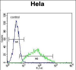
    LEP Antibody (N-term) (Cat. #AP52465PU-N) flow cytometric analysis of Hela cells (right histogram) compared to a negative control cell (left histogram).FITC-conjugated goat-anti-rabbit secondary antibodies were used for the analysis.

    LEP Antibody (N-term) (Cat. #AP52465PU-N) flow cytometric analysis of Hela cells (right histogram) compared to a negative control cell (left histogram).FITC-conjugated goat-anti-rabbit secondary antibodies were used for the analysis.
    Leptin (LEP) (N-term) Rabbit Polyclonal Antibody
    LEP Antibody (N-term).
    FC, IF, IHC, WB
    Human
    Unconjugated
    400 ul
    $645.00
    2 Weeks*
  • p53 (TP53) Mouse Monoclonal Antibody [Clone ID: UMAB55]

    Immunohistochemical staining of paraffin-embedded Human normal colon tissue and colonrectal cancer tissue using anti-TP53 mouse monoclonal antibody. (UM500049; heat-induced epitope retrieval by 10mM citric buffer, pH6.0, 120°C for 3min)

    Immunohistochemical staining of paraffin-embedded Human normal colon tissue and colonrectal cancer tissue using anti-TP53 mouse monoclonal antibody. (UM500049; heat-induced epitope retrieval by 10mM citric buffer, pH6.0, 120°C for 3min)
    p53 (TP53) Mouse Monoclonal Antibody [Clone ID: UMAB55]
    Purified P53 (TP53) mouse monoclonal antibody, clone UMAB55.
    IF, IHC, WB
    Human
    Unconjugated
    100 ul
    $610.00
    In Stock*
  • NDUFB10 Mouse Monoclonal Antibody [Clone ID: OTI2B4]

    Simple Western™ analysis of endogenous protein NDUFB10 from HELA lysates (0.1 mg/mL) using NDUFB10 Mouse Monoclonal Antibody #TA505114. The virtual lane view (left) shows the target (as indicated) at a 1:50 dilution of primary antibody. The corresponding electropherogram view (right) plots chemiluminescence by molecular weight along the capillary at a 1:50 dilution of primary antibody. This experiment was performed under reducing conditions on the Jess™ Simple Western instrument from ProteinSimple, a Bio-Techne brand, using the 12–230 kDa Separation Module.

    Simple Western™ analysis of endogenous protein NDUFB10 from HELA lysates (0.1 mg/mL) using NDUFB10 Mouse Monoclonal Antibody #TA505114. The virtual lane view (left) shows the target (as indicated) at a 1:50 dilution of primary antibody. The corresponding electropherogram view (right) plots chemiluminescence by molecular weight along the capillary at a 1:50  dilution of primary antibody. This experiment was performed under reducing conditions on the Jess™ Simple Western instrument from ProteinSimple, a Bio-Techne brand, using the 12–230 kDa Separation Module.
    NDUFB10 Mouse Monoclonal Antibody [Clone ID: OTI2B4]
    NDUFB10 mouse monoclonal antibody,clone OTI2B4.
    IF, IHC, Simple Western, WB
    Dog, Human, Monkey, Mouse, Rat
    Unconjugated
    100 ul
    $447.00
    In Stock*
  • Calpain 2 (CAPN2) Mouse Monoclonal Antibody [Clone ID: OTI3G1]

    Western blot analysis of extracts (35ug) from 9 different cell lines by using anti-CAPN2 monoclonal antibody (HepG2: human; HeLa: human; SVT2: mouse; A549: human; COS7: monkey; Jurkat: human; MDCK: canine; PC12: rat; MCF7: human) (1:500).

    Western blot analysis of extracts (35ug) from 9 different cell lines by using anti-CAPN2 monoclonal antibody (HepG2: human; HeLa: human; SVT2: mouse; A549: human; COS7: monkey; Jurkat: human; MDCK: canine; PC12: rat; MCF7: human) (1:500).
    Calpain 2 (CAPN2) Mouse Monoclonal Antibody [Clone ID: OTI3G1] 1 Citation
    CAPN2 (Calpain 2) mouse monoclonal antibody, clone OTI3G1 (formerly 3G1).
    IHC, WB
    Dog, Human, Mouse, Rat
    Unconjugated
    100 ul
    $447.00
    In Stock*
  • UBC6e (UBE2J1) Mouse Monoclonal Antibody [Clone ID: OTI3E6]

    Immunohistochemical staining of paraffin-embedded Adenocarcinoma of Human breast tissue using anti-UBE2J1 mouse monoclonal antibody. Heat-induced epitope retrieval by EDTA solution buffer pH 8.0 at 120°C for 3 min.

    Immunohistochemical staining of paraffin-embedded Adenocarcinoma of Human breast tissue using anti-UBE2J1 mouse monoclonal antibody. Heat-induced epitope retrieval by EDTA solution buffer pH 8.0 at 120°C for 3 min.
    UBC6e (UBE2J1) Mouse Monoclonal Antibody [Clone ID: OTI3E6]
    UBE2J1 mouse monoclonal antibody, clone OTI3E6 (formerly 3E6).
    FC, IHC
    Human, Mouse, Rat
    Unconjugated
    100 ul
    $447.00
    In Stock*