
Unlock Spatial Insights with Immuno-Oncology Antibody Panel
Understanding the tumor microenvironment is critical for advancing cancer research. The Immuno-Oncology Antibody Panel comprises 37 rigorously validated antibodies optimized for immunohistochemistry (IHC) in diverse normal and cancerous tissues. Designed for spatial proteome profiling, this panel enables researchers to investigate key immune and tumor markers with high specificity and reproducibility.
Comprehensive Target Coverage
The I/O Antibody Core Panel includes markers for:
- Immune Cell Subsets: CD3e, CD4, CD8a, CD20, CD45, CD56, CD68, CD79a, FOXP3, CD163, CD1c, CD16
- Macrophage & Stromal Markers: CD163, PDPN, CD1c, Fibronectin, CD39
- Checkpoint & Immunoregulation: PD-1, PD-L1, PD-L2, CTLA-4, LAG3, TIGIT, CD40
- Tumor & Oncoproteins: EPCAM, KRT8, KRT18, MKI67, p53, BRAF, HER2, EGFR, PARP1, CD138
- Angiogenesis & Microenvironment: CD31, B2M, STING, Fibronectin, ACTB
Each antibody has been rigorously validated for IHC, ensuring robust performance across multiple tissue types.
Immuno-Oncology Antibody Panel
ACTB
B2M
BRAF
CD138
CD16
CD163
CD1C
CD20
CD31
CD39
CD3e
CD4
CD40
CD45
CD56
CD68
CD79α
CD8a
CTLA4
EGFR
EPCAM
Fibronectin
FOXP3
GZMB
HER2
Keratin 18
KRT8
LAG3
MKI67
p53
PARP1
PD-1
PD-L1
PD-L2
PDPN
STING
TIGIT
Focused Functional Panels
For streamlined analysis, the Immuno-Oncology Antibody Core Panel is also available in the following targeted functional sub-panels:
-
Tumor-Infiltrating Lymphocyte Panels
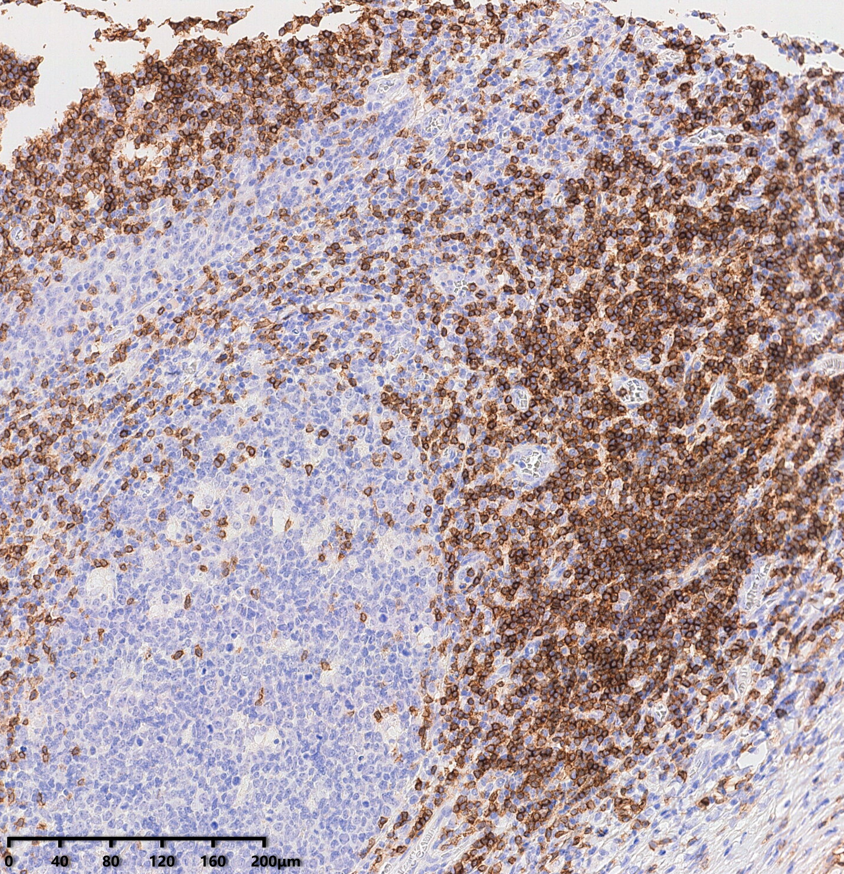
CD3e ()
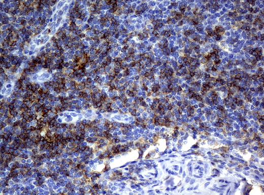
CD4 ()
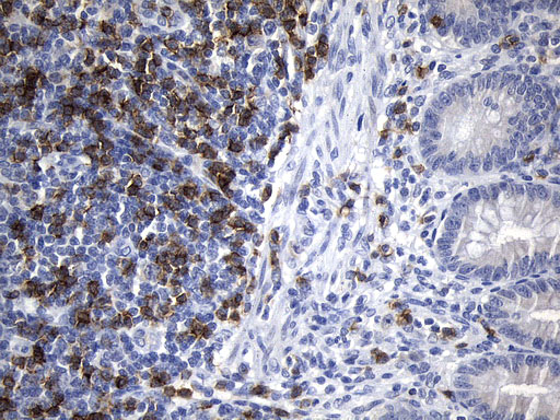
CD8a ()
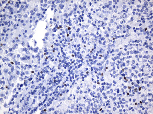
FOXP3 ()
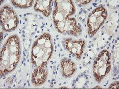
GZMB ()
T-cell infiltration is a key determinant of anti-tumor immunity. CD3e identifies total T cells, CD4 and CD8a distinguish helper and cytotoxic subsets, and FOXP3 marks regulatory T cells. GZMB (Granzyme B) and CD16 (Fc receptor) provide insights into cytotoxic and NK-cell activity.
This panel enables detailed immune profiling of T-cell responses in tumors.
-
B-Cell & NK Cell Immunity Panel (5-Plex):
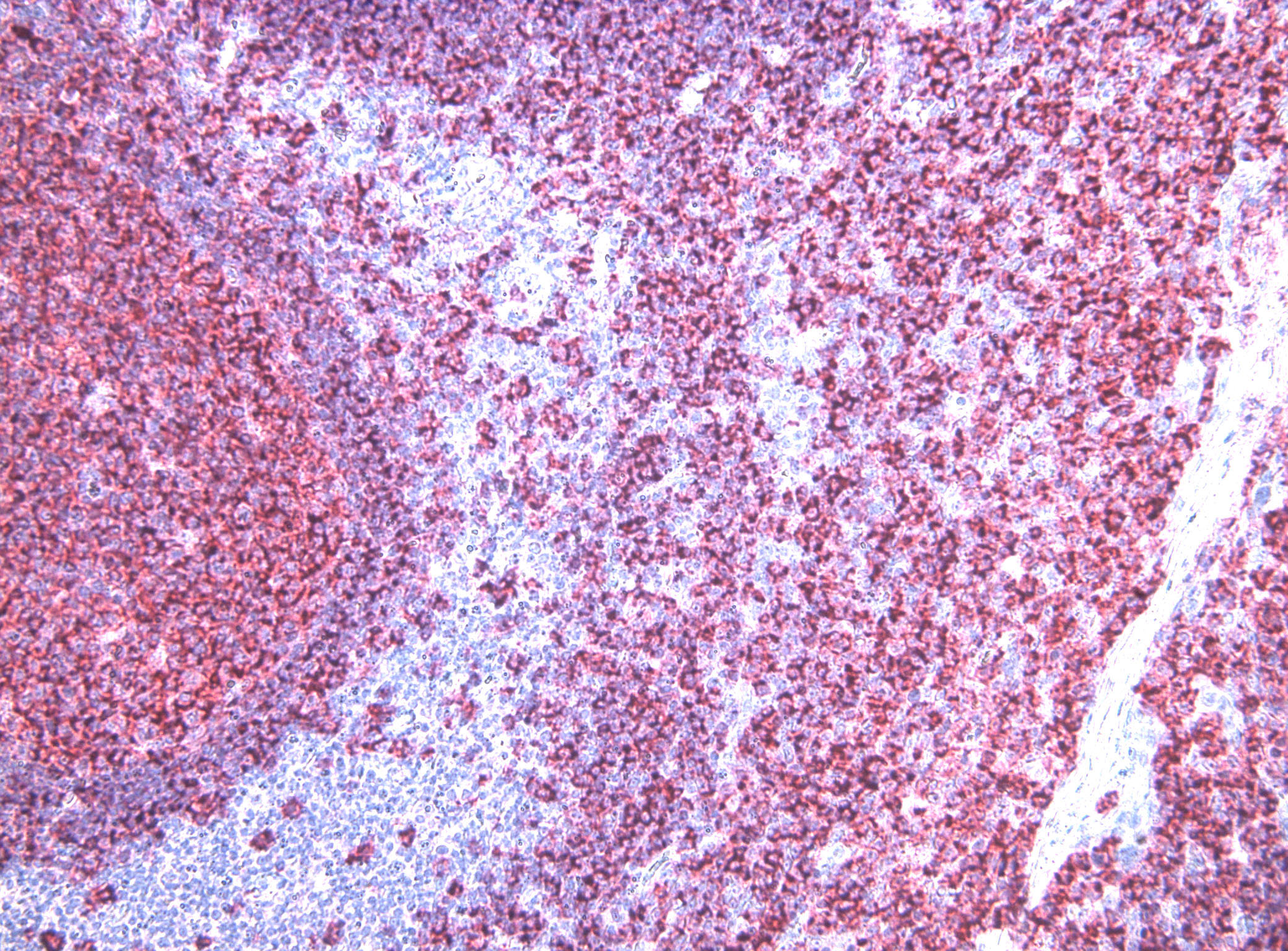
CD20 ()
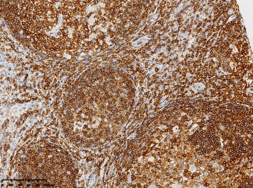
CD45 ()
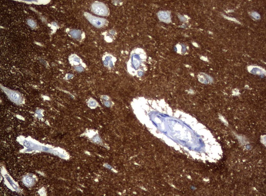
CD56 ()
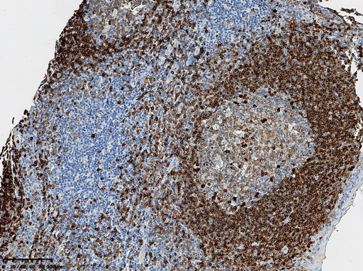
CD79a ()
B cells and NK cells contribute to adaptive and innate immune responses in tumors. CD20 and CD79a mark B-cell subsets, while CD56 identifies NK cells involved in tumor surveillance. CD45 serves as a pan-leukocyte marker, and Beta-2 Microglobulin is essential for antigen presentation via MHC class I. This panel aids in evaluating immune cell recruitment and tumor immune evasion.
-
Macrophage Polarization & Stromal Interaction Panel (6-Plex):
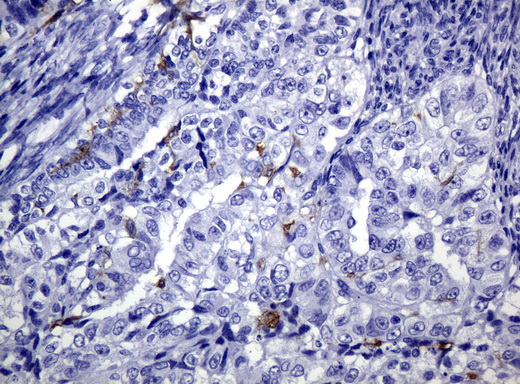
CD1c ()
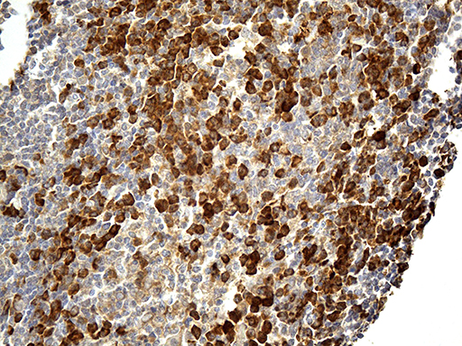
CD163 ()
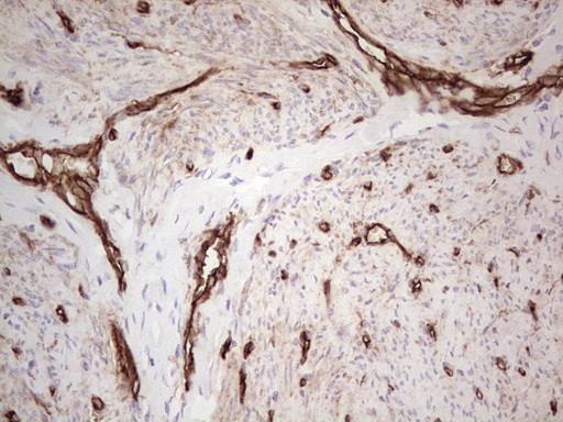
CD39 ()
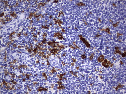
CD68 ()
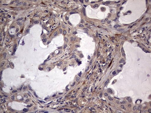
fibronectin ()
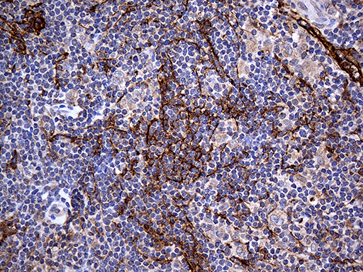
PDPN ()
Tumor-associated macrophages (TAMs) and stromal components play a critical role in immune suppression and metastasis. CD68 and CD163 distinguish macrophage subsets, while PDPN and Fibronectin highlight stromal cell contributions. CD1c identifies dendritic cells, and CD39 is associated with immunosuppressive metabolic pathways.
This panel enables characterization of macrophage and stromal crosstalk in the tumor microenvironment.
-
Checkpoint Immunoregulation Panel (8-Plex):
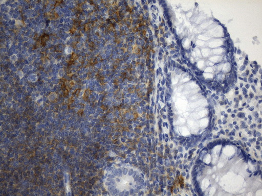
CD40 ()
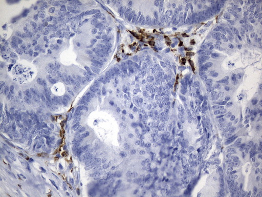
CTLA-4 ()
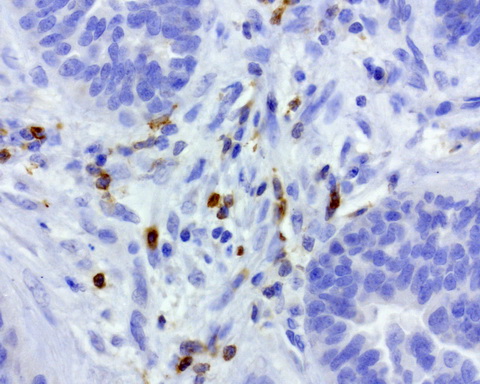
PD-1 ()
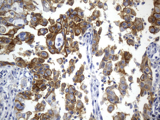
PD-L1 ()
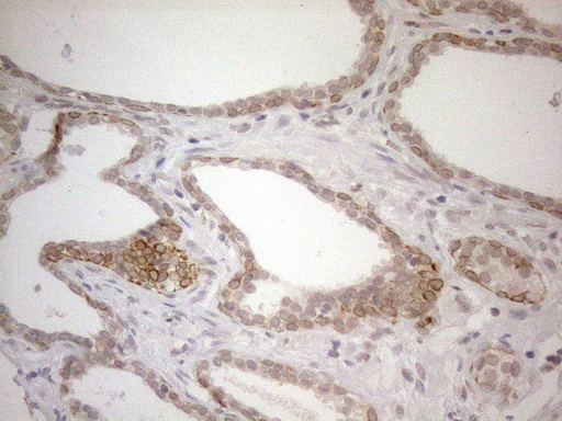
PD-L2 ()
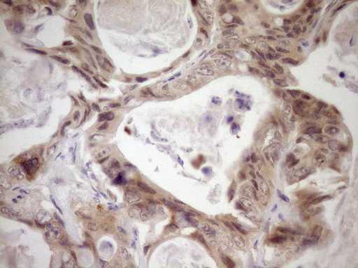
LAG3 ()
Immune checkpoint pathways regulate T-cell activation and immune escape mechanisms in tumors. PD-1 and PD-L1 are key modulators of immune evasion, influencing the efficacy of immune checkpoint blockade therapies. CD45 marks immune cell presence, and CD56 highlights NK cell-mediated cytotoxicity.
This panel facilitates the study of immune suppression and potential therapeutic targets in immuno-oncology.
-
Tumor Progression & Oncoprotein Panel (9-Plex):
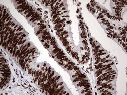
PARP1 ()
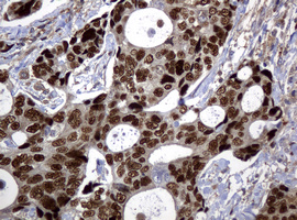
p53 ()
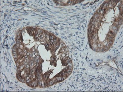
BRAF ()
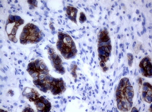
HER2 ()
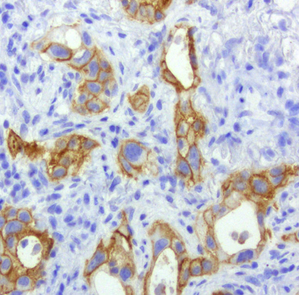
EGFR ()
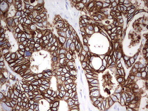
EPCAM ()
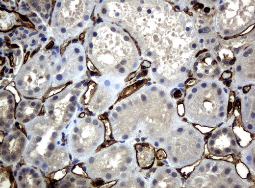
CD31 ()
Tumor growth, genetic instability, and microenvironmental interactions define cancer progression. MKI67 marks proliferating cells, PARP1 indicates DNA damage repair activity, and p53 serves as a tumor suppressor marker. Oncogenes such as BRAF, HER2, and EGFR contribute to tumor signaling. EPCAM identifies epithelial tumors, CD138 (SDC1) marks plasma cells and myeloma, and CD31 highlights tumor vascularization.
This panel enables characterization of tumor aggressiveness and therapeutic vulnerabilities.
-
Epithelial & Structural Marker Panel:
fibronectin ()
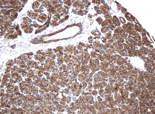
KRT8 ()
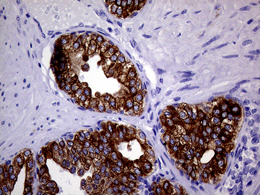
KRT18 ()
Tumor architecture and cellular integrity are defined by epithelial and cytoskeletal markers. KRT8 and KRT18 identify epithelial-derived cancers, ACTB (beta-actin) serves as a loading control and structural marker, and Fibronectin provides insights into extracellular matrix remodeling.
This panel supports studies on tumor organization and metastatic potential.
Antibodies Use Case in Spatial Proteomic Research Applications
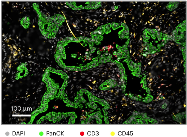
Shiau, Carina, et al. “Spatially resolved analysis of pancreatic cancer identifies therapy-associated remodeling of the tumor microenvironment.” Nature Genetics, Sept. 2024, doi:10.1038/s41588-024-01890-9.
A publication by Dr. Carina Shiau and Jingyi Cao from the Massachusetts General Hospital and Harvard Medical School in Nature Genetics analyzes pancreatic cancer's tumor microenvironment (TME) remodeling post-chemotherapy and radiotherapy.
UltraMAB CD3/CD3E Mouse Monoclonal Antibody () was part of a fluorophore-conjugated antibody cocktail used on FFPE tumor sections, helping identify T cells and enabling spatial immune cell analysis within the TME.
Enable Precision Oncology Research
The Immuno-Oncology Antibody Panels enables researchers with spatial biomarker insights, enabling high-resolution tumor profiling and immune landscape characterization. Enhance your IHC studies with our highly validated antibodies for precision oncology research.
