
TissueScan, Human Brain cDNA Array
SKU
HBRT101
24 samples covering all major human brain normal tissues at different locations. Available in 2, 5, 10 identical plates.

$600.00
In Stock*
Specifications
| Product Data | |
| TissueScan Type | Normal Tissue |
|---|---|
| Array Name | Human Brain cDNA Array |
| Diagnosis Category | Normal |
| Tissue | Brain |
| Shipping | Ambient |
| Description | 24 samples covering all major human brain normal tissues at different locations. Available in 2, 5, 10 identical plates. |
Reviews


Documents
| Product Manuals |
| FAQs |
| SDS |
Citations
Small mitochondrial protein NERCLIN regulates cardiolipin homeostasis and mitochondrial ultrastructure
Konovalova, Torregrosa-Muñumer, Manjunath et al
Proc Natl Acad Sci U S A (2023) 120 (30), e2210599120 DOI: 10.1073/pnas.2210599120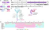
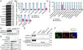
Konovalova, Torregrosa-Muñumer, Manjunath et al
Proc Natl Acad Sci U S A (2023) 120 (30), e2210599120 DOI: 10.1073/pnas.2210599120
Cited by 6
AI Summary: OriGene’s Human Normal Brain Tissue qPCR Array (HBRT101) was used in this study to analyze NERCLIN expression levels across various human brain tissues, aiding in the characterization of its tissue distribution.
Snippet: /13/03/00/15). All the cell lines were anonymized. DNA and RNA Analysis. For expression analysis, human multiple tissue cDNA panel (MTC Clontech, 636742) and human normal brain tissue qPCR array (OriGene Technologies, HBRT101) were used. Prior to their use, these samples were deidentified by manufacturers. Detailed methods are described in SI Appendix, Materials and Methods. Supplementary Ma
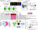
FIBCD1 is an endocytic GAG receptor associated with a novel neurodevelopmental disorder
Fell, Hagelkruys, Cicvaric et al
EMBO Mol Med (2022) 14 (9), e15829 DOI: 10.15252/emmm.202215829
Fell, Hagelkruys, Cicvaric et al
EMBO Mol Med (2022) 14 (9), e15829 DOI: 10.15252/emmm.202215829
Cited by 15
AI Summary: OriGene’s TissueScan Human Brain cDNA Array (#HBRT101) and Human Normal cDNA Array (#HMRT304) were used to analyze FIBCD1 expression in human tissues, contributing to the identification of its role as an endocytic GAG receptor linked to neurodevelopmental disorders.
Snippet: …Human cDNA panels were obtained from OriGene: TissueScan, Human Brain cDNA Array (#HBRT101) and Human Normal cDNA Array (#HMRT304). … In Silico modelling of FIBCD1 Docking solutions In silico;Human cDNA panels were obtained from OriGene: TissueScan, Human Brain cDNA Array (#HBRT101) and Human Normal cDNA Array (#HMRT304). …
Nitric-Oxide Synthase trafficking inducer (NOSTRIN) is an emerging negative regulator of colon cancer progression
Paul, Gope, Das et al
BMC Cancer (2022) 22 (1), 594 DOI: 10.1186/s12885-022-09670-6
Paul, Gope, Das et al
BMC Cancer (2022) 22 (1), 594 DOI: 10.1186/s12885-022-09670-6
Cited by 5
AI Summary: OriGene contributed to this study by providing the TissueScan Colon Cancer cDNA Array V (Cat No. HCRT105), which was used to analyze NOSTRIN expression across different stages of human colon cancer.
Snippet: …Methods Cell;- Clinical samples across all stages of the disease were acquired from Origene (USA) as Tissue scan colon cancer cDNA array V (Cat No. HCRT105). …
Calsyntenin-3 interacts with the sodium-dependent vitamin C transporter-2 to regulate vitamin C uptake
Subramanian, Teafatiller, Vidal et al
Int J Biol Macromol (2021) 192, 1178-1184 DOI: 10.1016/j.ijbiomac.2021.10.058
Subramanian, Teafatiller, Vidal et al
Int J Biol Macromol (2021) 192, 1178-1184 DOI: 10.1016/j.ijbiomac.2021.10.058
Cited by 6
AI Summary: OriGene's normal human brain cDNA was used in this study for RT-qPCR analysis to measure the expression levels of hSVCT2 and hCLSTN3, aiding in the investigation of their interaction and role in vitamin C uptake.
Snippet: The different regions of the normal human brain cDNA obtained from OriGene Biotechnology was directly subjected to RT-qPCR using hSVCT2 or hCLSTN3 specific primers (Table 1). β-Actin was used as a reference target to normalize relative expression values Save;- The different regions of the normal human brain cDNA obtained from OriGene Biotechnology was directly subjected to RT-qPCR using hSVC...
Small mitochondrial protein NERCLIN regulates cardiolipin homeostasis and mitochondrial ultrastructure
Konovalova, Torregrosa-Muñumer, Manjunath et al
bioRxiv (2021)
Konovalova, Torregrosa-Muñumer, Manjunath et al
bioRxiv (2021)
Snippet: DNA and RNA analysis For expression analysis human multiple tissue cDNA panel (MTC Clontech, 636742) and human normal brain tissue qPCR array (OriGene Technologies, HBRT101) were used. 1-3 ng of cDNA was used for real-time PCR assay
Mutations in FAM50A suggest that Armfield XLID syndrome is a spliceosomopathy
Lee, Khan, Armfield-Uhas et al
Nat Commun (2020) 11 (1), 3698 DOI: 10.1038/s41467-020-17452-6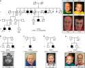
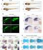
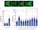
Lee, Khan, Armfield-Uhas et al
Nat Commun (2020) 11 (1), 3698 DOI: 10.1038/s41467-020-17452-6
Cited by 72
AI Summary: OriGene’s Rapid Scan Human Brain Panel was used in this study to assess FAM50A expression in various regions of the fetal brain, helping to demonstrate its presence in the cerebellum and hypothalamus and supporting the gene’s role in neurodevelopmental processes.
Snippet: ech). _FAM50A_ was expressed in all eight fetal tissues assessed, including brain (Supplementary Fig. 3a). Next, we evaluated _FAM50A_ expression in fetal brain using a Rapid Scan Human Brain Panel (Origene). _FAM50A_ was detectable in the fetal cerebellum and hypothalamus (Supplementary Fig. 3b). However, we observed low expression of _FAM50A_ in the temporal lobe and were unable to detect expr;- Intellectual disability (ID) is a heterogeneous clinical entity and includes an excess of males who harbor variants on the X-chromosome (XLID). We report rare FAM50A missense variants in the original Armfield XLID syndrome family localized in Xq28 and four additional;ght fetal tissues assessed, including brain (Supplementary Fig. 3a [/articles/s41467-020-17452-6#MOESM1]). Next, we evaluated _FAM50A_ expression in fetal brain using a Rapid Scan Human Brain Panel (Origene). _FAM50A_ was detectable in the fetal cerebellum and hypothalamus (Supplementary Fig. 3b [/articles/s41467-020-17452-6#MOESM1]). However, we observed low expression of _FAM50A_ in the tempor;lontech). FAM50A was expressed in all eight fetal tissues assessed, including brain (Supplementary Fig. 3a). Next, we evaluated FAM50A expression in fetal brain using a Rapid Scan Human Brain Panel (Origene). FAM50A was detectable in the fetal cerebellum and hypothalamus (Supplementary Fig. 3b). However, we observed low expression of FAM50A in the temporal lobe and were unable to detect expressi
Showing 1-6 of 42 papers.
Powered by BizGenius
*Delivery time may vary from web posted schedule. Occasional delays may occur due to unforeseen
complexities in the preparation of your product. International customers may expect an additional 1-2 weeks
in shipping.
Related Products
- Price:
- Actual Price:
Welcome to OriGene AI!
I'm your AI-powered guide to everything OriGene.
Ask me anything:
- Product specs
- Ordering help
- Technical questions
- How to find exactly what your experiment needs
Prefer a live agent? Connect with us by clicking the green chat icon, as seen in the image below or email our team at custsupport@origene.com.
