
Ready-to-image pooled line (puromycin-selected).
View productMito-Glow: Live-Cell
Mitochondrial Imaging
Genetically encoded reporters that label mitochondria in living cells: stable across passages, independent of membrane potential.
Lentivirus or stable cell lines · Multiplex-ready
Why Mito-Glow
Long-Term Mitochondrial Imaging
Mito-Glow uses stable expression of mitochondria-targeted fluorescent proteins to support live-cell imaging across passages, with promoter options suited to different cellular models.
Membrane Potential–Independent Labeling
Visualize mitochondrial morphology and distribution during changes in membrane potential, supporting studies of treatments and mutations that perturb mitochondrial function.
See the CCCP assayStable Expression Across Passages
Maintain mitochondrial labeling through continued reporter expression as cells divide, enabling repeated imaging and long-term experiments without additional dye loading.
See long-term imaging dataPromoter Options for Diverse Cell Models
Choose CMV for standard immortalized lines, EF1α for primary and iPSC-derived cells, or hSyn for neuron-selective expression.
Browse the product table
Product Family
Explore the Mito-Glow Product Family
Find the mitochondrial label, promoter, and delivery format suited to your cell model.
Products
No product matches these filters. Stable cell lines are currently available with the CMV promoter only — choose a lentivirus format for EF1α or hSyn.
| Product | Promoter | Format | Best for | Link |
|---|---|---|---|---|
| (not in current selection) | View product | |||
Each label is offered as lentivirus for your own transduction, and as a pooled stable cell line generated under puromycin selection (CMV promoter only). Product links are placeholders pending final catalog numbers.
Product Family
Explore the Mito-Glow Product Family
Find the mitochondrial label, promoter, and delivery format suited to your cell model.
Products
No product matches these filters. Stable cell lines are currently available with the CMV promoter only — choose a lentivirus format for EF1α or hSyn.
| Product | Promoter | Format | Best for | Link |
|---|---|---|---|---|
| (not in current selection) | View product Coming soon | |||
Ready-to-image pooled line (puromycin-selected).
View product Coming soon
Each label is offered as lentivirus for your own transduction, and as a pooled stable cell line generated under puromycin selection (CMV promoter only). Product links are placeholders pending final catalog numbers.
Validation Data
Confirmed mitochondrial localization, live and unfixed
Mito-Glow Persists Following Membrane Depolarization
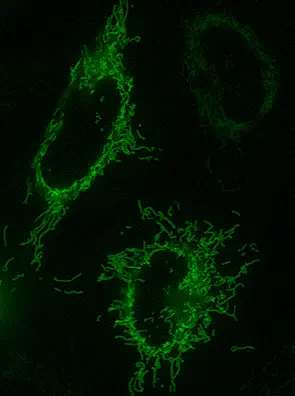
HeLa cells stably expressing a mitochondrial outer membrane label (TOMM20-mGGP - MT100005LV) were imaged at multiple passage numbers. Mitochondrial fluorescence and localization were maintained across the passages evaluated, supporting the use of Mito-Glow for longitudinal imaging studies.
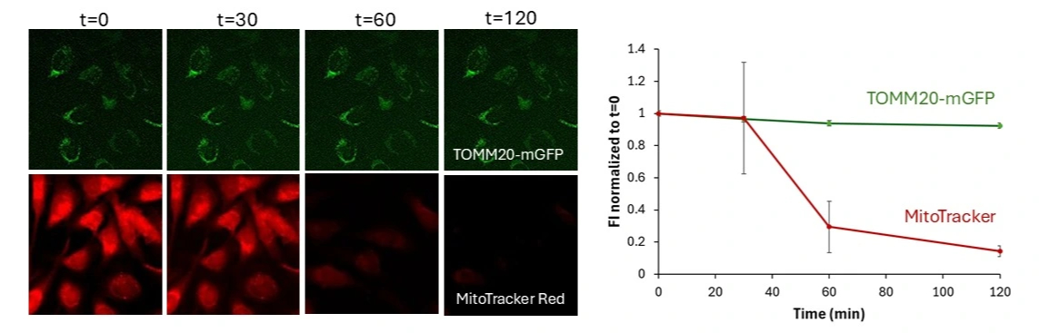
Mitochondrial morphology and distribution remain clearly labeled during membrane depolarization, demonstrating that Mito-Glow signal is independent of mitochondrial membrane potential.
Sustained Mitochondrial Labeling Across Cell Passages

HeLa cells stably expressing a mitochondrial outer membrane label (TOMM20-mGGP - MT100005LV) were treated with 20 μM CCCP to induce mitochondrial membrane depolarization and imaged at the indicated times. Mitochondrial labeling persisted throughout the treatment period, supporting membrane potential–independent visualization under the conditions tested.
Stable reporter expression maintains crisp mitochondrial labeling as cells divide, enabling repeated live-cell imaging across passages without additional dye loading.
