Knockdown Lysates

Items 1-50 of 514

Set Ascending Direction
  • Tag
    Expression Host
    Applications
    Size
    Delivery Time
  • TDP43 (TARDBP) Human Knockdown Lysate

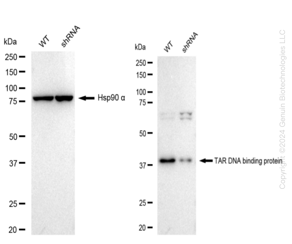
    Western blotting analysis. TARDBP protein expression in wild-type (WT) and shRNA knockdown (KD) HeLa cells was detected using Western blotting.  Hsp90 α served as a loading control. The blots were incubated with primary antibodies (Cat#61519, 1:5,000) against TARDBP and  Hsp90 α , respectively, followed by incubating with HRP-conjugated goat anti-rabbit secondary antibody (Cat#201, 1:20,000). Images were developed using FeQ™ ECL Substrate Kit (Cat#226).

    Western blotting analysis. TARDBP protein expression in wild-type (WT) and shRNA knockdown (KD) HeLa  cells was detected using Western blotting.  Hsp90 α served as a loading control. The blots were incubated with primary antibodies (Cat#61519, 1:5,000) against TARDBP and  Hsp90 α , respectively, followed by incubating with HRP-conjugated goat anti-rabbit secondary antibody (Cat#201, 1:20,000). Images were developed using FeQ™ ECL Substrate Kit (Cat#226).
    TDP43 (TARDBP) Human Knockdown Lysate
    WB-validated TARDBP Knockdown HeLa Cell Lysate.
    Tag Free
    HeLa
    100 ug
    $589.00
    5 Days*
  • Fibronectin (FN1) Human Knockdown Lysate

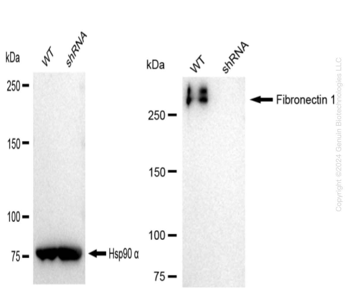
    Western blotting analysis. FN1 protein expression in wild-type (WT) and shRNA knockdown (KD) HeLa cells was detected using Western blotting.  Hsp90 α served as a loading control. The blots were incubated with primary antibodies (Cat#62510, 1:5,000) against FN1 and  Hsp90 α , respectively, followed by incubating with HRP-conjugated goat anti-rabbit secondary antibody (Cat#201, 1:20,000). Images were developed using FeQ™ ECL Substrate Kit (Cat#226).

    Western blotting analysis. FN1 protein expression in wild-type (WT) and shRNA knockdown (KD) HeLa cells was detected using Western blotting.  Hsp90 α served as a loading control. The blots were incubated with primary antibodies (Cat#62510, 1:5,000) against FN1 and  Hsp90 α , respectively, followed by incubating with HRP-conjugated goat anti-rabbit secondary antibody (Cat#201, 1:20,000). Images were developed using FeQ™ ECL Substrate Kit (Cat#226).
    Fibronectin (FN1) Human Knockdown Lysate
    WB-validated FN1 Knockdown HeLa Cell Lysate.
    Tag Free
    HeLa
    100 ug
    $589.00
    5 Days*
  • DDB2 Human Knockdown Lysate

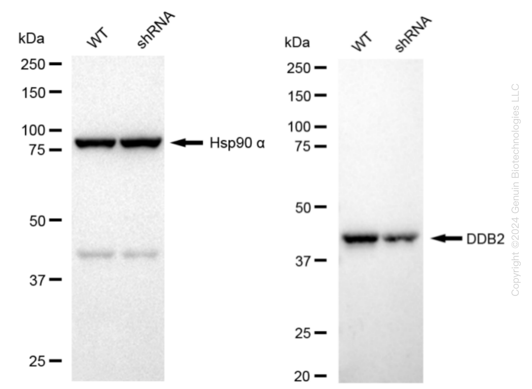
    Western blotting analysis. DDB2 protein expression in wild-type (WT) and shRNA knockdown (KD) HT-1080 cells was detected using Western blotting. Hsp90 α served as a loading control. The blots were incubated with primary antibodies against DDB2 and Hsp90 α, respectively, followed by incubating with HRP-conjugated goat anti-rabbit secondary antibody. Images were developed using FeQ™ ECL Substrate Kit.

    Western blotting analysis. DDB2 protein expression in wild-type (WT) and shRNA knockdown (KD) HT-1080 cells was detected using Western blotting. Hsp90 α served as a loading control. The blots were incubated with primary antibodies against DDB2 and Hsp90 α, respectively, followed by incubating with HRP-conjugated goat anti-rabbit secondary antibody. Images were developed using FeQ™ ECL Substrate Kit.
    DDB2 Human Knockdown Lysate
    WB-validated DDB2 Knockdown HT-1080 Cell Lysate.
    Tag Free
    100 ug
    $589.00
    5 Days*
  • ATG16L1 Human Knockdown Lysate

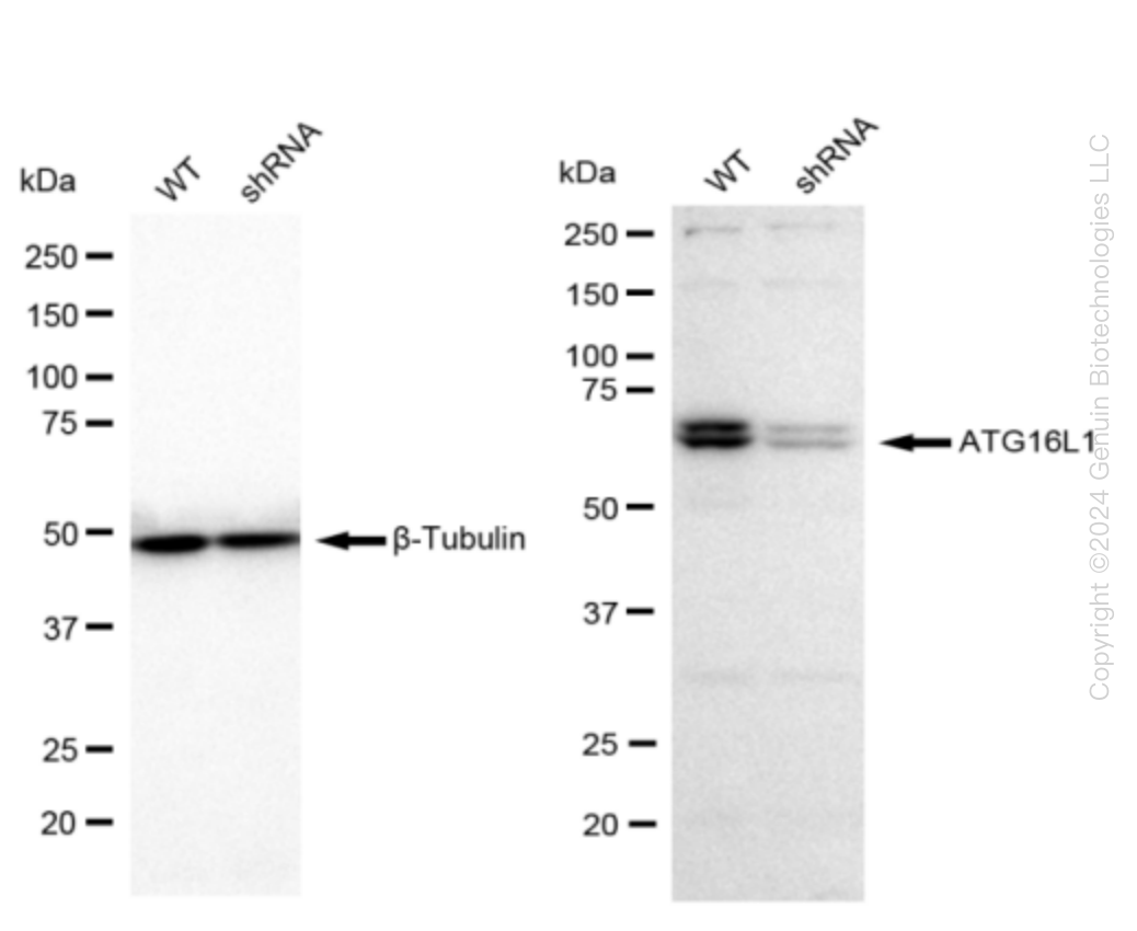
    Western blotting analysis. ATG16L1 protein expression in wild-type (WT) and shRNA knockdown (KD) 293T cells was detected using Western blotting. β-Tubulin served as a loading control. The blots were incubated with primary antibodies against ATG16L1 and β-Tubulin, respectively, followed by incubating with HRP-conjugated goat anti-rabbit secondary antibody. Images were developed using FeQ™ ECL Substrate Kit.

    Western blotting analysis. ATG16L1 protein expression in wild-type (WT) and shRNA knockdown (KD) 293T cells was detected using Western blotting. β-Tubulin served as a loading control. The blots were incubated with primary antibodies against ATG16L1 and β-Tubulin, respectively, followed by incubating with HRP-conjugated goat anti-rabbit secondary antibody. Images were developed using FeQ™ ECL Substrate Kit.
    ATG16L1 Human Knockdown Lysate
    WB-validated ATG16L1 Knockdown Cell Lysate.
    Tag Free
    293T
    100 ug
    $589.00
    5 Days*
  • CTP synthase (CTPS1) Human Knockdown Lysate

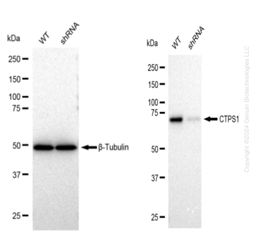
    Western blotting analysis. CTPS1 protein expression in wild-type (WT) and shRNA knockdown (KD) 293T cells was detected using Western blotting. β-Tubulin served as a loading control. The blots were incubated with primary antibodies against CTPS1 and β-Tubulin, respectively, followed by incubating with HRP-conjugated goat anti-rabbit secondary antibody. Images were developed using FeQ™ ECL Substrate Kit.

    Western blotting analysis. CTPS1 protein expression in wild-type (WT) and shRNA knockdown (KD) 293T cells was detected using Western blotting. β-Tubulin served as a loading control. The blots were incubated with primary antibodies against CTPS1 and β-Tubulin, respectively, followed by incubating with HRP-conjugated goat anti-rabbit secondary antibody. Images were developed using FeQ™ ECL Substrate Kit.
    CTP synthase (CTPS1) Human Knockdown Lysate
    WB-validated CTPS1 Knockdown Cell Lysate.
    Tag Free
    293T
    100 ug
    $589.00
    5 Days*
  • FOXO4 Human Knockdown Lysate

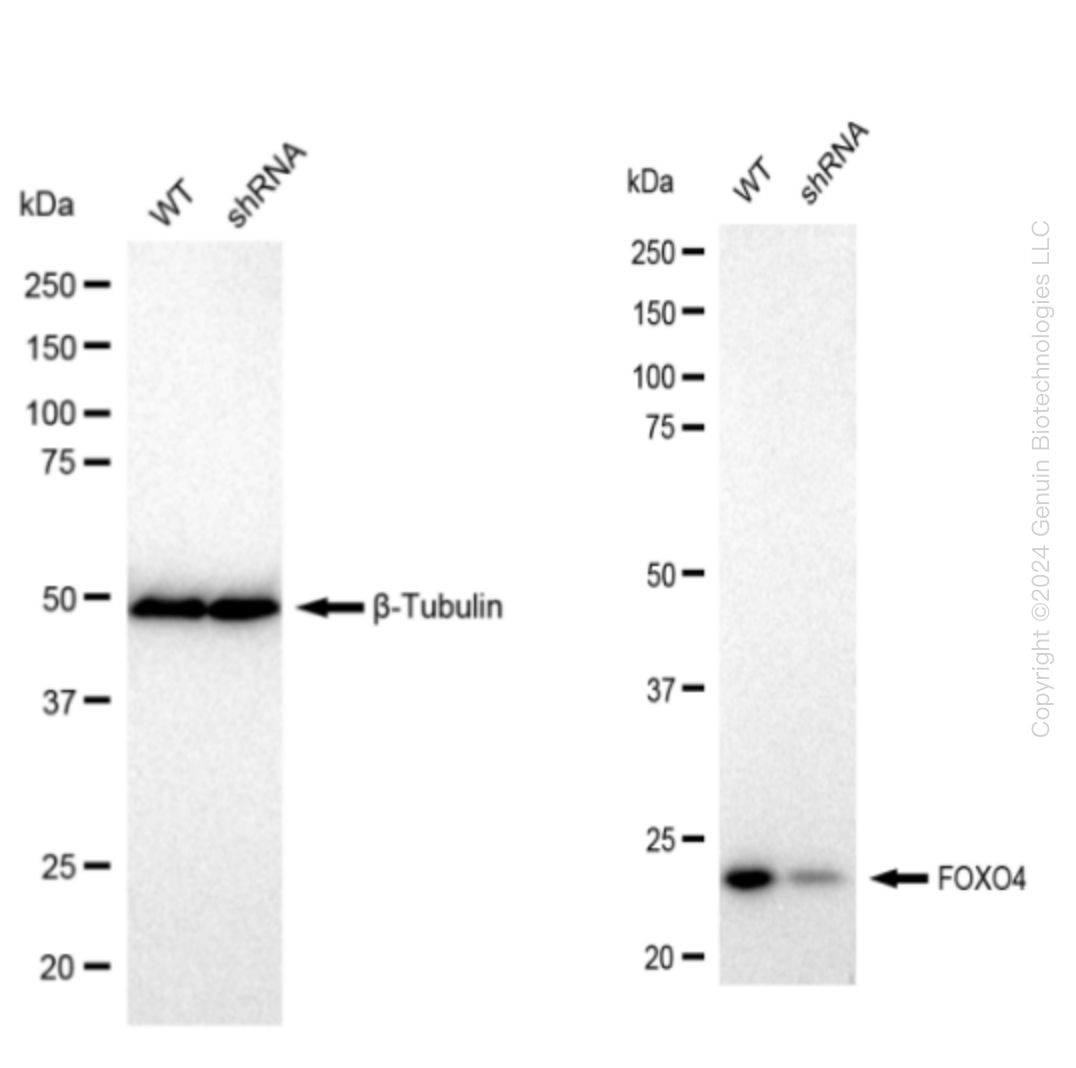
    Western blotting analysis. FOXO4 protein expression in wild-type (WT) and shRNA knockdown (KD) 293T cells was detected using Western blotting. β-Tubulin served as a loading control. The blots were incubated with primary antibodies against FOXO4 and β-Tubulin, respectively, followed by incubating with HRP-conjugated goat anti-rabbit secondary antibody. Images were developed using FeQ™ ECL Substrate Kit.

    Western blotting analysis. FOXO4 protein expression in wild-type (WT) and shRNA knockdown (KD) 293T cells was detected using Western blotting. β-Tubulin served as a loading control. The blots were incubated with primary antibodies against FOXO4 and β-Tubulin, respectively, followed by incubating with HRP-conjugated goat anti-rabbit secondary antibody. Images were developed using FeQ™ ECL Substrate Kit.
    FOXO4 Human Knockdown Lysate
    WB-validated FOXO4 Knockdown 293T Cell Lysate.
    Tag Free
    293T
    100 ug
    $589.00
    5 Days*
  • HSF1 Human Knockdown Lysate

    Western blotting analysis. HSF1 protein expression in wild-type (WT) and shRNA knockdown (KD) 293T cells was detected using Western blotting. β-Tubulin served as a loading control. The blots were incubated with primary antibodies against HSF1 and  β-Tubulin, respectively, followed by incubating with HRP-conjugated goat anti-rabbit secondary antibody. Images were developed using FeQ™ ECL Substrate Kit.

    Western blotting analysis. HSF1 protein expression in wild-type (WT) and shRNA knockdown (KD) 293T cells was detected using Western blotting. β-Tubulin served as a loading control. The blots were incubated with primary antibodies against HSF1 and  β-Tubulin, respectively, followed by incubating with HRP-conjugated goat anti-rabbit secondary antibody. Images were developed using FeQ™ ECL Substrate Kit.
    HSF1 Human Knockdown Lysate
    WB-validated HSF1 Knockdown 293T Cell Lysate.
    Tag Free
    293T
    100 ug
    $589.00
    5 Days*
  • HSP90AB1 Human Knockdown Lysate

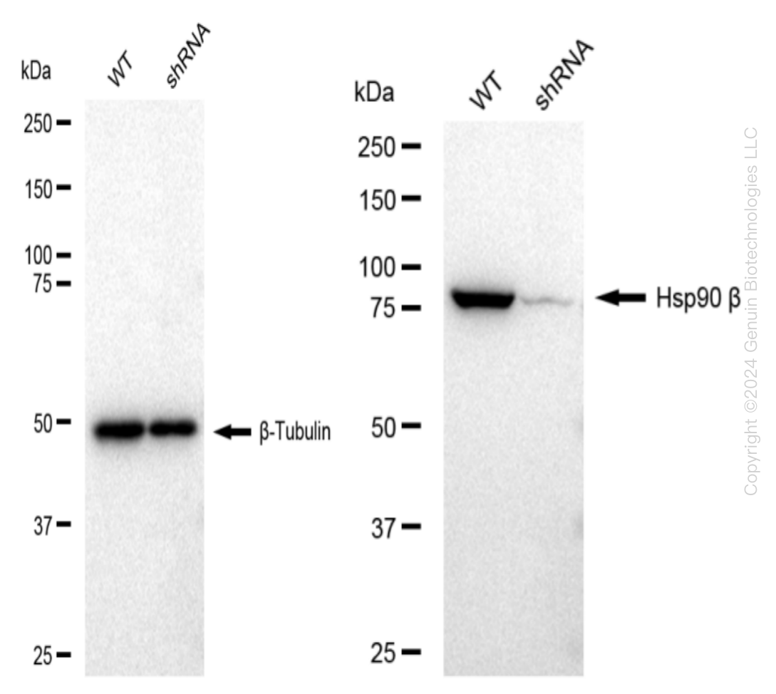
    Western blotting analysis. HSP90AB1 protein expression in wild-type (WT) and shRNA knockdown (KD) 293T cells was detected using Western blotting. β-Tubulin served as a loading control. The blots were incubated with primary antibodies against HSP90AB1 and β-Tubulin, respectively, followed by incubating with HRP-conjugated goat anti-rabbit secondary antibody. Images were developed using FeQ™ ECL Substrate Kit.

    Western blotting analysis. HSP90AB1 protein expression in wild-type (WT) and shRNA knockdown (KD) 293T cells was detected using Western blotting. β-Tubulin served as a loading control. The blots were incubated with primary antibodies against HSP90AB1 and β-Tubulin, respectively, followed by incubating with HRP-conjugated goat anti-rabbit secondary antibody. Images were developed using FeQ™ ECL Substrate Kit.
    HSP90AB1 Human Knockdown Lysate
    WB-validated HSP90AB1 Knockdown 293T Cell Lysate.
    Tag Free
    293T
    100 ug
    $589.00
    5 Days*
  • Hsc70 (HSPA8) Human Knockdown Lysate

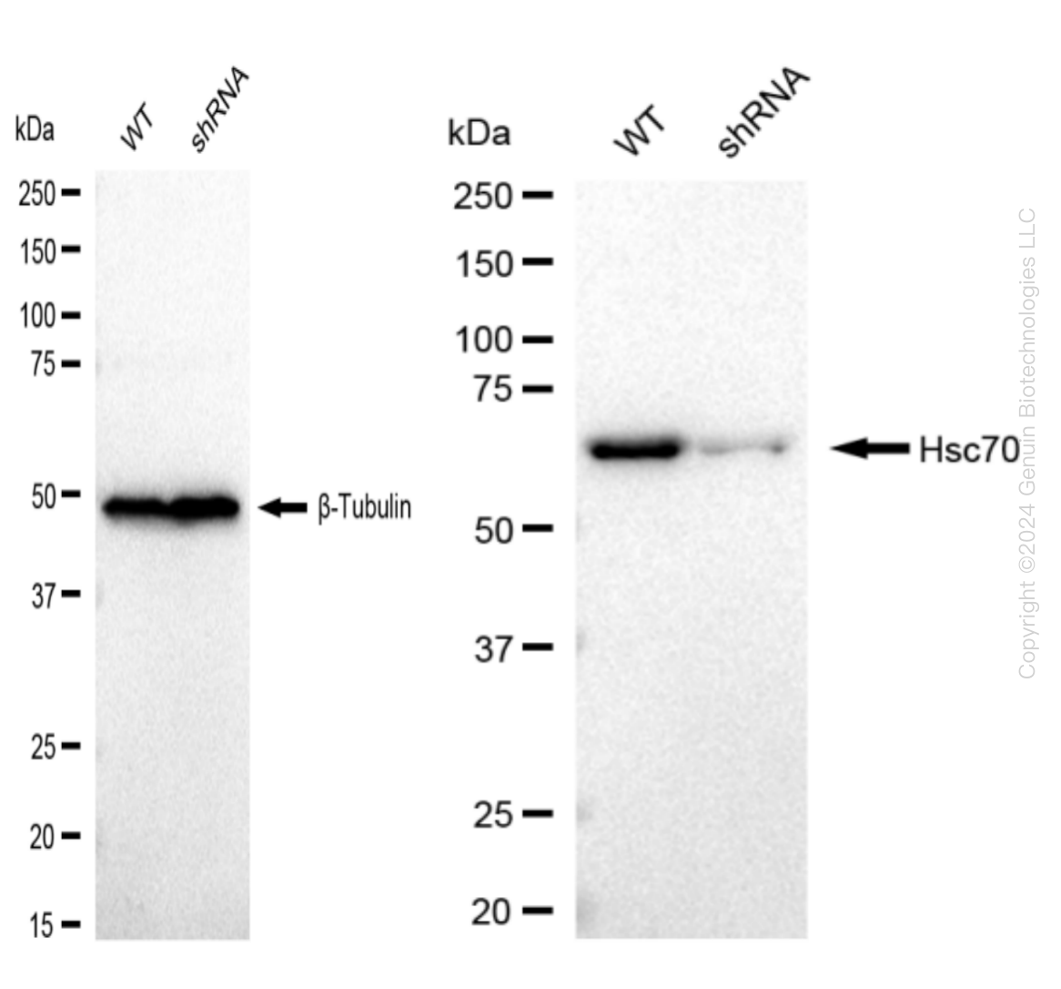
    Western blotting analysis. HSPA8 protein expression in wild-type (WT) and shRNA knockdown (KD) 293T cells was detected using Western blotting.  β-Tubulin served as a loading control. The blots were incubated with primary antibodies (Cat#61475, 1:5,000) against HSPA8 and  β-Tubulin , respectively, followed by incubating with HRP-conjugated goat anti-rabbit secondary antibody (Cat#201, 1:20,000). Images were developed using FeQ™ ECL Substrate Kit (Cat#226).

    Western blotting analysis. HSPA8 protein expression in wild-type (WT) and shRNA knockdown (KD) 293T cells was detected using Western blotting.  β-Tubulin served as a loading control. The blots were incubated with primary antibodies (Cat#61475, 1:5,000) against HSPA8 and  β-Tubulin , respectively, followed by incubating with HRP-conjugated goat anti-rabbit secondary antibody (Cat#201, 1:20,000). Images were developed using FeQ™ ECL Substrate Kit (Cat#226).
    Hsc70 (HSPA8) Human Knockdown Lysate
    WB-validated HSPA8 Knockdown 293T Cell Lysate.
    Tag Free
    293T
    100 ug
    $589.00
    5 Days*
  • IQGAP2 Human Knockdown Lysate

    Western blotting analysis. IQGAP2 protein expression in wild-type (WT) and shRNA knockdown (KD) 293T cells was detected using Western blotting. Hsp90 α served as a loading control. The blots were incubated with primary antibodies (Cat#61138, 1:5,000) against IQGAP2 and Hsp90 α, respectively, followed by incubating with HRP-conjugated goat anti-rabbit secondary antibody (Cat#201, 1:20,000). Images were developed using FeQ™ ECL Substrate Kit (Cat#226).

    Western blotting analysis. IQGAP2 protein expression in wild-type (WT) and shRNA knockdown (KD) 293T cells was detected using Western blotting. Hsp90 α served as a loading control. The blots were incubated with primary antibodies (Cat#61138, 1:5,000) against IQGAP2 and Hsp90 α, respectively, followed by incubating with HRP-conjugated goat anti-rabbit secondary antibody (Cat#201, 1:20,000). Images were developed using FeQ™ ECL Substrate Kit (Cat#226).
    IQGAP2 Human Knockdown Lysate
    WB-validated IQGAP2 Knockdown 293T Cell Lysate.
    Tag Free
    293T
    100 ug
    $589.00
    5 Days*
  • ATG7 Human Knockdown Lysate

    Western blotting analysis. ATG7 protein expression in wild-type (WT) and shRNA knockdown (KD) 293T cells was detected using Western blotting. β-Tubulin served as a loading control. The blots were incubated with primary antibodies against ATG7 and β-Tubulin, respectively, followed by incubating with HRP-conjugated goat anti-rabbit secondary antibody. Images were developed using FeQ™ ECL Substrate Kit.

    Western blotting analysis. ATG7 protein expression in wild-type (WT) and shRNA knockdown (KD) 293T cells was detected using Western blotting. β-Tubulin served as a loading control. The blots were incubated with primary antibodies against ATG7 and β-Tubulin, respectively, followed by incubating with HRP-conjugated goat anti-rabbit secondary antibody. Images were developed using FeQ™ ECL Substrate Kit.
    ATG7 Human Knockdown Lysate
    WB-validated ATG7 Knockdown 293T Cell Lysate.
    Tag Free
    293T
    100 ug
    $589.00
    5 Days*
  • Cullin 1 (CUL1) Human Knockdown Lysate

    Western blotting analysis. CUL1 protein expression in wild-type (WT) and shRNA knockdown (KD) 293T cells was detected using Western blotting. β-Tubulin served as a loading control. The blots were incubated with primary antibodies against CUL1 and β-Tubulin, respectively, followed by incubating with HRP-conjugated goat anti-rabbit secondary antibody. Images were developed using FeQ™ ECL Substrate Kit.

    Western blotting analysis. CUL1 protein expression in wild-type (WT) and shRNA knockdown (KD) 293T cells was detected using Western blotting. β-Tubulin served as a loading control. The blots were incubated with primary antibodies against CUL1 and β-Tubulin, respectively, followed by incubating with HRP-conjugated goat anti-rabbit secondary antibody. Images were developed using FeQ™ ECL Substrate Kit.
    Cullin 1 (CUL1) Human Knockdown Lysate
    WB-validated CUL1 Knockdown 293T Cell Lysate.
    Tag Free
    293T
    100 ug
    $589.00
    5 Days*
  • GRP78 (HSPA5) Human Knockdown Lysate

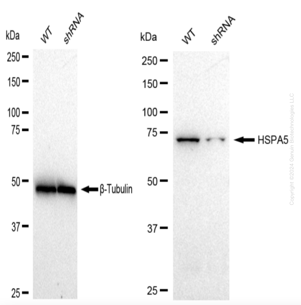
    Western blotting analysis. HSPA5 protein expression in wild-type (WT) and shRNA knockdown (KD) 293T cells was detected using Western blotting. β-Tubulin served as a loading control. The blots were incubated with primary antibodies against HSPA5 and β-Tubulin, respectively, followed by incubating with HRP-conjugated goat anti-rabbit secondary antibody. Images were developed using FeQ™ ECL Substrate Kit.

    Western blotting analysis. HSPA5 protein expression in wild-type (WT) and shRNA knockdown (KD) 293T cells was detected using Western blotting. β-Tubulin served as a loading control. The blots were incubated with primary antibodies against HSPA5 and β-Tubulin, respectively, followed by incubating with HRP-conjugated goat anti-rabbit secondary antibody. Images were developed using FeQ™ ECL Substrate Kit.
    GRP78 (HSPA5) Human Knockdown Lysate
    WB-validated HSPA5 Knockdown 293T Cell Lysate.
    Tag Free
    293T
    100 ug
    $589.00
    5 Days*
  • Hsp22 (HSPB8) Human Knockdown Lysate

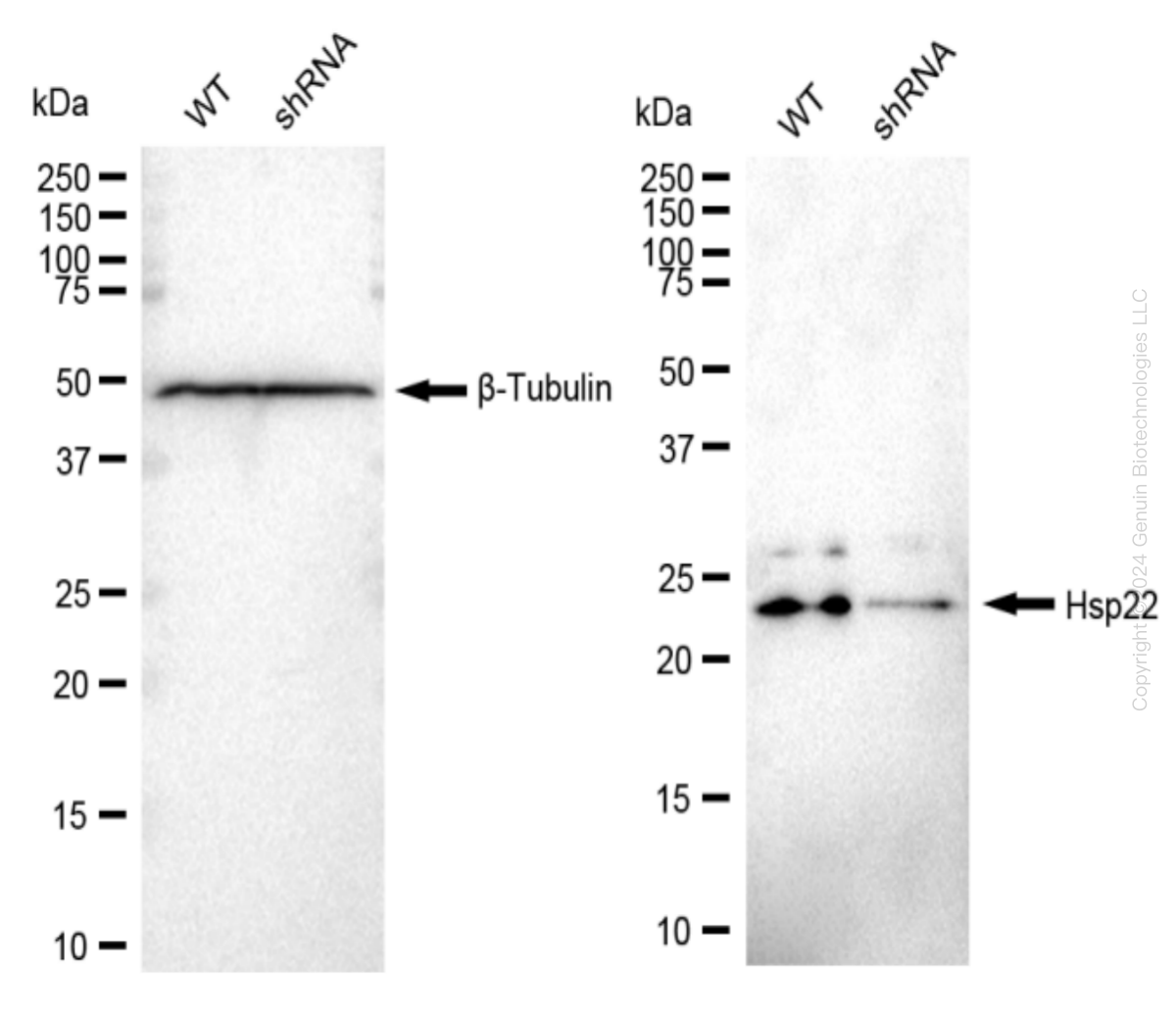
    Western blotting analysis. HSPB8 protein expression in wild-type (WT) and shRNA knockdown (KD) 293T cells was detected using Western blotting. β-Tubulin served as a loading control. The blots were incubated with primary antibodies against HSPB8 and β-Tubulin, respectively, followed by incubating with HRP-conjugated goat anti-rabbit secondary antibody. Images were developed using FeQ™ ECL Substrate Kit.

    Western blotting analysis. HSPB8 protein expression in wild-type (WT) and shRNA knockdown (KD) 293T cells was detected using Western blotting. β-Tubulin served as a loading control. The blots were incubated with primary antibodies against HSPB8 and β-Tubulin, respectively, followed by incubating with HRP-conjugated goat anti-rabbit secondary antibody. Images were developed using FeQ™ ECL Substrate Kit.
    Hsp22 (HSPB8) Human Knockdown Lysate
    WB-validated HSPB8 Knockdown 293T Cell Lysate.
    Tag Free
    293T
    100 ug
    $589.00
    5 Days*
  • Desmin (DES) Mouse Knockdown Lysate

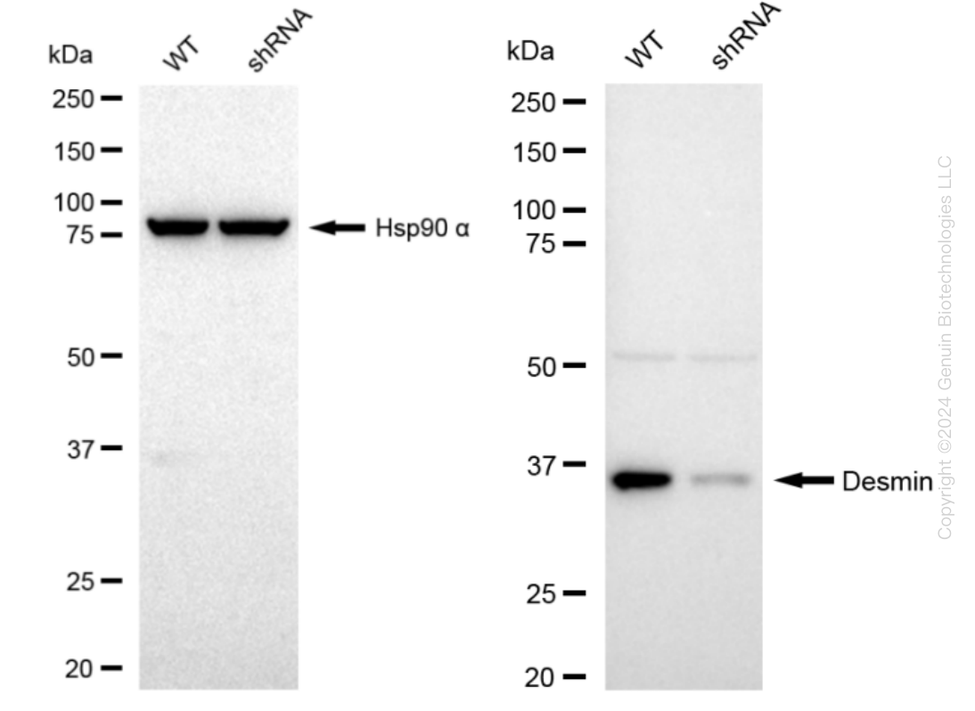
    Western blotting analysis. DES protein expression in wild-type (WT) and shRNA knockdown (KD) C2C12 cells was detected using Western blotting. Hsp90 α served as a loading control. The blots were incubated with primary antibodies against DES and Hsp90 α, respectively, followed by incubating with HRP-conjugated goat anti-rabbit secondary antibody. Images were developed using FeQ™ ECL Substrate Kit.

    Western blotting analysis. DES protein expression in wild-type (WT) and shRNA knockdown (KD) C2C12 cells was detected using Western blotting. Hsp90 α served as a loading control. The blots were incubated with primary antibodies against DES and Hsp90 α, respectively, followed by incubating with HRP-conjugated goat anti-rabbit secondary antibody. Images were developed using FeQ™ ECL Substrate Kit.
    Desmin (DES) Mouse Knockdown Lysate
    WB-validated DES Knockdown C2C12 Cell Lysate.
    Tag Free
    100 ug
    $589.00
    5 Days*
  • AAMP Human Knockdown Lysate

    Western blotting analysis. AAMP protein expression in wild-type (WT) and shRNA knockdown (KD) HeLa cells was detected using Western blotting. GAPDH served as a loading control. The blots were incubated with primary antibodies against AAMP and GAPDH, respectively, followed by incubating with HRP-conjugated goat anti-rabbit secondary antibody. Images were developed using FeQ™ ECL Substrate Kit.

    Western blotting analysis. AAMP protein expression in wild-type (WT) and shRNA knockdown (KD) HeLa cells was detected using Western blotting. GAPDH served as a loading control. The blots were incubated with primary antibodies against AAMP and GAPDH, respectively, followed by incubating with HRP-conjugated goat anti-rabbit secondary antibody. Images were developed using FeQ™ ECL Substrate Kit.
    AAMP Human Knockdown Lysate
    WB-validated AAMP Knockdown HeLa Cell Lysate.
    Tag Free
    HeLa
    100 ug
    $589.00
    5 Days*
  • ABCD1 Human Knockdown Lysate

    Western blotting analysis. ABCD1 protein expression in wild-type (WT) and shRNA knockdown (KD) HeLa cells was detected using Western blotting. β-Tubulin served as a loading control. The blots were incubated with primary antibodies against ABCD1 and β-Tubulin, respectively, followed by incubating with HRP-conjugated goat anti-rabbit secondary antibody. Images were developed using FeQ™ ECL Substrate Kit.

    Western blotting analysis. ABCD1 protein expression in wild-type (WT) and shRNA knockdown (KD) HeLa cells was detected using Western blotting. β-Tubulin served as a loading control. The blots were incubated with primary antibodies against ABCD1 and β-Tubulin, respectively, followed by incubating with HRP-conjugated goat anti-rabbit secondary antibody. Images were developed using FeQ™ ECL Substrate Kit.
    ABCD1 Human Knockdown Lysate
    WB-validated ABCD1 Knockdown HeLa Cell Lysate.
    Tag Free
    HeLa
    100 ug
    $589.00
    5 Days*
  • ABHD5 Human Knockdown Lysate

    Western blotting analysis. ABHD5 protein expression in wild-type (WT) and shRNA knockdown (KD) HeLa cells was detected using Western blotting. Hsp90 α served as a loading control. The blots were incubated with primary antibodies against ABHD5 and Hsp90 α, respectively, followed by incubating with HRP-conjugated goat anti-rabbit secondary antibody. Images were developed using FeQ™ ECL Substrate Kit.

    Western blotting analysis. ABHD5 protein expression in wild-type (WT) and shRNA knockdown (KD) HeLa cells was detected using Western blotting. Hsp90 α served as a loading control. The blots were incubated with primary antibodies against ABHD5 and Hsp90 α, respectively, followed by incubating with HRP-conjugated goat anti-rabbit secondary antibody. Images were developed using FeQ™ ECL Substrate Kit.
    ABHD5 Human Knockdown Lysate
    WB-validated ABHD5 Knockdown HeLa Cell Lysate.
    Tag Free
    HeLa
    100 ug
    $589.00
    5 Days*
  • ACAA2 Human Knockdown Lysate

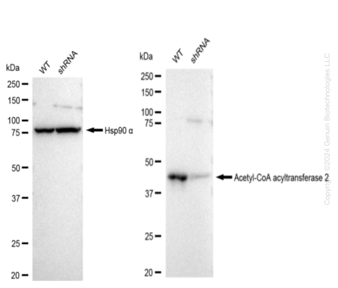
    Western blotting analysis. ACAA2 protein expression in wild-type (WT) and shRNA knockdown (KD) HeLa cells was detected using Western blotting.  Hsp90 α served as a loading control. The blots were incubated with primary antibodies (Cat#61563, 1:5,000) against ACAA2 and Hsp90 α , respectively, followed by incubating with HRP-conjugated goat anti-rabbit secondary antibody (Cat#201, 1:20,000). Images were developed using FeQ™ ECL Substrate Kit (Cat#226).

    Western blotting analysis. ACAA2 protein expression in wild-type (WT) and shRNA knockdown (KD) HeLa cells was detected using Western blotting.  Hsp90 α served as a loading control. The blots were incubated with primary antibodies (Cat#61563, 1:5,000) against ACAA2 and Hsp90 α , respectively, followed by incubating with HRP-conjugated goat anti-rabbit secondary antibody (Cat#201, 1:20,000). Images were developed using FeQ™ ECL Substrate Kit (Cat#226).
    ACAA2 Human Knockdown Lysate
    WB-validated ACAA2 Knockdown HeLa Cell Lysate.
    Tag Free
    HeLa
    100 ug
    $589.00
    5 Days*
  • ACADVL Human Knockdown Lysate

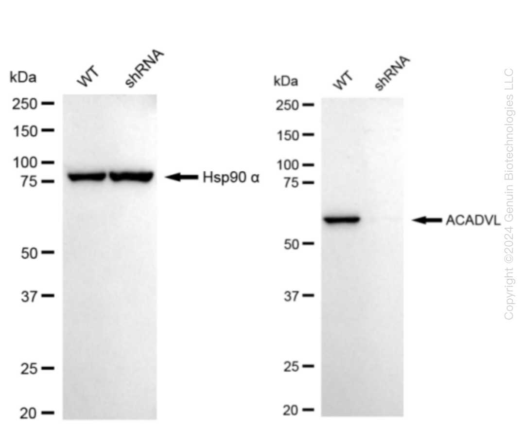
    Western blotting analysis. ACADVL protein expression in wild-type (WT) and shRNA knockdown (KD) HeLa cells was detected using Western blotting. Hsp90 α served as a loading control. The blots were incubated with primary antibodies against ACADVL and  Hsp90 α, respectively, followed by incubating with HRP-conjugated goat anti-rabbit secondary antibody. Images were developed using FeQ™ ECL Substrate Kit.

    Western blotting analysis. ACADVL protein expression in wild-type (WT) and shRNA knockdown (KD) HeLa cells was detected using Western blotting. Hsp90 α served as a loading control. The blots were incubated with primary antibodies against ACADVL and  Hsp90 α, respectively, followed by incubating with HRP-conjugated goat anti-rabbit secondary antibody. Images were developed using FeQ™ ECL Substrate Kit.
    ACADVL Human Knockdown Lysate
    WB-validated ACADVL Knockdown HeLa Cell Lysate.
    Tag Free
    HeLa
    100 ug
    $589.00
    5 Days*
  • Centaurin beta 2 (ACAP2) Human Knockdown Lysate

    Western blotting analysis. ACAP2 protein expression in wild-type (WT) and shRNA knockdown (KD) HeLa cells was detected using Western blotting. GAPDH served as a loading control. The blots were incubated with primary antibodies against ACAP2 and GAPDH, respectively, followed by incubating with HRP-conjugated goat anti-rabbit secondary antibody. Images were developed using FeQ™ ECL Substrate Kit.

    Western blotting analysis. ACAP2 protein expression in wild-type (WT) and shRNA knockdown (KD) HeLa cells was detected using Western blotting. GAPDH served as a loading control. The blots were incubated with primary antibodies against ACAP2 and GAPDH, respectively, followed by incubating with HRP-conjugated goat anti-rabbit secondary antibody. Images were developed using FeQ™ ECL Substrate Kit.
    Centaurin beta 2 (ACAP2) Human Knockdown Lysate
    WB-validated ACAP2 Knockdown HeLa Cell Lysate.
    Tag Free
    HeLa
    100 ug
    $589.00
    5 Days*
  • ACAT1 Human Knockdown Lysate

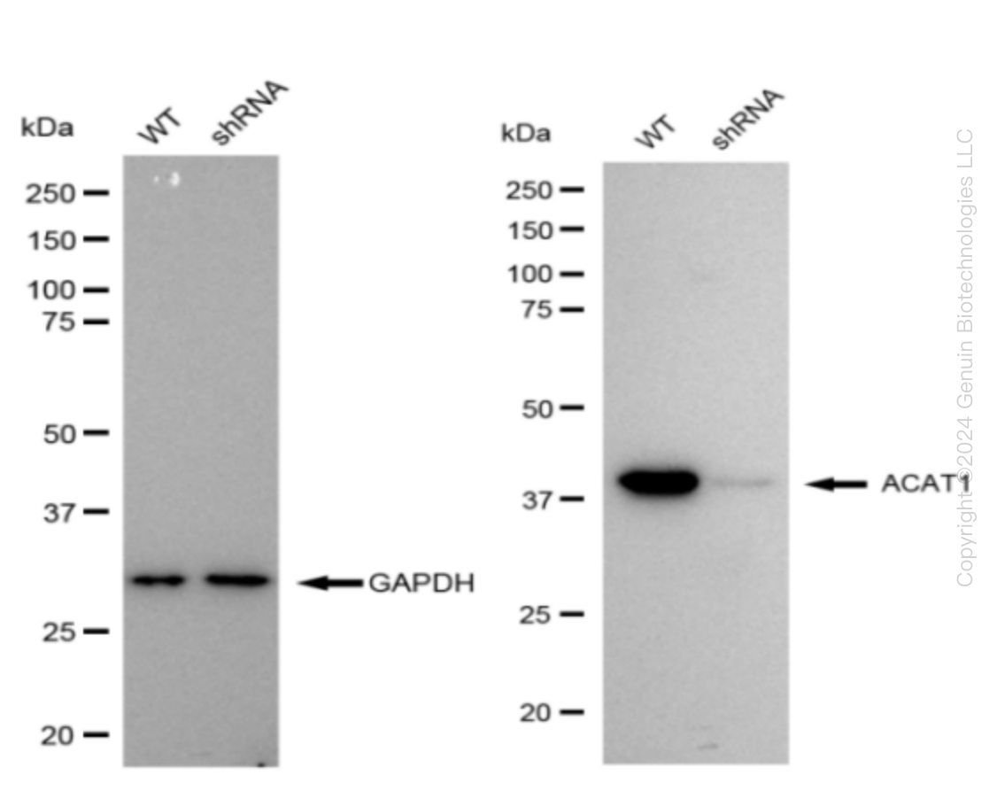
    Western blotting analysis. ACAT1 protein expression in wild-type (WT) and shRNA knockdown (KD) HeLa cells was detected using Western blotting. GAPDH served as a loading control. The blots were incubated with primary antibodies (Cat#69117, 1:5,000) against ACAT1 and GAPDH, respectively, followed by incubating with HRP-conjugated goat anti-rabbit secondary antibody (Cat#201, 1:20,000). Images were developed using FeQ™ ECL Substrate Kit (Cat#226).

    Western blotting analysis. ACAT1 protein expression in wild-type (WT) and shRNA knockdown (KD) HeLa cells was detected using Western blotting. GAPDH served as a loading control. The blots were incubated with primary antibodies (Cat#69117, 1:5,000) against ACAT1 and GAPDH, respectively, followed by incubating with HRP-conjugated goat anti-rabbit secondary antibody (Cat#201, 1:20,000). Images were developed using FeQ™ ECL Substrate Kit (Cat#226).
    ACAT1 Human Knockdown Lysate
    WB-validated ACAT1 Knockdown HeLa Cell Lysate.
    Tag Free
    HeLa
    100 ug
    $589.00
    5 Days*
  • ACAT2 Human Knockdown Lysate

    Western blotting analysis. ACAT2 protein expression in wild-type (WT) and shRNA knockdown (KD) HeLa cells was detected using Western blotting. Hsp90 α served as a loading control. The blots were incubated with primary antibodies against ACAT2 and  Hsp90 α, respectively, followed by incubating with HRP-conjugated goat anti-rabbit secondary antibody. Images were developed using FeQ™ ECL Substrate Kit.

    Western blotting analysis. ACAT2 protein expression in wild-type (WT) and shRNA knockdown (KD) HeLa cells was detected using Western blotting. Hsp90 α served as a loading control. The blots were incubated with primary antibodies against ACAT2 and  Hsp90 α, respectively, followed by incubating with HRP-conjugated goat anti-rabbit secondary antibody. Images were developed using FeQ™ ECL Substrate Kit.
    ACAT2 Human Knockdown Lysate
    WB-validated ACAT2 Knockdown HeLa Cell Lysate.
    Tag Free
    HeLa
    100 ug
    $589.00
    5 Days*
  • ACBD3 Human Knockdown Lysate

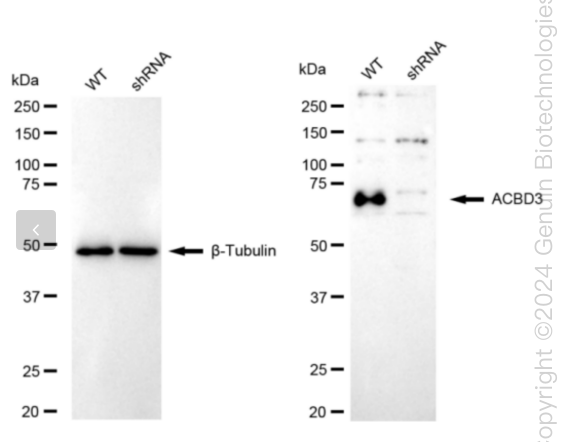
    Western blotting analysis. ACBD3 protein expression in wild-type (WT) and shRNA knockdown (KD) HeLa cells was detected using Western blotting. β-Tubulin served as a loading control. The blots were incubated with primary antibodies against ACBD3 and β-Tubulin, respectively, followed by incubating with HRP-conjugated goat anti-rabbit secondary antibody. Images were developed using FeQ™ ECL Substrate Kit.

    Western blotting analysis. ACBD3 protein expression in wild-type (WT) and shRNA knockdown (KD) HeLa cells was detected using Western blotting. β-Tubulin served as a loading control. The blots were incubated with primary antibodies against ACBD3 and β-Tubulin, respectively, followed by incubating with HRP-conjugated goat anti-rabbit secondary antibody. Images were developed using FeQ™ ECL Substrate Kit.
    ACBD3 Human Knockdown Lysate
    WB-validated ACBD3 Knockdown HeLa Cell Lysate.
    Tag Free
    HeLa
    100 ug
    $589.00
    5 Days*
  • ATP citrate lyase (ACLY) Human Knockdown Lysate

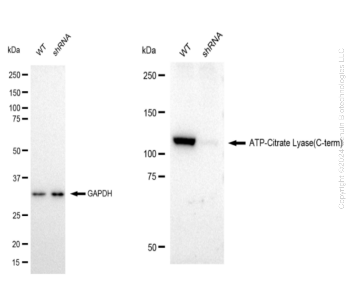
    Western blotting analysis. ACLY protein expression in wild-type (WT) and shRNA knockdown (KD) HeLa cells was detected using Western blotting. β-Tubulin served as a loading control. The blots were incubated with primary antibodies (Cat#68149, 1:5,000) against ACLY and β-Tubulin, respectively, followed by incubating with HRP-conjugated goat anti-rabbit secondary antibody (Cat#201, 1:20,000). Images were developed using FeQ™ ECL Substrate Kit (Cat#226).

    Western blotting analysis. ACLY protein expression in wild-type (WT) and shRNA knockdown (KD) HeLa cells was detected using Western blotting. β-Tubulin served as a loading control. The blots were incubated with primary antibodies (Cat#68149, 1:5,000) against ACLY and β-Tubulin, respectively, followed by incubating with HRP-conjugated goat anti-rabbit secondary antibody (Cat#201, 1:20,000). Images were developed using FeQ™ ECL Substrate Kit (Cat#226).
    ATP citrate lyase (ACLY) Human Knockdown Lysate
    WB-validated ACLY Knockdown HeLa Cell Lysate.
    Tag Free
    HeLa
    100 ug
    $589.00
    5 Days*
  • FACL4 (ACSL4) Human Knockdown Lysate

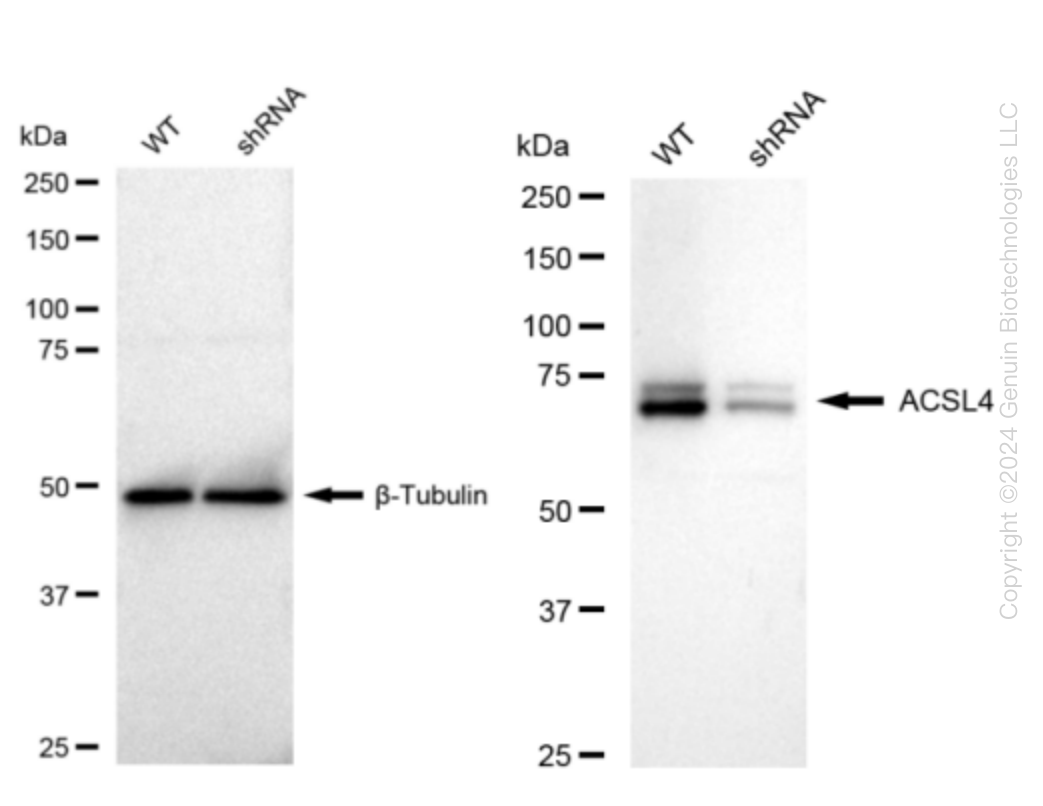
    Western blotting analysis. ACSL4 protein expression in wild-type (WT) and shRNA knockdown (KD) HeLa cells was detected using Western blotting. β-Tubulin served as a loading control. The blots were incubated with primary antibodies against ACSL4 and β-Tubulin, respectively, followed by incubating with HRP-conjugated goat anti-rabbit secondary antibody. Images were developed using FeQ™ ECL Substrate Kit.

    Western blotting analysis. ACSL4 protein expression in wild-type (WT) and shRNA knockdown (KD) HeLa cells was detected using Western blotting. β-Tubulin served as a loading control. The blots were incubated with primary antibodies against ACSL4 and β-Tubulin, respectively, followed by incubating with HRP-conjugated goat anti-rabbit secondary antibody. Images were developed using FeQ™ ECL Substrate Kit.
    FACL4 (ACSL4) Human Knockdown Lysate
    WB-validated ACSL4 Knockdown HeLa Cell Lysate.
    Tag Free
    HeLa
    100 ug
    $589.00
    5 Days*
  • BAF53A (ACTL6A) Human Knockdown Lysate

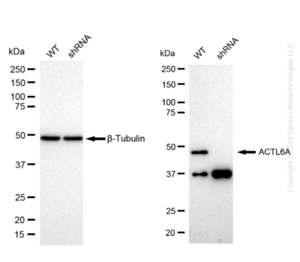
    Western blotting analysis. ACTL6A protein expression in wild-type (WT) and shRNA knockdown (KD) HeLa cells was detected using Western blotting. β-Tubulin served as a loading control. The blots were incubated with primary antibodies against ACTL6A and β-Tubulin, respectively, followed by incubating with HRP-conjugated goat anti-rabbit secondary antibody. Images were developed using FeQ™ ECL Substrate Kit.

    Western blotting analysis. ACTL6A protein expression in wild-type (WT) and shRNA knockdown (KD) HeLa cells was detected using Western blotting. β-Tubulin served as a loading control. The blots were incubated with primary antibodies against ACTL6A and β-Tubulin, respectively, followed by incubating with HRP-conjugated goat anti-rabbit secondary antibody. Images were developed using FeQ™ ECL Substrate Kit.
    BAF53A (ACTL6A) Human Knockdown Lysate
    WB-validated ACTL6A Knockdown HeLa Cell Lysate.
    Tag Free
    HeLa
    100 ug
    $589.00
    5 Days*
  • Arp3 (ACTR3) Human Knockdown Lysate

    Western blotting analysis. ACTR3 protein expression in wild-type (WT) and shRNA knockdown (KD) HeLa cells was detected using Western blotting. HSP90-Alpha served as a loading control. The blots were incubated with primary antibodies (Cat#69173, 1:5,000) against ACTR3 and HSP90-Alpha, respectively, followed by incubating with HRP-conjugated goat anti-rabbit secondary antibody (Cat#201, 1:20,000). Images were developed using FeQ™ ECL Substrate Kit (Cat#226).

    Western blotting analysis. ACTR3 protein expression in wild-type (WT) and shRNA knockdown (KD) HeLa cells was detected using Western blotting. HSP90-Alpha served as a loading control. The blots were incubated with primary antibodies (Cat#69173, 1:5,000) against ACTR3 and HSP90-Alpha, respectively, followed by incubating with HRP-conjugated goat anti-rabbit secondary antibody (Cat#201, 1:20,000). Images were developed using FeQ™ ECL Substrate Kit (Cat#226).
    Arp3 (ACTR3) Human Knockdown Lysate
    WB-validated ACTR3 Knockdown HeLa Cell Lysate.
    Tag Free
    HeLa
    100 ug
    $589.00
    5 Days*
  • Adiponectin Receptor 1 (ADIPOR1) Human Knockdown Lysate

    Western blotting analysis. ADIPOR1 protein expression in wild-type (WT) and shRNA knockdown (KD) HeLa cells was detected using Western blotting.  Hsp90 α α served as a loading control. The blots were incubated with primary antibodies (Cat#61573, 1:5,000) against ADIPOR1 and Hsp90 α , respectively, followed by incubating with HRP-conjugated goat anti-rabbit secondary antibody (Cat#201, 1:20,000). Images were developed using FeQ™ ECL Substrate Kit (Cat#226).

    Western blotting analysis. ADIPOR1 protein expression in wild-type (WT) and shRNA knockdown (KD) HeLa cells was detected using Western blotting.  Hsp90 α α served as a loading control. The blots were incubated with primary antibodies (Cat#61573, 1:5,000) against ADIPOR1 and Hsp90 α , respectively, followed by incubating with HRP-conjugated goat anti-rabbit secondary antibody (Cat#201, 1:20,000). Images were developed using FeQ™ ECL Substrate Kit (Cat#226).
    Adiponectin Receptor 1 (ADIPOR1) Human Knockdown Lysate
    WB-validated ADIPOR1 Knockdown HeLa Cell Lysate.
    Tag Free
    HeLa
    100 ug
    $589.00
    5 Days*
  • AdSS 2 (ADSS) Human Knockdown Lysate

    Western blotting analysis. ADSS2 protein expression in wild-type (WT) and shRNA knockdown (KD) HeLa cells was detected using Western blotting. GAPDH served as a loading control. The blots were incubated with primary antibodies (Cat#61272, 1:5,000) against ADSS2 and GAPDH, respectively, followed by incubating with HRP-conjugated goat anti-rabbit secondary antibody (Cat#201, 1:20,000). Images were developed using FeQ™ ECL Substrate Kit (Cat#226).

    Western blotting analysis. ADSS2 protein expression in wild-type (WT) and shRNA knockdown (KD) HeLa cells was detected using Western blotting. GAPDH served as a loading control. The blots were incubated with primary antibodies (Cat#61272, 1:5,000) against ADSS2 and GAPDH, respectively, followed by incubating with HRP-conjugated goat anti-rabbit secondary antibody (Cat#201, 1:20,000). Images were developed using FeQ™ ECL Substrate Kit (Cat#226).
    AdSS 2 (ADSS) Human Knockdown Lysate
    WB-validated ADSS2 Knockdown HeLa Cell Lysate.
    Tag Free
    HeLa
    100 ug
    $589.00
    5 Days*
  • AGL Human Knockdown Lysate

    Western blotting analysis. AGL protein expression in wild-type (WT) and shRNA knockdown (KD) HeLa cells was detected using Western blotting. Hsp90 α served as a loading control. The blots were incubated with primary antibodies against AGL and Hsp90 α, respectively, followed by incubating with HRP-conjugated goat anti-rabbit secondary antibody. Images were developed using FeQ™ ECL Substrate Kit.

    Western blotting analysis. AGL protein expression in wild-type (WT) and shRNA knockdown (KD) HeLa cells was detected using Western blotting. Hsp90 α  served as a loading control. The blots were incubated with primary antibodies against AGL and Hsp90 α, respectively, followed by incubating with HRP-conjugated goat anti-rabbit secondary antibody. Images were developed using FeQ™ ECL Substrate Kit.
    AGL Human Knockdown Lysate
    WB-validated AGL Knockdown HeLa Cell Lysate.
    Tag Free
    HeLa
    100 ug
    $589.00
    5 Days*
  • Argonaute 2 (AGO2) Human Knockdown Lysate

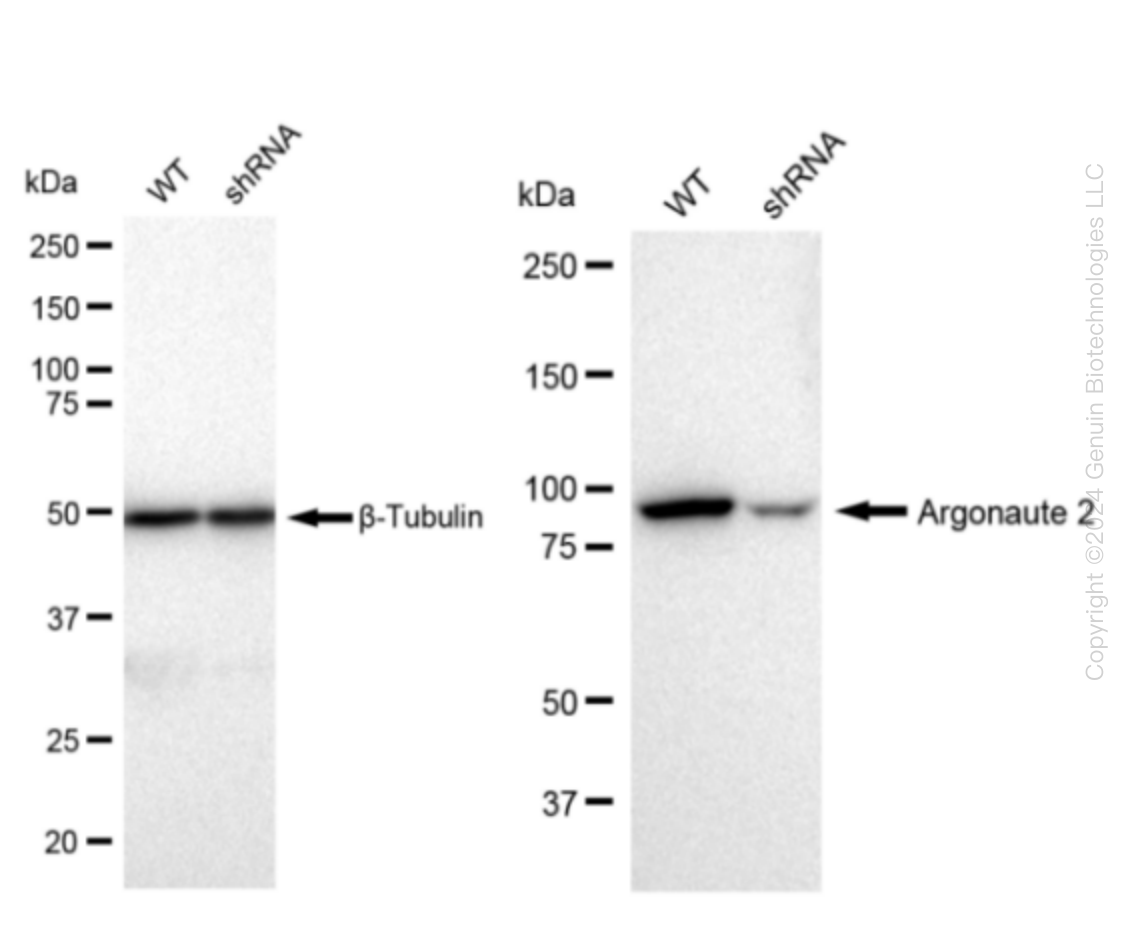
    Western blotting analysis. AGO2 protein expression in wild-type (WT) and shRNA knockdown (KD) HeLa cells was detected using Western blotting. β-Tubulin served as a loading control. The blots were incubated with primary antibodies against AGO2 and β-Tubulin, respectively, followed by incubating with HRP-conjugated goat anti-rabbit secondary antibody. Images were developed using FeQ™ ECL Substrate Kit.

    Western blotting analysis. AGO2 protein expression in wild-type (WT) and shRNA knockdown (KD) HeLa cells was detected using Western blotting. β-Tubulin served as a loading control. The blots were incubated with primary antibodies against AGO2 and β-Tubulin, respectively, followed by incubating with HRP-conjugated goat anti-rabbit secondary antibody. Images were developed using FeQ™ ECL Substrate Kit.
    Argonaute 2 (AGO2) Human Knockdown Lysate
    WB-validated AGO2 Knockdown HeLa Cell Lysate.
    Tag Free
    HeLa
    100 ug
    $589.00
    5 Days*
  • Aryl hydrocarbon Receptor (AHR) Human Knockdown Lysate

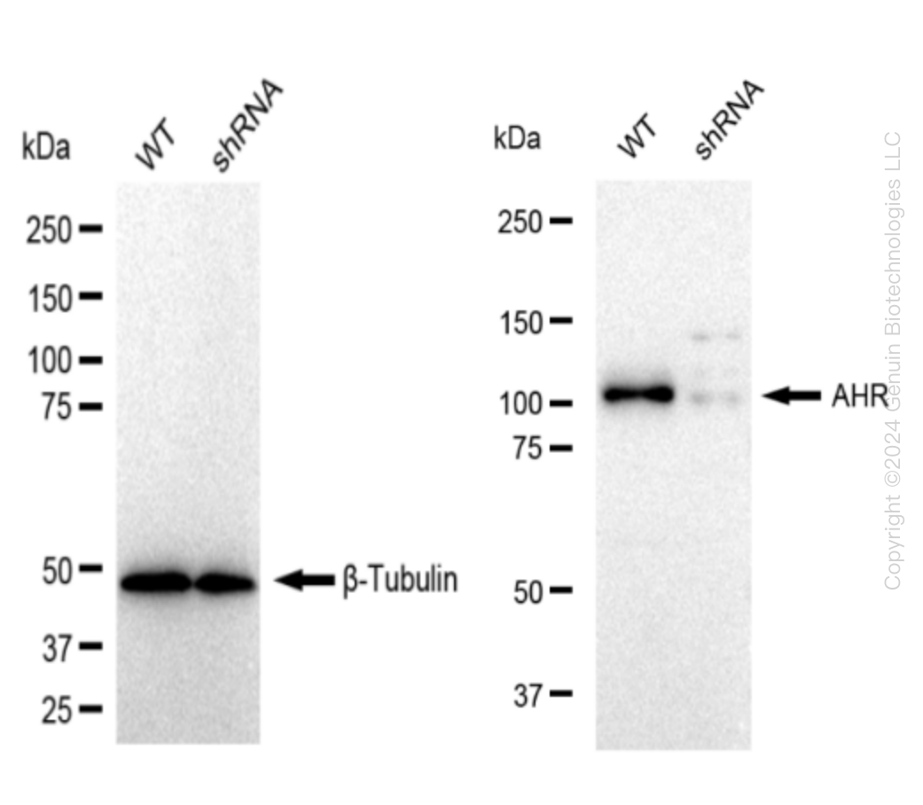
    Western blotting analysis. AHR protein expression in wild-type (WT) and shRNA knockdown (KD) HeLa cells was detected using Western blotting. β-Tubulin served as a loading control. The blots were incubated with primary antibodies against AHR and β-Tubulin, respectively, followed by incubating with HRP-conjugated goat anti-rabbit secondary antibody. Images were developed using FeQ™ ECL Substrate Kit.

    Western blotting analysis. AHR protein expression in wild-type (WT) and shRNA knockdown (KD) HeLa cells was detected using Western blotting. β-Tubulin served as a loading control. The blots were incubated with primary antibodies against AHR and β-Tubulin, respectively, followed by incubating with HRP-conjugated goat anti-rabbit secondary antibody. Images were developed using FeQ™ ECL Substrate Kit.
    Aryl hydrocarbon Receptor (AHR) Human Knockdown Lysate
    WB-validated AHR Knockdown HeLa Cell Lysate.
    Tag Free
    HeLa
    100 ug
    $589.00
    5 Days*
  • AK3L1 (AK4) Human Knockdown Lysate

    Western blotting analysis. AK4 protein expression in wild-type (WT) and shRNA knockdown (KD) HeLa cells was detected using Western blotting. Hsp90 α served as a loading control. The blots were incubated with primary antibodies against AK4 and Hsp90 α, respectively, followed by incubating with HRP-conjugated goat anti-rabbit secondary antibody. Images were developed using FeQ™ ECL Substrate Kit.

    Western blotting analysis. AK4 protein expression in wild-type (WT) and shRNA knockdown (KD) HeLa cells was detected using Western blotting. Hsp90 α served as a loading control. The blots were incubated with primary antibodies against AK4 and Hsp90 α, respectively, followed by incubating with HRP-conjugated goat anti-rabbit secondary antibody. Images were developed using FeQ™ ECL Substrate Kit.
    AK3L1 (AK4) Human Knockdown Lysate
    WB-validated AK4 Knockdown HeLa Cell Lysate.
    Tag Free
    HeLa
    100 ug
    $589.00
    5 Days*
  • PRAS40 (AKT1S1) Human Knockdown Lysate

    Western blotting analysis. AKT1S1 protein expression in wild-type (WT) and shRNA knockdown (KD) HeLa cells was detected using Western blotting. Hsp90 α served as a loading control. The blots were incubated with primary antibodies against AKT1S1 and  Hsp90 α, respectively, followed by incubating with HRP-conjugated goat anti-rabbit secondary antibody. Images were developed using FeQ™ ECL Substrate Kit.

    Western blotting analysis. AKT1S1 protein expression in wild-type (WT) and shRNA knockdown (KD) HeLa cells was detected using Western blotting. Hsp90 α served as a loading control. The blots were incubated with primary antibodies against AKT1S1 and  Hsp90 α, respectively, followed by incubating with HRP-conjugated goat anti-rabbit secondary antibody. Images were developed using FeQ™ ECL Substrate Kit.
    PRAS40 (AKT1S1) Human Knockdown Lysate
    WB-validated AKT1S1 Knockdown HeLa Cell Lysate.
    Tag Free
    HeLa
    100 ug
    $589.00
    5 Days*
  • CD166 (ALCAM) Human Knockdown Lysate

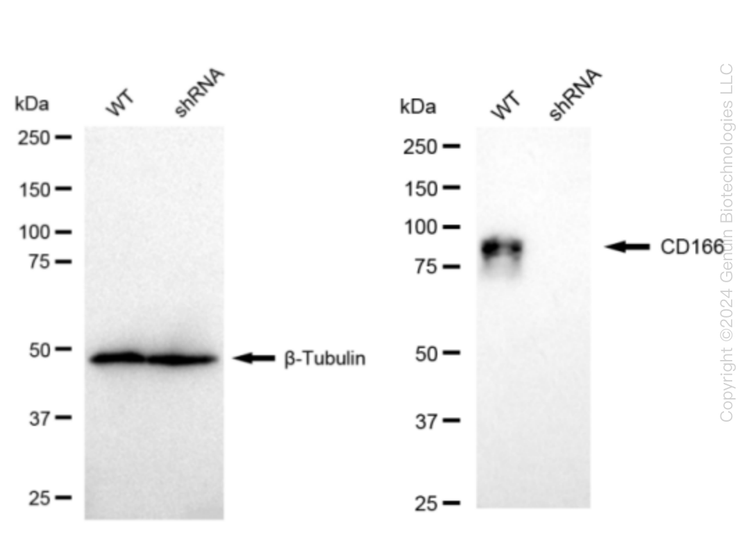
    Western blotting analysis. ALCAM protein expression in wild-type (WT) and shRNA knockdown (KD) HeLa cells was detected using Western blotting. β-Tubulin served as a loading control. The blots were incubated with primary antibodies against ALCAM and β-Tubulin, respectively, followed by incubating with HRP-conjugated goat anti-rabbit secondary antibody. Images were developed using FeQ™ ECL Substrate Kit.

    Western blotting analysis. ALCAM protein expression in wild-type (WT) and shRNA knockdown (KD) HeLa cells was detected using Western blotting. β-Tubulin served as a loading control. The blots were incubated with primary antibodies against ALCAM and β-Tubulin, respectively, followed by incubating with HRP-conjugated goat anti-rabbit secondary antibody. Images were developed using FeQ™ ECL Substrate Kit.
    CD166 (ALCAM) Human Knockdown Lysate
    WB-validated ALCAM Knockdown HeLa Cell Lysate.
    Tag Free
    HeLa
    100 ug
    $589.00
    5 Days*
  • CD13 (ANPEP) Human Knockdown Lysate

    Western blotting analysis. ANPEP protein expression in wild-type (WT) and shRNA knockdown (KD) HeLa cells was detected using Western blotting. β-Tubulin served as a loading control. The blots were incubated with primary antibodies against ANPEP and β-Tubulin, respectively, followed by incubating with HRP-conjugated goat anti-rabbit secondary antibody. Images were developed using FeQ™ ECL Substrate Kit.

    Western blotting analysis. ANPEP protein expression in wild-type (WT) and shRNA knockdown (KD) HeLa cells was detected using Western blotting. β-Tubulin served as a loading control. The blots were incubated with primary antibodies against ANPEP and β-Tubulin, respectively, followed by incubating with HRP-conjugated goat anti-rabbit secondary antibody. Images were developed using FeQ™ ECL Substrate Kit.
    CD13 (ANPEP) Human Knockdown Lysate
    WB-validated ANPEP Knockdown HeLa Cell Lysate.
    Tag Free
    HeLa
    100 ug
    $589.00
    5 Days*
  • Annexin A10 (ANXA10) Human Knockdown Lysate

    Western blotting analysis. ANXA10 protein expression in wild-type (WT) and shRNA knockdown (KD) HeLa cells was detected using Western blotting.  β-Tubulin served as a loading control. The blots were incubated with primary antibodies (Cat#61578, 1:5,000) against ANXA10 and β-Tubulin , respectively, followed by incubating with HRP-conjugated goat anti-rabbit secondary antibody (Cat#201, 1:20,000). Images were developed using FeQ™ ECL Substrate Kit (Cat#226).

    Western blotting analysis. ANXA10 protein expression in wild-type (WT) and shRNA knockdown (KD) HeLa cells was detected using Western blotting.  β-Tubulin served as a loading control. The blots were incubated with primary antibodies (Cat#61578, 1:5,000) against ANXA10 and β-Tubulin , respectively, followed by incubating with HRP-conjugated goat anti-rabbit secondary antibody (Cat#201, 1:20,000). Images were developed using FeQ™ ECL Substrate Kit (Cat#226).
    Annexin A10 (ANXA10) Human Knockdown Lysate
    WB-validated ANXA10 Knockdown HeLa Cell Lysate.
    Tag Free
    HeLa
    100 ug
    $589.00
    5 Days*
  • Annexin V (ANXA5) Human Knockdown Lysate

    Western blotting analysis. ANXA5 protein expression in wild-type (WT) and shRNA knockdown (KD) HeLa cells was detected using Western blotting. GAPDH served as a loading control. The blots were incubated with primary antibodies (Cat#69585, 1:10,000) against ANXA5 and GAPDH, respectively, followed by incubating with HRP-conjugated goat anti-rabbit secondary antibody (Cat#201, 1:20,000). Images were developed using FeQ™ ECL Substrate Kit (Cat#226).

    Western blotting analysis. ANXA5 protein expression in wild-type (WT) and shRNA knockdown (KD) HeLa cells was detected using Western blotting. GAPDH served as a loading control. The blots were incubated with primary antibodies (Cat#69585, 1:10,000) against ANXA5 and GAPDH, respectively, followed by incubating with HRP-conjugated goat anti-rabbit secondary antibody (Cat#201, 1:20,000). Images were developed using FeQ™ ECL Substrate Kit (Cat#226).
    Annexin V (ANXA5) Human Knockdown Lysate
    WB-validated ANXA5 Knockdown HeLa Cell Lysate.
    Tag Free
    HeLa
    100 ug
    $589.00
    5 Days*
  • Annexin VII (ANXA7) Human Knockdown Lysate

    Western blotting analysis. ANXA7 protein expression in wild-type (WT) and shRNA knockdown (KD) HeLa cells was detected using Western blotting. Hsp90 α served as a loading control. The blots were incubated with primary antibodies against ANXA7 and Hsp90 α, respectively, followed by incubating with HRP-conjugated goat anti-rabbit secondary antibody. Images were developed using FeQ™ ECL Substrate Kit.

    Western blotting analysis. ANXA7 protein expression in wild-type (WT) and shRNA knockdown (KD) HeLa cells was detected using Western blotting. Hsp90 α served as a loading control. The blots were incubated with primary antibodies against ANXA7 and Hsp90 α, respectively, followed by incubating with HRP-conjugated goat anti-rabbit secondary antibody. Images were developed using FeQ™ ECL Substrate Kit.
    Annexin VII (ANXA7) Human Knockdown Lysate
    WB-validated ANXA7 Knockdown HeLa Cell Lysate.
    Tag Free
    HeLa
    100 ug
    $589.00
    5 Days*
  • AP2M1 Human Knockdown Lysate

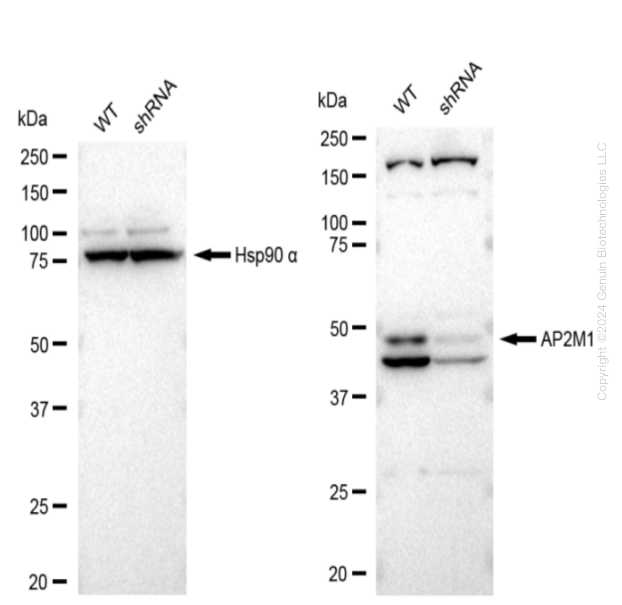
    Western blotting analysis. AP2M1 protein expression in wild-type (WT) and shRNA knockdown (KD) HeLa cells was detected using Western blotting.  Hsp90 α served as a loading control. The blots were incubated with primary antibodies (Cat#61579, 1:5,000) against AP2M1 and Hsp90 α , respectively, followed by incubating with HRP-conjugated goat anti-rabbit secondary antibody (Cat#201, 1:20,000). Images were developed using FeQ™ ECL Substrate Kit (Cat#226).

    Western blotting analysis. AP2M1 protein expression in wild-type (WT) and shRNA knockdown (KD) HeLa cells was detected using Western blotting.  Hsp90 α served as a loading control. The blots were incubated with primary antibodies (Cat#61579, 1:5,000) against AP2M1 and Hsp90 α , respectively, followed by incubating with HRP-conjugated goat anti-rabbit secondary antibody (Cat#201, 1:20,000). Images were developed using FeQ™ ECL Substrate Kit (Cat#226).
    AP2M1 Human Knockdown Lysate
    WB-validated AP2M1 Knockdown HeLa Cell Lysate.
    Tag Free
    HeLa
    100 ug
    $589.00
    5 Days*
  • Desmoplakin (DSP) Human Knockdown Lysate

    Western blotting analysis. DSP protein expression in wild-type (WT) and shRNA knockdown (KD) HeLa cells was detected using Western blotting. Hsp90 α served as a loading control. The blots were incubated with primary antibodies against DSP and Hsp90 α, respectively, followed by incubating with HRP-conjugated goat anti-rabbit secondary antibody. Images were developed using FeQ™ ECL Substrate Kit.

    Western blotting analysis. DSP protein expression in wild-type (WT) and shRNA knockdown (KD) HeLa cells was detected using Western blotting. Hsp90 α served as a loading control. The blots were incubated with primary antibodies against DSP and Hsp90 α, respectively, followed by incubating with HRP-conjugated goat anti-rabbit secondary antibody. Images were developed using FeQ™ ECL Substrate Kit.
    Desmoplakin (DSP) Human Knockdown Lysate
    WB-validated DSP Knockdown HeLa Cell Lysate.
    Tag Free
    HeLa
    100 ug
    $589.00
    5 Days*
  • Dynamin 1 (DNM1) Human Knockdown Lysate

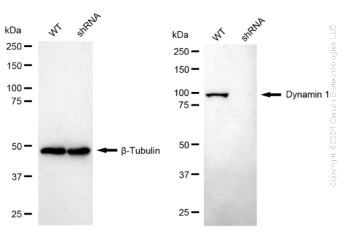
    Western blotting analysis. DNM1 protein expression in wild-type (WT) and shRNA knockdown (KD) HeLa cells was detected using Western blotting. β-Tubulin served as a loading control. The blots were incubated with primary antibodies against DNM1 and β-Tubulin, respectively, followed by incubating with HRP-conjugated goat anti-rabbit secondary antibody. Images were developed using FeQ™ ECL Substrate Kit.

    Western blotting analysis. DNM1 protein expression in wild-type (WT) and shRNA knockdown (KD) HeLa cells was detected using Western blotting. β-Tubulin served as a loading control. The blots were incubated with primary antibodies against DNM1 and β-Tubulin, respectively, followed by incubating with HRP-conjugated goat anti-rabbit secondary antibody. Images were developed using FeQ™ ECL Substrate Kit.
    Dynamin 1 (DNM1) Human Knockdown Lysate
    WB-validated DNM1 Knockdown HeLa Cell Lysate.
    Tag Free
    HeLa
    100 ug
    $589.00
    5 Days*
  • DPP3 Human Knockdown Lysate

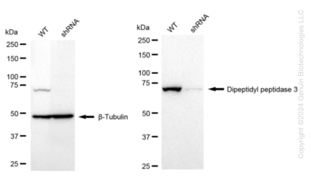
    Western blotting analysis. DPP3 protein expression in wild-type (WT) and shRNA knockdown (KD) HeLa cells was detected using Western blotting. β-Tubulin served as a loading control. The blots were incubated with primary antibodies against DPP3 and β-Tubulin, respectively, followed by incubating with HRP-conjugated goat anti-rabbit secondary antibody. Images were developed using FeQ™ ECL Substrate Kit.

    Western blotting analysis. DPP3 protein expression in wild-type (WT) and shRNA knockdown (KD) HeLa cells was detected using Western blotting. β-Tubulin served as a loading control. The blots were incubated with primary antibodies against DPP3 and β-Tubulin, respectively, followed by incubating with HRP-conjugated goat anti-rabbit secondary antibody. Images were developed using FeQ™ ECL Substrate Kit.
    DPP3 Human Knockdown Lysate
    WB-validated DPP3 Knockdown HeLa Cell Lysate.
    Tag Free
    HeLa
    100 ug
    $589.00
    5 Days*
  • DPYD Human Knockdown Lysate

    Western blotting analysis. DPYD protein expression in wild-type (WT) and shRNA knockdown (KD) HeLa cells was detected using Western blotting. β-Tubulin served as a loading control. The blots were incubated with primary antibodies against DPYD and β-Tubulin, respectively, followed by incubating with HRP-conjugated goat anti-rabbit secondary antibody. Images were developed using FeQ™ ECL Substrate Kit.

    Western blotting analysis. DPYD protein expression in wild-type (WT) and shRNA knockdown (KD) HeLa cells was detected using Western blotting. β-Tubulin served as a loading control. The blots were incubated with primary antibodies against DPYD and β-Tubulin, respectively, followed by incubating with HRP-conjugated goat anti-rabbit secondary antibody. Images were developed using FeQ™ ECL Substrate Kit.
    DPYD Human Knockdown Lysate
    WB-validated DPYD Knockdown HeLa Cell Lysate.
    Tag Free
    HeLa
    100 ug
    $589.00
    5 Days*
  • DNAJC12 Human Knockdown Lysate

    Western blotting analysis. DNAJC12 protein expression in wild-type (WT) and shRNA knockdown (KD) HeLa cells was detected using Western blotting. β-Tubulin served as a loading control. The blots were incubated with primary antibodies against DNAJC12 and β-Tubulin, respectively, followed by incubating with HRP-conjugated goat anti-rabbit secondary antibody. Images were developed using FeQ™ ECL Substrate Kit.

    Western blotting analysis. DNAJC12 protein expression in wild-type (WT) and shRNA knockdown (KD) HeLa cells was detected using Western blotting. β-Tubulin served as a loading control. The blots were incubated with primary antibodies against DNAJC12 and β-Tubulin, respectively, followed by incubating with HRP-conjugated goat anti-rabbit secondary antibody. Images were developed using FeQ™ ECL Substrate Kit.
    DNAJC12 Human Knockdown Lysate
    WB-validated DNAJC12 Knockdown HeLa Cell Lysate.
    Tag Free
    HeLa
    100 ug
    $589.00
    5 Days*
  • Desmocollin 1 (DSC1) Human Knockdown Lysate

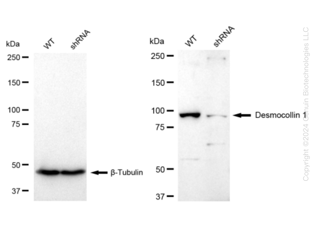
    Western blotting analysis. DSC1 protein expression in wild-type (WT) and shRNA knockdown (KD) HeLa cells was detected using Western blotting. β-Tubulin served as a loading control. The blots were incubated with primary antibodies against DSC1 and β-Tubulin, respectively, followed by incubating with HRP-conjugated goat anti-rabbit secondary antibody. Images were developed using FeQ™ ECL Substrate Kit.

    Western blotting analysis. DSC1 protein expression in wild-type (WT) and shRNA knockdown (KD) HeLa cells was detected using Western blotting. β-Tubulin served as a loading control. The blots were incubated with primary antibodies against DSC1 and β-Tubulin, respectively, followed by incubating with HRP-conjugated goat anti-rabbit secondary antibody. Images were developed using FeQ™ ECL Substrate Kit.
    Desmocollin 1 (DSC1) Human Knockdown Lysate
    WB-validated DSC1 Knockdown HeLa Cell Lysate.
    Tag Free
    HeLa
    100 ug
    $589.00
    5 Days*
  • AP2S1 Human Knockdown Lysate

    Western blotting analysis. AP2S1 protein expression in wild-type (WT) and shRNA knockdown (KD) HeLa cells was detected using Western blotting.  β-Tubulin α served as a loading control. The blots were incubated with primary antibodies (Cat#61329, 1:5,000) against AP2S1 and β-Tubulin , respectively, followed by incubating with HRP-conjugated goat anti-rabbit secondary antibody (Cat#201, 1:20,000). Images were developed using FeQ™ ECL Substrate Kit (Cat#226).

    Western blotting analysis. AP2S1 protein expression in wild-type (WT) and shRNA knockdown (KD) HeLa cells was detected using Western blotting.  β-Tubulin α served as a loading control. The blots were incubated with primary antibodies (Cat#61329, 1:5,000) against AP2S1 and β-Tubulin , respectively, followed by incubating with HRP-conjugated goat anti-rabbit secondary antibody (Cat#201, 1:20,000). Images were developed using FeQ™ ECL Substrate Kit (Cat#226).
    AP2S1 Human Knockdown Lysate
    WB-validated AP2S1 Knockdown HeLa Cell Lysate.
    Tag Free
    HeLa
    100 ug
    $589.00
    5 Days*
  • DIAPH1 Human Knockdown Lysate

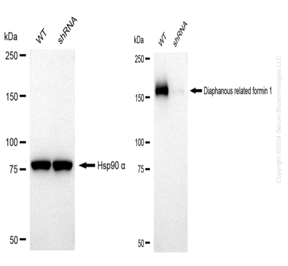
    Western blotting analysis. DIAPH1 protein expression in wild-type (WT) and shRNA knockdown (KD) HeLa cells was detected using Western blotting. Hsp90 α served as a loading control. The blots were incubated with primary antibodies against DIAPH1 and  Hsp90 α, respectively, followed by incubating with HRP-conjugated goat anti-rabbit secondary antibody. Images were developed using FeQ™ ECL Substrate Kit.

    Western blotting analysis. DIAPH1 protein expression in wild-type (WT) and shRNA knockdown (KD) HeLa cells was detected using Western blotting. Hsp90 α served as a loading control. The blots were incubated with primary antibodies against DIAPH1 and  Hsp90 α, respectively, followed by incubating with HRP-conjugated goat anti-rabbit secondary antibody. Images were developed using FeQ™ ECL Substrate Kit.
    DIAPH1 Human Knockdown Lysate
    WB-validated DIAPH1 Knockdown HeLa Cell Lysate.
    Tag Free
    HeLa
    100 ug
    $589.00
    5 Days*
  • Pyruvate Dehydrogenase E2 (DLAT) Human Knockdown Lysate

    Western blotting analysis. DLAT protein expression in wild-type (WT) and shRNA knockdown (KD) HeLa cells was detected using Western blotting. GAPDH served as a loading control. The blots were incubated with primary antibodies against DLAT and GAPDH, respectively, followed by incubating with HRP-conjugated goat anti-rabbit secondary antibody. Images were developed using FeQ™ ECL Substrate Kit.

    Western blotting analysis. DLAT protein expression in wild-type (WT) and shRNA knockdown (KD) HeLa cells was detected using Western blotting. GAPDH served as a loading control. The blots were incubated with primary antibodies against DLAT and GAPDH, respectively, followed by incubating with HRP-conjugated goat anti-rabbit secondary antibody. Images were developed using FeQ™ ECL Substrate Kit.
    Pyruvate Dehydrogenase E2 (DLAT) Human Knockdown Lysate
    WB-validated DLAT Knockdown HeLa Cell Lysate.
    Tag Free
    HeLa
    100 ug
    $589.00
    5 Days*