Phospho-Akt

19 Items

Set Ascending Direction
  • Applications
    Reactivities
    Conjugation
    Size
    Delivery Time
  • Phospho-AKT1 Rabbit Monoclonal Antibody

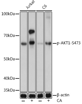
    Western blot analysis of various lysates using Phospho-AKT1-S473 mAb (TA420054) at 1:1000 dilution. Both Jurkat cells and C6 cells were treated by Calyculin A (100 nM) at 37℃ for 30 minutes.Secondary antibody: HRP-conjugated Goat anti-Rabbit IgG (H+L) at 1:10000 dilution.Lysates/proteins: 25μg per lane.Blocking buffer: 3% nonfat dry milk in TBST.Detection: ECL Basic Kit .Exposure time: 1s.

    Western blot analysis of various lysates using Phospho-AKT1-S473  mAb (TA420054) at 1:1000 dilution. Both Jurkat cells and C6 cells were treated by Calyculin A (100 nM) at 37℃ for 30 minutes.Secondary antibody: HRP-conjugated Goat anti-Rabbit IgG (H+L) at 1:10000 dilution.Lysates/proteins: 25μg per lane.Blocking buffer: 3% nonfat dry milk in TBST.Detection: ECL Basic Kit .Exposure time: 1s.
    Phospho-AKT1 Rabbit Monoclonal Antibody -
    Phospho-AKT1-S473 Rabbit mAb.
    ELISA, IHC, WB
    Human, Mouse, Rat
    Unconjugated
    100 ul
    $475.00
    3 Weeks*
  • AKT1 Mouse Monoclonal Antibody [Clone ID: 17F6.B11]

    Immunofluorescence Microscopy of Mouse Anti-AKTpS473 antibody using STED nanoscopy to evaluate AKT activation and migration. Tissue: A431 cells. Antigen retrieval: Panel A: serum starved,unstimulated cells. Panel B: serum starved, EGF stimulated for 15 mins. A massive increase in AKT-pS473 activation, as measured by intensity signal, peaked at 15 minutes and was associated with depolymerized tubulin. Staining: Panel A shows STED data (AKT-pS473, red channel) collected simultaneously with confocal signal (a-tubulin, green channel). Upon stimulation of cells with EGF, a rapid activation of AKT is observed (Panel B) along with a coincident change in the tubulin organization (yellow signal), as well as an extensive cell shape-change (cell membrane folding) and accumulation of AKTpS473 at the cell periphery.

    Immunofluorescence Microscopy of Mouse Anti-AKTpS473 antibody using STED nanoscopy to evaluate AKT activation and migration. Tissue: A431 cells. Antigen retrieval: Panel A: serum starved,unstimulated cells. Panel B: serum starved, EGF stimulated for 15 mins.
A massive increase in AKT-pS473 activation, as measured by intensity signal, peaked at 15 minutes and was associated with depolymerized tubulin.
Staining: Panel A shows STED data (AKT-pS473, red channel) collected simultaneously with confocal signal (a-tubulin, green channel). Upon stimulation of cells with EGF, a rapid activation of AKT is observed (Panel B) along with a coincident change in the tubulin organization (yellow signal), as well as an extensive cell shape-change (cell membrane folding) and accumulation of AKTpS473 at the cell periphery.
    AKT1 Mouse Monoclonal Antibody [Clone ID: 17F6.B11] -
    AKT phospho S473 Antibody.
    ELISA, FC, IF, IHC, WB
    Human, Mouse
    Unconjugated
    1 mg
    $4,200.00
    5 Days*
  • Phospho AKT (Thr308) Rabbit Polyclonal Antibody

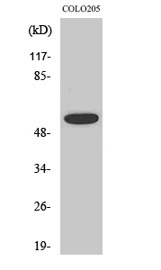
    Western blot analysis of Phospho-AKT (Thr308) in COLO205 lysates using Phospho-AKT (Thr308) antibody.

    Western blot analysis of Phospho-AKT (Thr308) in COLO205 lysates using Phospho-AKT (Thr308) antibody.
    Phospho AKT (Thr308) Rabbit Polyclonal Antibody -
    Phospho-AKT (Thr308) Rabbit polyclonal Antibody.
    ELISA, ICC, IHC, WB
    Human, Mouse, Rat
    Unconjugated
    100 ul
    $380.00
    4 Weeks*
  • AKT1 Mouse Monoclonal Antibody [Clone ID: 18F3.H11]

    Immunohistochemistry of Mouse anti-AKT pT308 antibody. Tissue: human brain cerebellum tissue (40X). Fixation: formalin fixed paraffin embedded. Antigen retrieval: not required. Primary antibody: AKT pT308 antibody at 20 µg/mL for 1 h at RT. Secondary antibody: Peroxidase rabbit secondary antibody at 1:10,000 for 45 min at RT. Localization: staining of Purkinje neurons and cell processes in the cerebellum, cytosolic as well as occasionally nuclear. Staining: AKT pT308 as precipitated red signal with hematoxylin purple nuclear counterstain.

    Immunohistochemistry of Mouse anti-AKT pT308 antibody. Tissue: human brain cerebellum tissue (40X). Fixation: formalin fixed paraffin embedded. Antigen retrieval: not required.
Primary antibody: AKT pT308 antibody at 20 µg/mL for 1 h at RT.
Secondary antibody: Peroxidase rabbit secondary antibody at 1:10,000 for 45 min at RT.
Localization: staining of Purkinje neurons and cell processes in the cerebellum, cytosolic as well as occasionally nuclear.
Staining: AKT pT308 as precipitated red signal with hematoxylin purple nuclear counterstain.
    AKT1 Mouse Monoclonal Antibody [Clone ID: 18F3.H11] -
    AKT phospho T308 Antibody.
    ELISA, FC, IHC, WB
    Human, Mouse
    Unconjugated
    1 mg
    $4,200.00
    5 Days*
  • Phospho-AKT1 Rabbit Monoclonal Antibody

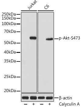
    Western blot analysis of various lysates using Phospho-Akt-S473 Rabbit mAb (TA420035) at 1:1000 dilution. Both Jurkat and C6 cells were treated by Calyculin A (100 nM) at 37℃ for 30 minutes after serum-starvation overnight.Secondary antibody: HRP-conjugated Goat anti-Rabbit IgG (H+L) at 1:10000 dilution.Lysates/proteins: 25μg per lane.Blocking buffer: 3% nonfat dry milk in TBST.Detection: ECL Basic Kit .Exposure time: 1s.

    Western blot analysis of various lysates using Phospho-Akt-S473 Rabbit mAb (TA420035) at 1:1000 dilution. Both Jurkat and C6 cells were treated by Calyculin A (100 nM) at 37℃ for 30 minutes after serum-starvation overnight.Secondary antibody: HRP-conjugated Goat anti-Rabbit IgG (H+L) at 1:10000 dilution.Lysates/proteins: 25μg per lane.Blocking buffer: 3% nonfat dry milk in TBST.Detection: ECL Basic Kit .Exposure time: 1s.
    Phospho-AKT1 Rabbit Monoclonal Antibody -
    Phospho-Akt-S473 Rabbit mAb.
    ELISA, WB
    Human, Mouse, Rat
    Unconjugated
    100 ul
    $475.00
    3 Weeks*
  • Phospho Akt (Ser473) Rabbit Polyclonal Antibody

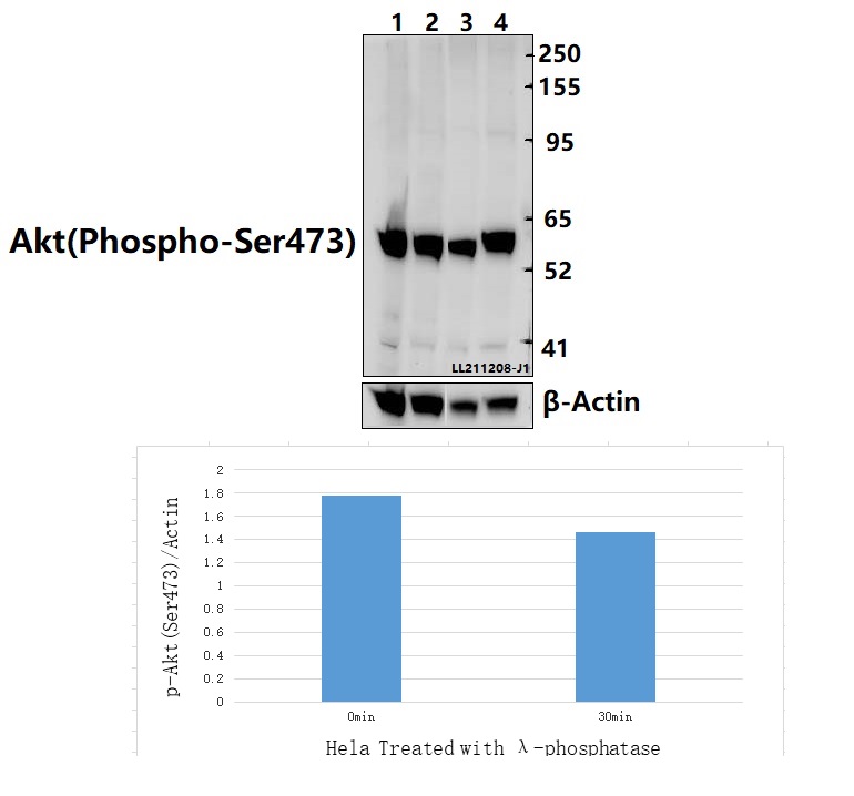
    Western blot (WB) analysis of Akt (Phospho-Ser473) polyclonal antibody at 1:5000 dilution Lane1:C6 whole cell lysate(40ug) Lane2:BV2 whole cell lysate(40ug) Lane3:Hela treated with λ-phosphatase whole cell lysate(40ug) Lane4:Hela whole cell lysate(40ug)

    Western blot (WB) analysis of Akt (Phospho-Ser473) polyclonal antibody at 1:5000 dilution
Lane1:C6 whole cell lysate(40ug)
Lane2:BV2 whole cell lysate(40ug)
Lane3:Hela treated with λ-phosphatase whole cell lysate(40ug)
Lane4:Hela whole cell lysate(40ug)
    Phospho Akt (Ser473) Rabbit Polyclonal Antibody -
    Akt (Phospho-Ser473) polyclonal antibody.
    IF, WB
    Human, Mouse, Rat
    Unconjugated
    100 ul
    $340.00
    7 Weeks*
  • Phospho Akt (Thr450) Rabbit Polyclonal Antibody

    Western blot (WB) analysis of AKT (phospho-T450) polyclonal antibody at 1:500 dilution Lane1:L02 whole cell lysate Lane2:L02 treated with EGF(0.1ng/ml,5min) whole cell lysate Lane3:L02 treated with EGF(0.1ng/ml,10min) whole cell lysate Lane4:L02 treated with EGF(0.1ng/ml,15min) whole cell lysate

    Western blot (WB) analysis of AKT (phospho-T450) polyclonal antibody at 1:500 dilution
Lane1:L02 whole cell lysate
Lane2:L02 treated with EGF(0.1ng/ml,5min) whole cell lysate
Lane3:L02 treated with EGF(0.1ng/ml,10min) whole cell lysate
Lane4:L02 treated with EGF(0.1ng/ml,15min) whole cell lysate
    Phospho Akt (Thr450) Rabbit Polyclonal Antibody -
    AKT (phospho-T450) polyclonal antibody.
    WB
    Human, Mouse, Rat
    Unconjugated
    100 ul
    $340.00
    7 Weeks*
  • Phospho-AKT1 Rabbit Monoclonal Antibody

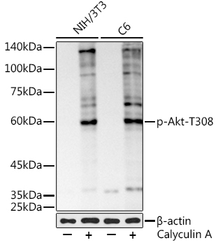
    Western blot analysis of various lysates using Phospho-Akt-T308 Rabbit mAb (TA420036) at 1:1000 dilution. NIH/3T3 and C6 cells were treated by Calyculin A (100 nM) at 37℃ for 30 minutes after serum-starvation overnight.Secondary antibody: HRP-conjugated Goat anti-Rabbit IgG (H+L) at 1:10000 dilution.Lysates/proteins: 25μg per lane.Blocking buffer: 3% nonfat dry milk in TBST.Detection: ECL Basic Kit .Exposure time: 180s.

    Western blot analysis of various lysates using Phospho-Akt-T308 Rabbit mAb (TA420036) at 1:1000 dilution. NIH/3T3 and C6 cells were treated by Calyculin A (100 nM) at 37℃ for 30 minutes after serum-starvation overnight.Secondary antibody: HRP-conjugated Goat anti-Rabbit IgG (H+L) at 1:10000 dilution.Lysates/proteins: 25μg per lane.Blocking buffer: 3% nonfat dry milk in TBST.Detection: ECL Basic Kit .Exposure time: 180s.
    Phospho-AKT1 Rabbit Monoclonal Antibody -
    Phospho-Akt-T308 Rabbit mAb.
    ELISA, WB
    Mouse, Rat
    Unconjugated
    100 ul
    $475.00
    3 Weeks*
  • Phospho AKT1 (Ser124) Rabbit Monoclonal Antibody [Clone ID: OTIRA0624]

    Western blot analysis of Phospho-AKT1 (S124) expression in (1) MCF-7 cell treated with Alkaline Phosphatase lysate; (2) MCF-7 cell lysate.

    Western blot analysis of Phospho-AKT1 (S124) expression in (1) MCF-7 cell treated with Alkaline Phosphatase  lysate; (2) MCF-7 cell lysate.
    Phospho AKT1 (Ser124) Rabbit Monoclonal Antibody [Clone ID: OTIRA0624] -
    Phospho-AKT1 (S124) Antibody.
    IP, WB
    Human, Mouse, Rat
    Unconjugated
    100 ul
    $480.00
    4 Weeks*
  • Phospho AKT1 (Thr450) Rabbit Monoclonal Antibody [Clone ID: R07-4A7]

    Western blot analysis of Phospho-AKT1 (Thr450) in U2OS, C6 lysates using Phospho-AKT1 (Thr450) antibody.

    Western blot analysis of Phospho-AKT1 (Thr450) in U2OS, C6 lysates using Phospho-AKT1 (Thr450) antibody.
    Phospho AKT1 (Thr450) Rabbit Monoclonal Antibody [Clone ID: R07-4A7] -
    Phospho-AKT1 (Thr450) Rabbit mAb.
    WB
    Human, Mouse, Rat
    Unconjugated
    100 ul
    $320.00
    4 Weeks*
  • Phospho AKT (Thr308) Rabbit Polyclonal Antibody

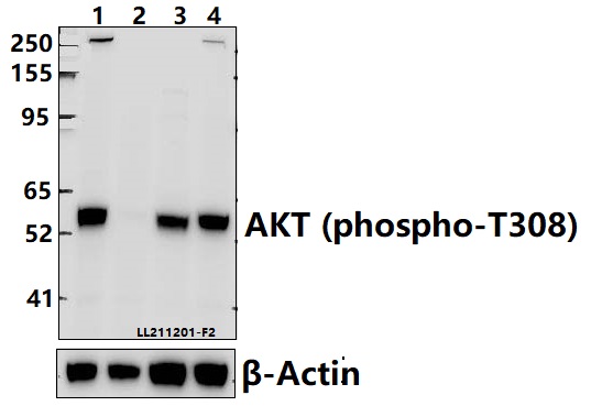
    Western blot (WB) analysis of AKT (phospho-T308) polyclonal antibody at 1:5000 dilution Lane1:HeLa whole cell lysate(40ug) Lane2:Hela treated with λ-phosphatase whole cell lysate(40ug) Lane3:PMVEC whole cell lysate(40ug) Lane4:BV2 whole cell lysate(40ug)

    Western blot (WB) analysis of AKT (phospho-T308) polyclonal antibody at 1:5000 dilution
Lane1:HeLa whole cell lysate(40ug)
Lane2:Hela treated with λ-phosphatase whole cell lysate(40ug)
Lane3:PMVEC whole cell lysate(40ug)
Lane4:BV2 whole cell lysate(40ug)
    Phospho AKT (Thr308) Rabbit Polyclonal Antibody -
    AKT (phospho-T308) polyclonal antibody.
    WB
    Human, Mouse, Rat
    Unconjugated
    100 ul
    $340.00
    7 Weeks*
  • Phospho AKT (Ser473) Rabbit Monoclonal Antibody [Clone ID: R01-4H6]

    Western blot analysis of Phospho-AKT (Ser473) in C6, 3T3, Hela lysates using Phospho-AKT (Ser473) antibody.

    Western blot analysis of Phospho-AKT (Ser473) in C6, 3T3, Hela lysates using Phospho-AKT (Ser473) antibody.
    Phospho AKT (Ser473) Rabbit Monoclonal Antibody [Clone ID: R01-4H6] -
    Phospho-AKT (Ser473) Rabbit monoclonal Antibody.
    WB
    Human, Mouse, Rat
    Unconjugated
    100 ul
    $380.00
    4 Weeks*
  • Phospho Akt (Ser473) Rabbit Polyclonal Antibody

    Western blot (WB) analysis of Akt (Phospho-S473) polyclonal antibody at 1:2000 dilution Lane1:Jurkat whole cell lysate(40ug) Lane2:Jurkat treated with Calyculin A(100 nM,10 minutes) whole cell lysate(40ug) Lane3:Jurkat treated with Calyculin A(100 nM,30 minutes) whole cell lysate(40ug)

    Western blot (WB) analysis of Akt (Phospho-S473) polyclonal antibody at 1:2000 dilution
Lane1:Jurkat whole cell lysate(40ug)
Lane2:Jurkat treated with Calyculin A(100 nM,10 minutes) whole cell lysate(40ug)
Lane3:Jurkat treated with Calyculin A(100 nM,30 minutes) whole cell lysate(40ug)
    Phospho Akt (Ser473) Rabbit Polyclonal Antibody -
    Akt (Phospho-S473) polyclonal antibody.
    WB
    Human
    Unconjugated
    100 ul
    $340.00
    7 Weeks*
  • Phospho Akt (Ser473) Rabbit Polyclonal Antibody

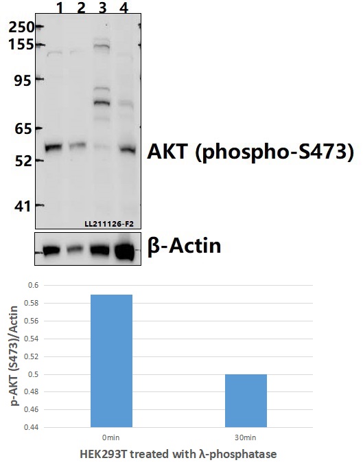
    Western blot (WB) analysis of AKT (phospho-S473) polyclonal antibody at 1:2000 dilution Lane1:HEK293T whole cell lysate(40ug) Lane2:HEK293T treated with λ-phosphatase whole cell lysate(40ug) Lane3:BV2 whole cell lysate(40ug) Lane4:C6 whole cell lysate(40ug)

    Western blot (WB) analysis of AKT (phospho-S473) polyclonal antibody at 1:2000 dilution
Lane1:HEK293T whole cell lysate(40ug)
Lane2:HEK293T treated with λ-phosphatase whole cell lysate(40ug)
Lane3:BV2 whole cell lysate(40ug)
Lane4:C6 whole cell lysate(40ug)
    Phospho Akt (Ser473) Rabbit Polyclonal Antibody -
    AKT (phospho-S473) polyclonal antibody.
    WB
    Human, Mouse, Rat
    Unconjugated
    100 ul
    $340.00
    7 Weeks*
  • Phospho Akt (Thr450) Rabbit Polyclonal Antibody

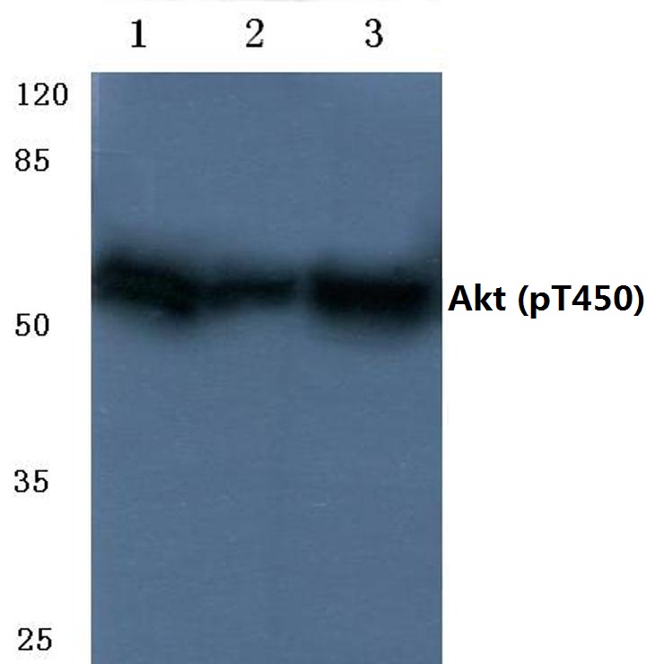
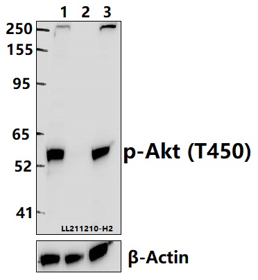
    Western blot (WB) analysis of Akt (Phospho-T450) polyclonal antibody at 1:2000 dilution Lane1:HeLa whole cell lysate(40ug) Lane2:Hela treated with λ-phosphatase whole cell lysate(40ug) Lane3:BV2 whole cell lysate(40ug)

    Western blot (WB) analysis of Akt (Phospho-T450) polyclonal antibody at 1:2000 dilution
Lane1:HeLa whole cell lysate(40ug)
Lane2:Hela treated with λ-phosphatase whole cell lysate(40ug)
Lane3:BV2 whole cell lysate(40ug)
    Phospho Akt (Thr450) Rabbit Polyclonal Antibody -
    Akt (Phospho-T450) polyclonal antibody.
    WB
    Human, Mouse, Rat
    Unconjugated
    100 ul
    $340.00
    7 Weeks*
  • Phospho AKT (Thr450) Rabbit Polyclonal Antibody

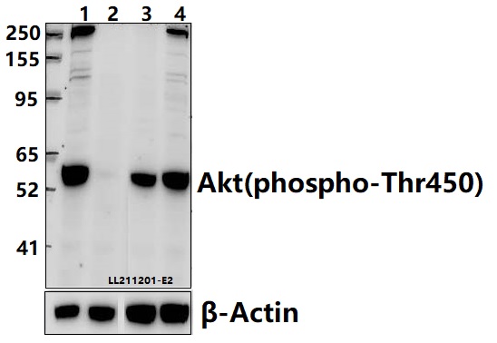
    Western blot (WB) analysis of Akt (phospho-Thr450) polyclonal antibody at 1:5000 dilution Lane1:HeLa whole cell lysate(40ug) Lane2:Hela treated with λ-phosphatase whole cell lysate(40ug) Lane3:The Liver tissue lysate of Mouse(40ug) Lane4:PMVEC whole cell lysate(40ug)

    Western blot (WB) analysis of Akt (phospho-Thr450) polyclonal antibody at 1:5000 dilution
Lane1:HeLa whole cell lysate(40ug)
Lane2:Hela treated with λ-phosphatase whole cell lysate(40ug)
Lane3:The Liver tissue lysate of Mouse(40ug)
Lane4:PMVEC whole cell lysate(40ug)
    Phospho AKT (Thr450) Rabbit Polyclonal Antibody -
    AKT (phospho-Thr450) polyclonal antibody.
    WB
    Human, Mouse, Rat
    Unconjugated
    100 ul
    $340.00
    7 Weeks*
  • Phospho AKT1 (Ser129) Rabbit Monoclonal Antibody [Clone ID: OTIRA0610]

    Western blot analysis of Phospho-AKT1 (S129) expression in (1) MCF-7 cell lysate treated with Alkaline Phosphatase; (2) MCF-7 cell lysate.

    Western blot analysis of Phospho-AKT1 (S129) expression in (1) MCF-7 cell lysate treated with Alkaline Phosphatase; (2) MCF-7 cell lysate.
    Phospho AKT1 (Ser129) Rabbit Monoclonal Antibody [Clone ID: OTIRA0610] -
    Phospho-AKT1 (S129) Antibody.
    WB
    Human, Mouse, Rat
    Unconjugated
    100 ul
    $480.00
    4 Weeks*
  • Phospho Akt (Ser473) AKT1 (S473) Rabbit Monoclonal Antibody [Clone ID: RM251] -
    Rabbit monoclonal to phospho-Akt (Ser473), AKT1 (phospho S473).
    IHC, WB
    Human, Mouse, Rat
    Unconjugated
    100 ul
    $605.00
    5 Days*
  • Phospho-AKT1 (pThr342) Rabbit Polyclonal Antibody -
    Anti-AKT (Thr342) Antibody.
    WB
    Drosophila
    Unconjugated
    100 ul
    $610.00
    5 Days*

19 Items