
GFP Rabbit Polyclonal Antibody

Specifications
| Product Data | |
| Application | ELISA, IP, WB |
|---|---|
| Recommended Dilution | ELISA. Western blot: 1/5,000. Immunoprecipitation: 1/200-1/500. The antibody has successfully been used in Immunohistochemistry on Vibratome sections and in Filter Binding Assays. |
| Reactivity | A. victoria |
| Antibody Host | Rabbit |
| Clonality | Polyclonal |
| Immunogen | E.coli expressed full-length GFP (Green Fluorescent Protein). |
| Specificity | This antibody reacts with wild-type GFP, and its variants such as EGFP and EBFP. |
| Buffer | State: Purified State: Lyophilized purified IgG fraction Preservative: 0.09% Sodium Azide |
| Reconstitution Method | Restore to 1 mg/ml by adding 0.2 ml water. |
| Purification | Protein A Chromatography |
| Conjugation | Unconjugated |
| Storage | Store the antibody undiluted at 2-8°C for one month or (in small aliquots) at -20°C for longer. Avoid repeated freezing and thawing. Shelf life: one year from despatch |
| Shipping | Ambient |
| Background | Green fluorescent protein (GFP) is a spontaneously fluorescent protein isolated from the Pacific jellyfish, Aequorea victoria. It transduces the blue chemiluminescence into green fluorescent light. Since the molecular cloning of GFP cDNA1 and demonstration that GFP can be expressed as a functional transgene2, GFP has become a new tool with exciting applications in developmental, cell and molecular biology3. GFP is an ideal fluorescent probe: its fluorescence is not species specific and can be expressed in bacteria, yeast, plant and mammalian cells; it can fuse with proteins of interest without interfering significantly with their assembly or function. |
| Synonyms | Green fluorescent protein, GFP-Tag |
| Note | This product was originally produced by Torrey Pine Biolabs |
| Reference Data | |
Reviews


Documents
| Product Manuals |
| FAQs |
Citations
Transcription factor clusters enhance gene expression in vivo without modulating bursting dynamics
Cadar, Birnie, Marsman et al
Mol Cell (2026) 86 (20), 4234-4249.e9 DOI: 10.1016/j.molcel.2026.09.011
Cadar, Birnie, Marsman et al
Mol Cell (2026) 86 (20), 4234-4249.e9 DOI: 10.1016/j.molcel.2026.09.011
Snippet: Rabbit polyclonal anti-GFP Acris Antibodies Cat #TP401;RRID: AB_10013661 Mouse monoclonal anti-Dorsal 7A4 Developmental Studies Hybridoma Bank RRID: AB_528204
Cancer cachexia overrides sex-specific traits in Drosophila
Rivero, Techer, Ammar et al
bioRxiv (2026)
Rivero, Techer, Ammar et al
bioRxiv (2026)
Snippet: iton X100 (PBT). Following a 5 min wash in PBT, tissues were blocked in PBT containing 1% BSA for 30 min. Primary antibodies were incubated overnight at 4 °C, using the following dilution: anti-GFP (Acris Antibodies Cat#TP401) 1:200, anti-phospho-Histone H3 (Upstate Cat#TAKMA312B) 1:2000, anti-Disc-large (DSHB Cat#4F3) 1:100, anti-Mef2 (a gift from Eileen Furlong, Heidelberg, Germany) 1:200. Alex
Degrade Green Fluorescent Protein (deGradFP) Method in Trypanosoma brucei
Ishii, Akiyoshi
Methods Mol Biol (2026) 3069, 203-212 DOI: 10.1007/978-1-0716-5508-5_14
Ishii, Akiyoshi
Methods Mol Biol (2026) 3069, 203-212 DOI: 10.1007/978-1-0716-5508-5_14
AI Summary: OriGene’s anti-GFP antibody (TP401) was used in this study to detect YFP-KKT4 fusion proteins, facilitating the assessment of protein degradation efficiency in the deGradFP method applied to Trypanosoma brucei.
Snippet: YFP-KKT4 was detected with primary antibody anti-GFP (TP401, OriGene) and secondary antibody IRDye anti-rabbit 800 (LI-COR). A loading control (PFR2) was detected
Identification of feeding apparatus components in a heterotrophic marine flagellate
Clifford, Taylor, Ishii et al
bioRxiv (2026)
Clifford, Taylor, Ishii et al
bioRxiv (2026)
AI Summary: OriGene's rabbit polyclonal anti-GFP antibody (TP401) was used in this study to detect GFP-tagged proteins during immunostaining, enabling the localization of feeding apparatus components in Diplonema papillatum.
Snippet: Gels were incubated with 600 µL of primary antibodies (rabbit polyclonal anti-GFP antibody (OriGene, TP401, 1 mg/mL, used at 1:500 dilution) and mouse monoclonal anti-alpha tubulin antibody (Merck, T5168, used at 1:200 dilution) diluted in PBS with 1% BSA) overnight on a rocker at room temperature. Gels were washed with 4 mL PBS for 15 min, three times, followed by
Beat II and Side IV keep migrating longitudinal visceral muscle precursor cells on their substratum in Drosophila
Huang, Kinold, Weiß et al
J Cell Sci (2026) 139 (3) DOI: 10.1242/jcs.264157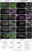
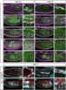
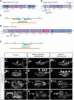
Huang, Kinold, Weiß et al
J Cell Sci (2026) 139 (3) DOI: 10.1242/jcs.264157
AI Summary: OriGene products were not mentioned or used in this paper, as no OriGene product names appear in the methods or antibody lists provided.
Snippet: Dilutions of primary antibodies were as follows: rabbit anti-GFP 1:1000 (cat. no. TP401, Acris Antibodies, Herford, Germany), mouse anti-GFP 1:100 [clone 1D2, AB 2617419;Developmental Studies Hybridoma Bank (DSHB), Iowa City, IA, USA],
Atypical contribution of caspase-3 to melanoma cancer cell motility by regulation of coronin 1B activity
Berthenet, Diazzi, Jamard et al
Cell Death Dis (2025) 16 (1), 690 DOI: 10.1038/s41419-025-07952-y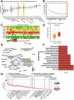
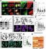
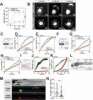
Berthenet, Diazzi, Jamard et al
Cell Death Dis (2025) 16 (1), 690 DOI: 10.1038/s41419-025-07952-y
Cited by 2
AI Summary: In this study, OriGene’s GFP antibody (TP401) was used to detect GFP-tagged proteins, contributing to the analysis of protein interactions and localization involved in caspase-3–mediated melanoma cell motility.
Snippet: sc-56053;Cell Signaling, 9662S;Euromedex, GTX110543), CASP7 (Cell Signaling, 12827S), SP1 (Cell Signaling, 9389S), CORO1B (Thermo Fisher Scientific, CF501573), P-CORO1B (Euromedex, EC-CP2621), GFP (Origene, TP401), Myc-tag (Thermo Fisher Scientific, 13-2500), BAX (Cell Signaling, 2772S), BAK (Cell Signaling, 12105S), P-PKCα (Invitrogen, 44-962G), PKCα (Cell Signaling, 2056T), ARP2/ARP3 (Bioss
Showing 1-6 of 33 papers.
Powered by BizGenius
*Delivery time may vary from web posted schedule. Occasional delays may occur due to unforeseen
complexities in the preparation of your product. International customers may expect an additional 1-2 weeks
in shipping.
Related Products
- Price:
- Actual Price:
Welcome to OriGene AI!
I'm your AI-powered guide to everything OriGene.
Ask me anything:
- Product specs
- Ordering help
- Technical questions
- How to find exactly what your experiment needs
Prefer a live agent? Connect with us by clicking the green chat icon, as seen in the image below or email our team at custsupport@origene.com.
