
PD-L1 (CD274) Mouse Monoclonal Antibody [Clone ID: OTI2C7]


$447.00
In Stock*
Specifications
| Product Data | |
| Clone Name | OTI2C7 |
|---|---|
| Application | FC, IF, IHC, LMNX, WB |
| Recommended Dilution | WB 1:200~2000, IF 1:100 |
| Reactivity | Human (Does not react with: Mouse) |
| Antibody Host | Mouse |
| Isotype | IgG1 |
| Clonality | Monoclonal |
| Immunogen | Full length human recombinant protein of human CD274(NP_054862) produced in HEK293T cell. |
| Buffer | PBS (pH 7.3) containing 1% BSA, 50% glycerol and 0.02% sodium azide |
| Concentration | 1 mg/ml |
| Purification | Purified from mouse ascites fluids or tissue culture supernatant by affinity chromatography (protein A/G) |
| Conjugation | Unconjugated |
| Storage | Store at -20°C as received. |
| Stability | Stable for 12 months from date of receipt. |
| Shipping | Blue Ice |
| Predicted Protein Size | 31 kDa |
| Gene Name | CD274 molecule |
| Database Link | |
| Synonyms | B7-H; B7H1; PD-L1; PDCD1L1; PDCD1LG1; PDL1 |
| Reference Data | |
| Protein Categories | ADC Targets, CAR-T Targets, CD Markers, Cytokines, Membrane Proteins |
| Protein Families | Druggable Genome, Transmembrane |
| Protein Pathways | Cell adhesion molecules (CAMs) |
Reviews


Documents
| Product Manuals |
| FAQs |
| SDS |
Resources
Other "OTI2C7" antibodies (4)
| SKU | Description | Size | Price | |
|---|---|---|---|---|
| CF507087 | Carrier-free (BSA/glycerol-free) CD274 mouse monoclonal antibody, clone OTI2C7 (formerly 2C7) | 100 ug |
$620.00
|
|
| TA507087AM | PD-L1 / CD274 mouse monoclonal antibody, clone OTI2C7 (formerly 2C7), Biotinylated | 100 ul |
$509.00
|
|
| TA507087BM | PD-L1 / CD274 mouse monoclonal antibody, clone OTI2C7 (formerly 2C7), HRP conjugated | 100 ul |
$509.00
|
|
| TA507087S | PD-L1 / CD274 mouse monoclonal antibody, clone OTI2C7 (formerly 2C7) | 30 ul |
$200.00
|
|
Citations
RNF138 promotes cisplatin resistance and PD-L1-mediated immune evasion via JAK2/STAT3 activation in nasopharyngeal carcinoma
He, Lou, Lei et al
Cell Rep (2026) 45 (6), 117547 DOI: 10.1016/j.celrep.2026.117547
He, Lou, Lei et al
Cell Rep (2026) 45 (6), 117547 DOI: 10.1016/j.celrep.2026.117547
Vimentin, inversely correlating with infiltration of CD8 + T lymphocytes, promotes nuclear translocation of PD-L1 in esophageal squamous cell carcinoma
Liang, He, Liu et al
Biochim Biophys Acta Mol Cell Res (2024) 1871 (7), 119781 DOI: 10.1016/j.bbamcr.2024.119781
Liang, He, Liu et al
Biochim Biophys Acta Mol Cell Res (2024) 1871 (7), 119781 DOI: 10.1016/j.bbamcr.2024.119781
Cited by 21
AI Summary: In this study, the OriGene anti-PD-L1 antibody (TA507087) was used to detect PD-L1 protein levels, contributing to the investigation of the relationship between vimentin and PD-L1 in esophageal squamous cell carcinoma.
Snippet: was blocked with 5 % BSA/TBST buffer for 1.5 h at room temperature and incubated with anti-β-actin, anti-vimentin (ab62194;dilution at 1:1000;Abcam), and anti-PD-L1 (TA507087;dilution at 1:2000;Origene, USA) antibodies diluted in TBST buffer overnight at 4 °C. Then, the membranes were incubated with horseradish peroxidase (HRP)-conjugated anti-mouse or anti-rabbit IgG (bs-0295G-AP;Bioss, China)
PD-L1 detection on circulating tumor-derived extracellular vesicles (T-EVs) from patients with lung cancer
Wu, Gu, Kang et al
Transl Lung Cancer Res (2021) 10 (6), 2441-2451 DOI: 10.21037/tlcr-20-1277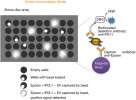
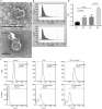
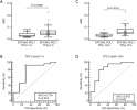
Wu, Gu, Kang et al
Transl Lung Cancer Res (2021) 10 (6), 2441-2451 DOI: 10.21037/tlcr-20-1277
Cited by 40
AI Summary: In this study, OriGene products—specifically the A549 (Cat# CL-0016) and SK-MES1 (Cat# CL-0007) lung cancer cell lines—were used as sources of tumor-derived exosomes to validate the newly developed Simoa Epcam-PD-L1 extracellular vesicle detection assay.
Clinicopathological analysis of epithelioid inflammatory myofibroblastic sarcoma
Du, Gao, Zhao et al
Oncol Lett (2018) 15 (6), 9317-9326 DOI: 10.3892/ol.2018.8530
Du, Gao, Zhao et al
Oncol Lett (2018) 15 (6), 9317-9326 DOI: 10.3892/ol.2018.8530
Cited by 26
Clinicopathological analysis of epithelioid inflammatory myofibroblastic sarcoma
Du, Gao, Zhao et al
Oncol Lett (2018)
Du, Gao, Zhao et al
Oncol Lett (2018)
Showing 1-5 of 5 papers.
Powered by BizGenius
*Delivery time may vary from web posted schedule. Occasional delays may occur due to unforeseen
complexities in the preparation of your product. International customers may expect an additional 1-2 weeks
in shipping.
Related Products
- Price:
- Actual Price:
Welcome to OriGene AI!
I'm your AI-powered guide to everything OriGene.
Ask me anything:
- Product specs
- Ordering help
- Technical questions
- How to find exactly what your experiment needs
Prefer a live agent? Connect with us by clicking the green chat icon, as seen in the image below or email our team at custsupport@origene.com.
