
GFP (Ads. to Hu, Ms, Rt Serum Proteins) Rabbit Polyclonal Antibody

Specifications
| Product Data | |
| Application | ELISA, FC, IF, IHC, IP, WB |
|---|---|
| Recommended Dilution | WB: 1:500 - 1:5,000 IHC: 1:200 - 1:3,000 IF: 1:500 - 1:5,000 FC: User Optimized ELISA: 1:20,000 - 1:120,000 IP: User Optimized |
| Reactivity | eGFP, GFP, rGFP, RS-GFP, S65T-GFP, YFP |
| Antibody Host | Rabbit |
| Isotype | IgG |
| Clonality | Polyclonal |
| Immunogen | The immunogen is a Green Fluorescent Protein (GFP) fusion protein corresponding to the full length amino acid sequence (246aa) derived from the jellyfish Aequorea victoria. |
| Specificity | Anti-GFP antibody was prepared from monospecific antiserum by immunoaffinity chromatography using Green Fluorescent Protein (Aequorea victoria) coupled to agarose beads followed by solid phase adsorption(s) to remove any unwanted reactivities. Assay by immunoelectrophoresis resulted in a single precipitin arc against anti-Rabbit Serum and purified and partially purified Green Fluorescent Protein (Aequorea victoria). No reaction was observed against Human, Mouse or Rat serum proteins. |
| Buffer | 0.02 M Potassium Phosphate, 0.15 M Sodium Chloride, pH 7.2 Stabilizer: None Preservative: 0.01% (w/v) Sodium Azide |
| Concentration | 1.25 mg/mL - lot specific |
| Purification | This product was prepared from monospecific antiserum by immunoaffinity chromatography using Green Fluorescent Protein (Aequorea victoria) coupled to agarose beads followed by solid phase adsorption(s) to remove any unwanted reactivities |
| Conjugation | Unconjugated |
| Storage | Store Anti-GFP Antibody at -20° C prior to opening. Aliquot contents and freeze at -20° C or below for extended storage. Avoid cycles of freezing and thawing. Centrifuge product if not completely clear after standing at room temperature. GFP antibody is stable for several weeks at 4° C as an undiluted liquid. Dilute only prior to immediate use. |
| Stability | Expiration date is one (1) year from date of receipt. |
| Shipping | Blue Ice |
| Predicted Protein Size | 27 kDa |
| Database Link | |
| Background | Green Fluorescent Protein (GFP) is a 27 kDa protein produced from the jellyfish Aequorea victoria, which emits green light (emission peak at a wavelength of 509nm) when excited by blue light. GFP is an important tool in cell biology research. GFP is widely used enabling researchers to visualize and localize GFP-tagged proteins within living cells without the need for chemical staining. GFP Antibody is ideal for Cell Biology, Neuroscience and Cancer research. |
| Synonyms | rabbit anti-GFP antibody, Green Fluorescent Protein, GFP antibody, Green Fluorescent Protein antibody, EGFP, enhanced Green Fluorescent Protein, Aequorea victoria, Jellyfish |
| Note | Anti-GFP antibody is designed to detect GFP and its variants. GFP antibody has been tested by western blot and ELISA. This product can be used to detect GFP by ELISA (sandwich or capture) for the direct binding of antigen and recognizes wild type, recombinant and enhanced forms of GFP. Biotin conjugated polyclonal anti-GFP used in a sandwich ELISA is well suited to titrate GFP in solution using this antibody in combination with Rockland's monoclonal anti-GFP (600-301-215) using either form of the antibody as the capture or detection antibodies. However, use the monoclonal form only for the detection of wild type or recombinant GFP as this form does not sufficiently detect 'enhanced' GFP. The detection antibody is typically conjugated to biotin and subsequently reacted with streptavidin conjugated HRP (code # S000-03). Fluorochrome conjugated polyclonal anti-GFP can be used to detect GFP by immunofluorescence microscopy in prokaryotic (E.coli) and eukaryotic (CHO cells) expression systems and can detect GFP containing inserts. Significant amplification of signal is achieved using fluorochrome conjugated polyclonal anti-GFP relative to the fluorescence of GFP alone. For immunoblotting use either alkaline phosphatase or peroxidase conjugated polyclonal anti-GFP to detect GFP or GFP containing proteins on western blots. Optimal titers for applications should be determined by the researcher. |
| Reference Data | |
Reviews


Documents
| Product Manuals |
| FAQs |
| SDS |
Citations
SMC5/6 acts jointly with Fanconi anemia factors to support DNA repair and genome stability
Rossi, Helbling-Leclerc, Kawasumi et al
EMBO Rep. (2019) 21 (2), e48222 DOI: 10.15252/embr.201948222
Rossi, Helbling-Leclerc, Kawasumi et al
EMBO Rep. (2019) 21 (2), e48222 DOI: 10.15252/embr.201948222
Cited by 30
AI Summary: OriGene’s anti-GFP rabbit polyclonal antibody (Cat. No. TP401) was used for both immunoprecipitation and Western blotting in TK6 cells to study protein interactions relevant to DNA repair and genome stability in this paper.
Snippet: and anti‐FANCJ rabbit polyclonal antibody, 1/1,000 (Cat No. NB100‐416, Novus Biologicals). For Co‐IP of SMC5, SMC6, NSMCE2, and FANCI, an antibody against FANCI home‐made by Weidong Wang's Lab at NIH/NIA was used. For Co‐IP in TK6 cells, anti‐GFP rabbit polyclonal (Cat. No. TP401, OriGene) was used for both IP (2.5 μg/sample) and WB (1:1,000)
Retinal and anterior eye compartments derive from a common progenitor pool in the avian optic cup
Venters, Cuenca, Hyer
Mol Vis (2011) 17, 3347-63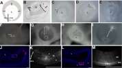

Venters, Cuenca, Hyer
Mol Vis (2011) 17, 3347-63
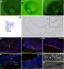
AI Summary: In this study, OriGene’s anti-GFP antibody (Cat# TA150041) was used to detect GFP-labeled cells, enabling the researchers to track the lineage and fate of progenitor cells in the avian optic cup during eye development.
Vaccinia extracellular virions enter cells by macropinocytosis and acid-activated membrane rupture
Schmidt, Bleck, Helenius et al
EMBO J (2011) 30 (17), 3647-61 DOI: 10.1038/emboj.2011.245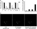
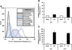
Schmidt, Bleck, Helenius et al
EMBO J (2011) 30 (17), 3647-61 DOI: 10.1038/emboj.2011.245
Cited by 128
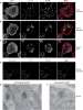
AI Summary: OriGene’s anti-β-actin antibody (TA-09) was used in this study to detect cellular actin in immunoblotting experiments, helping to analyze actin rearrangements during vaccinia extracellular virion entry.
The syntaxin 4 N terminus regulates its basolateral targeting by munc18c-dependent and -independent mechanisms
Torres, Funk, Zegers et al
J Biol Chem (2011) 286 (12), 10834-46 DOI: 10.1074/jbc.M110.186668
Torres, Funk, Zegers et al
J Biol Chem (2011) 286 (12), 10834-46 DOI: 10.1074/jbc.M110.186668
AI Summary: OriGene products, specifically the human syntaxin 4 cDNA clone (OriGene, SC119192) and syntaxin 4 shRNA (OriGene, TF312563), were used in this study to manipulate syntaxin 4 expression levels and analyze its role in basolateral targeting and interaction with munc18c in epithelial cells.
Splice variants of the dual specificity tyrosine phosphorylation-regulated kinase 4 (DYRK4) differ in their subcellular localization and catalytic activity
Papadopoulos, Arato, Lilienthal et al
J Biol Chem (2011) 286 (7), 5494-505 DOI: 10.1074/jbc.M110.157909
Papadopoulos, Arato, Lilienthal et al
J Biol Chem (2011) 286 (7), 5494-505 DOI: 10.1074/jbc.M110.157909
AI Summary: In this study, OriGene products—including the DYRK4 cDNA clones and DYRK4-specific antibodies—were used to express and detect DYRK4 splice variants, enabling the authors to analyze their subcellular localization and catalytic activity.
Dermal-type macrophages expressing CD209/DC-SIGN show inherent resistance to dengue virus growth
Kwan, Navarro-Sanchez, Dumortier et al
PLoS Negl Trop Dis (2008) 2 (10), e311 DOI: 10.1371/journal.pntd.0000311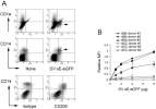
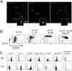
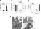
Kwan, Navarro-Sanchez, Dumortier et al
PLoS Negl Trop Dis (2008) 2 (10), e311 DOI: 10.1371/journal.pntd.0000311
Cited by 49
AI Summary: OriGene’s recombinant envelope E glycoprotein fused to green fluorescent protein was used to demonstrate binding and internalization of dengue virus by dermal macrophages, enabling the study of their inherent resistance to viral growth.
Showing 1-6 of 6 papers.
Powered by BizGenius
*Delivery time may vary from web posted schedule. Occasional delays may occur due to unforeseen
complexities in the preparation of your product. International customers may expect an additional 1-2 weeks
in shipping.
Related Products
- Price:
- Actual Price:
Welcome to OriGene AI!
I'm your AI-powered guide to everything OriGene.
Ask me anything:
- Product specs
- Ordering help
- Technical questions
- How to find exactly what your experiment needs
Prefer a live agent? Connect with us by clicking the green chat icon, as seen in the image below or email our team at custsupport@origene.com.
