
CD8A Mouse Monoclonal Antibody (HRP conjugated) [Clone ID: OTI3H6]


$509.00
5 Days*
Specifications
| Product Data | |
| Clone Name | OTI3H6 |
|---|---|
| Application | IHC, WB |
| Recommended Dilution | WB 1:2000, IHC 1:150 |
| Reactivity | Human |
| Antibody Host | Mouse |
| Isotype | IgG1 |
| Clonality | Monoclonal |
| Immunogen | Human recombinant protein fragment corresponding to amino acids 22-182 of human CD8A (NP_001759) produced in E.coli. |
| Buffer | PBS (pH 7.3) containing 1% BSA and 50% glycerol |
| Concentration | 0.5 mg/ml |
| Purification | Purified from mouse ascites fluids or tissue culture supernatant by affinity chromatography (protein A/G) |
| Conjugation | HRP |
| Storage | Store at -20°C as received. |
| Stability | Stable for 12 months from date of receipt. |
| Shipping | Blue Ice |
| Predicted Protein Size | 23.5 kDa |
| Gene Name | CD8a molecule |
| Database Link | |
| Background | The CD8 antigen is a cell surface glycoprotein found on most cytotoxic T lymphocytes that mediates efficient cell-cell interactions within the immune system. The CD8 antigen acts as a coreceptor with the T-cell receptor on the T lymphocyte to recognize antigens displayed by an antigen presenting cell in the context of class I MHC molecules. The coreceptor functions as either a homodimer composed of two alpha chains or as a heterodimer composed of one alpha and one beta chain. Both alpha and beta chains share significant homology to immunoglobulin variable light chains. This gene encodes the CD8 alpha chain. Multiple transcript variants encoding different isoforms have been found for this gene. provided by RefSeq, Nov 2011 |
| Synonyms | CD8; Leu2; MAL; p32 |
| Reference Data | |
| Protein Categories | ADC Targets, CD Markers, Immune system diseases, Intracellular Proteins, Membrane Proteins |
| Protein Families | Adult stem cells, Druggable Genome, ES Cell Differentiation/IPS, Secreted Protein, Transmembrane |
| Protein Pathways | Antigen processing and presentation, Cell adhesion molecules (CAMs), Hematopoietic cell lineage, Primary immunodeficiency, T cell receptor signaling pathway |
Reviews


Documents
| Product Manuals |
| FAQs |
| SDS |
Resources
Other "OTI3H6" antibodies (4)
| SKU | Description | Size | Price | |
|---|---|---|---|---|
| CF802079 | Carrier-free (BSA/glycerol-free) CD8A mouse monoclonal antibody,clone OTI3H6 | 100 ug |
$620.00
|
|
| TA802079 | CD8A mouse monoclonal antibody,clone OTI3H6 | 100 ul |
$447.00
|
|
| TA802079AM | CD8A mouse monoclonal antibody,clone 3H6, Biotinylated | 100 ul |
$509.00
|
|
| TA802079S | CD8A mouse monoclonal antibody,clone OTI3H6 | 30 ul |
$200.00
|
|
Citations
TIGIT agonism as a therapeutic strategy to suppress inflammation in hidradenitis suppurativa
Kidacki, Jaiswal, Cho et al
Front Immunol (2026) 17, 1761782 DOI: 10.3389/fimmu.2026.1761782
Kidacki, Jaiswal, Cho et al
Front Immunol (2026) 17, 1761782 DOI: 10.3389/fimmu.2026.1761782
AI Summary: OriGene's CD8 antibody (catalog number CF802079) was used in this study to identify and analyze CD8+ cells in tissue samples, contributing to the characterization of immune cell populations involved in hidradenitis suppurativa.
Snippet: CD8 antibody used CF802079 from Origene. Images were processed in QuPath, where cells were segmented using the built-in cell detection algorithm and PVR, TIGIT, or
Prostate tumor immune microenvironment changes following immunotherapy shared by patients who developed anti-tumor response or immune-related adverse events
Rastogi, McNeel
Oncoimmunology (2025) 14 (1), 2595788 DOI: 10.1080/2162402X.2025.2595788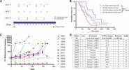
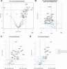
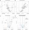
Rastogi, McNeel
Oncoimmunology (2025) 14 (1), 2595788 DOI: 10.1080/2162402X.2025.2595788
AI Summary: OriGene contributed to this study by providing the anti-human CD8a monoclonal antibody (Clone ID: OTI3H6, Cat# CF802079), which was used for spatial protein analysis of tumor biopsies to assess immune cell infiltration following immunotherapy.
Snippet: pression. Antibodies Syto13-FITC (nucleic acid stain) and PanCK-Cy3 were part of the Nanostring GeoMx kit. An anti-human CD8a (Clone ID: OTI3H6) monoclonal antibody (Cat# CF802079) was purchased from Origene and conjugated with AF647 using Alexa Fluor 647 antibody labeling kit (Cat# A20186, Thermo Fisher Scientific) following the manufacturer’s protocol. Nanostring GeoMx proteomics analysis Slid
Discovering molecular signatures in kidney transplant biopsies with borderline changes and isolated V-lesions: single-cell RNA-sequencing analysis of human blood and tissue Spatial transcriptomics
Song, Kang, Jang et al
Sci Rep (2025) 15 (1), 23770 DOI: 10.1038/s41598-025-05191-x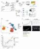
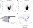
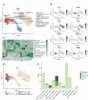
Song, Kang, Jang et al
Sci Rep (2025) 15 (1), 23770 DOI: 10.1038/s41598-025-05191-x
Cited by 2
AI Summary: OriGene’s anti-CD8 antibody (CF802079) was used for immunohistochemistry and immunofluorescence to identify CD8+ T cells in kidney allograft tissue, helping validate key findings in the study.
Snippet: perature and then washed three times with 1X PBS. To block non-specific binding sites, the slides were incubated in 5% BSA in PBS for 1 h at room temperature. Primary antibodies, anti-CD8 (CF802079;OriGene, dilution 1:100, [OTI3H6]) and anti-pSTAT1 (9167 S;Cell Signaling TECHNOLOGY, dilution 1:50, [58D6]) were diluted in 2% BSA in PBS. Subsequently the slides were incubated in a humid chamber
Integrated multi-omics profiling to dissect the spatiotemporal evolution of metastatic hepatocellular carcinoma
Sun, Wu, Zhang et al
Cancer Cell (2023) 42 (1), 135-156.e17 DOI: 10.1016/j.ccell.2023.11.010
Sun, Wu, Zhang et al
Cancer Cell (2023) 42 (1), 135-156.e17 DOI: 10.1016/j.ccell.2023.11.010
Cited by 125
AI Summary: OriGene’s Anti-CD8 antibody (Cat# CF802079) was used for digital spatial profiling (DSP) to analyze CD8+ T cells in tissue samples, contributing to the study’s investigation of immune environments in metastatic hepatocellular carcinoma.
Distribution of CD8 T Cells and NK Cells in the Stroma in Relation to Recurrence or Metastasis of Nasopharyngeal Carcinoma
Li, Dong, Dong et al
Cancer Manag Res (2022) 14, 2913-2926 DOI: 10.2147/CMAR.S365230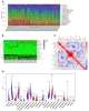
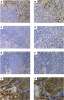
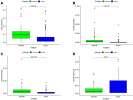
Li, Dong, Dong et al
Cancer Manag Res (2022) 14, 2913-2926 DOI: 10.2147/CMAR.S365230
AI Summary: OriGene products contributed to this study by providing specific monoclonal antibodies—anti-human CD8 (TA802079S), anti-human CD56 (TA805376S), anti-human CD68 (TA802949S), and anti-human CD138—which were used for immunohistochemical staining to evaluate the distribution of immune cells in nasopharyngeal carcinoma tissues.
Snippet: s (CD8), NK cells (CD56), macrophages (CD68) and plasma (CD138). These TIICs were stained with different antibodies as follows: an anti-human CD8 mouse monoclonal antibody (TA802079S, dilution 1:150, Origene), an anti-human CD56 mouse monoclonal antibody (TA805376S, dilution 1:150, Origene), an anti-human CD68 mouse monoclonal antibody (TA802949S, dilution 1:500, Origene), and an anti-human CD138;- Origene), an anti-human CD56 mouse monoclonal antibody (TA805376S, dilution 1:150, Origene (TA802949S, dilution 1:500, Origene), and an anti-human CD138 mouse Save Cached
Adaptive transcriptomic and immune infiltrate responses in the tumor immune microenvironment following neoadjuvant chemotherapy in high grade serous ovarian cancer reveal novel prognostic associations and activation of pro-tumorigenic pathways
James, Woodman, De La Cruz et al
Front Immunol (2022) 13, 965331 DOI: 10.3389/fimmu.2022.965331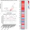
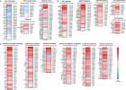
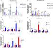
James, Woodman, De La Cruz et al
Front Immunol (2022) 13, 965331 DOI: 10.3389/fimmu.2022.965331
Cited by 17
AI Summary: OriGene’s CD8 antibody (TA802079) was used for immunofluorescence staining to identify and quantify CD8+ immune cell infiltrates in tumor tissue, contributing to the study’s analysis of immune changes in the tumor microenvironment following chemotherapy.
Snippet: … Primary antibodies and dilutions used were as follows: ATF3 (Novus Biologicals, NBP1-85816, [1:50]) AREG (Proteintech, 16036-1-AP, [1:50]) CD8 (Origene, TA802079, [1:50]) Microscopy Images were obtained from a Zeiss Axio Imager M1 and were acquired using diode lasers 402, 488, and 561. To obtain images for CD8+ cell counting, five randomly s;Primary antibodies and dilutions used were as follows: ATF3 (Novus Biologicals, NBP1-85816, [1:50]) AREG (Proteintech, 16036-1-AP, [1:50]) CD8 (Origene, TA802079, [1:50]) Microscopy
Showing 1-6 of 10 papers.
Powered by BizGenius
*Delivery time may vary from web posted schedule. Occasional delays may occur due to unforeseen
complexities in the preparation of your product. International customers may expect an additional 1-2 weeks
in shipping.
Related Products
- Price:
- Actual Price:
Welcome to OriGene AI!
I'm your AI-powered guide to everything OriGene.
Ask me anything:
- Product specs
- Ordering help
- Technical questions
- How to find exactly what your experiment needs
Prefer a live agent? Connect with us by clicking the green chat icon, as seen in the image below or email our team at custsupport@origene.com.
