
PLEKHO1 Rabbit Polyclonal Antibody

Specifications
| Product Data | |
| Application | IHC, WB |
|---|---|
| Recommended Dilution | WB: 200-1000 WB positive control: Hela cells, mouse muscle and kidney tissue IHC: 100-300 Positive control: Human thyroid cancer Predicted cell location: Cytoplasm |
| Reactivity | Human, Mouse, Rat |
| Antibody Host | Rabbit |
| Isotype | IgG |
| Clonality | Polyclonal |
| Immunogen | Fusion protein of human PLEKHO1 |
| Buffer | pH7.4 PBS, 0.05% NaN3, 40% Glyceroln |
| Concentration | lot specific |
| Purification | Antigen affinity purification |
| Conjugation | Unconjugated |
| Storage | Store at -20°C as received. |
| Stability | Stable for 12 months from date of receipt. |
| Shipping | Blue Ice |
| Predicted Protein Size | 46 kDa |
| Gene Name | pleckstrin homology domain containing O1 |
| Database Link | |
| Background | Pleckstrin homology domain-containing family O member 1 is a protein that in humans is encoded by thePLEKHO1 gene. Plays a role in the regulation of the actin cytoskeleton through its interactions with actin capping protein (CP). May function to target CK2 to the plasma membrane thereby serving as an adapter to facilitate the phosphorylation of CP by protein kinase 2 (CK2). Appears to target ATM to the plasma membrane. Appears to also inhibit tumor cell growth by inhibiting AKT-mediated cell-survival. |
| Synonyms | CKIP-1; CKIP1; JBP; OC120 |
| Reference Data | |
| Protein Categories | Intracellular Proteins |
Reviews


Documents
| Product Manuals |
| FAQs |
| SDS |
Citations
microRNA-494 Favors HO-1 Expression in Neuroblastoma Cells Exposed to Oxidative Stress in a Bach1-Independent Way
Piras, Furfaro, Caggiano et al
Front Oncol (2018) 8, 199 DOI: 10.3389/fonc.2018.00199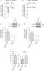
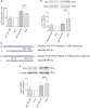
Piras, Furfaro, Caggiano et al
Front Oncol (2018) 8, 199 DOI: 10.3389/fonc.2018.00199
AI Summary: In this study, OriGene's rabbit anti-HO-1 antibody was used to detect HO-1 protein levels by immunodetection, enabling the authors to assess how miR-494 influences HO-1 expression in neuroblastoma cells under oxidative stress.
Snippet: mmunodetection was performed using mouse anti-hnRNPQ (1:1,000, Santa Cruz), rabbit anti-PTEN (1:1,000, Cell Signaling Technology, MA, USA), rabbit anti-Bach1 (1:4,000, Bethyl Lab, Montgomery, TX, USA), and rabbit anti-HO-1 (1:2,000, Origene, Herford, Germany) and specific secondary antibodies (GE Healthcare). The membranes were re-probed with the loading control antibodies, rabbit anti-GAPDH (1:1,...
Showing 1-1 of 1 papers.
Powered by BizGenius
*Delivery time may vary from web posted schedule. Occasional delays may occur due to unforeseen
complexities in the preparation of your product. International customers may expect an additional 1-2 weeks
in shipping.
Related Products
- Price:
- Actual Price:
Welcome to OriGene AI!
I'm your AI-powered guide to everything OriGene.
Ask me anything:
- Product specs
- Ordering help
- Technical questions
- How to find exactly what your experiment needs
Prefer a live agent? Connect with us by clicking the green chat icon, as seen in the image below or email our team at custsupport@origene.com.
