
Phospho Smad2/3 (Thr8) Rabbit Polyclonal Antibody

$335.00
4 Weeks*
Specifications
| Product Data | |
| Application | WB |
|---|---|
| Recommended Dilution | WB: 1:500-1:2000 |
| Reactivity | Human, Mouse, Rat |
| Modification | Phospho-specific |
| Antibody Host | Rabbit |
| Isotype | IgG |
| Clonality | Polyclonal |
| Immunogen | The antiserum was produced against A synthesized peptide derived from human Smad2/3 around the phosphorylation site of Threonine 8 |
| Buffer | Rabbit IgG in phosphate buffered saline , pH 7.4, 150mM NaCl, 0.02% sodium azide and 50% glycerol. |
| Concentration | lot specific |
| Purification | The antibody was affinity-purified from rabbit antiserum by affinity-chromatography using epitope-specific peptide. |
| Conjugation | Unconjugated |
| Storage | Store at -20°C as received. |
| Stability | Stable for 12 months from date of receipt. |
| Shipping | Blue Ice |
| Predicted Protein Size | 48/60 kDa |
| Gene Name | SMAD family member 3 |
| Database Link | |
| Background | Smad2 ubiquitously expressed transcription factor phosphorylated and activated by TGF-beta receptor-type kinases. Participates in a wide range of critical processes including morphogenesis, cell-fate determination, proliferation, differentiation and apoptosis. |
| Synonyms | HSPC193; HsT17436; JV15-2; LDS1C; LDS3; MADH3 |
| Reference Data | |
| Protein Categories | Growth Factors, Intracellular Proteins, Transciption Factors |
| Protein Families | Cancer stem cells, Druggable Genome, Embryonic stem cells, ES Cell Differentiation/IPS, Stem cell relevant signaling - JAK/STAT signaling pathway, Stem cell relevant signaling - TGFb/BMP signaling pathway, Transcription Factors |
| Protein Pathways | Adherens junction, Cell cycle, Chronic myeloid leukemia, Colorectal cancer, Pancreatic cancer, Pathways in cancer, TGF-beta signaling pathway, Wnt signaling pathway |
Reviews


Documents
| Product Manuals |
| FAQs |
| SDS |
Citations
Long-Term Protective Effects of Succinate Dehydrogenase Inhibition during Reperfusion with Malonate on Post-Infarction Left Ventricular Scar and Remodeling in Mice
Valls-Lacalle, Consegal, Ganse et al
Int J Mol Sci (2024) 25 (8) DOI: 10.3390/ijms25084366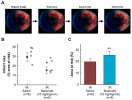
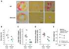
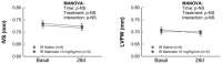
Valls-Lacalle, Consegal, Ganse et al
Int J Mol Sci (2024) 25 (8) DOI: 10.3390/ijms25084366
Cited by 8
AI Summary: In this study, the OriGene antibody pSMAD2/3 (Thr8) (#TA325852) was used to assess the activation of fibrotic signaling pathways, contributing to the evaluation of pro-inflammatory and fibrotic responses following myocardial infarction and malonate treatment in mice.
Snippet: of markers involved in the pro-inflammatory and fibrotic responses, including NF-κB p65 (#ab16502, abcam, 1:1000), Smad2/3 (#sc-398844, Santa Cruz Biotechnology, 1:500), pSMAD2/3 (Thr8) (#TA325852, OriGene, 1:1000), transforming growth factor β1 (TGFβ1) (#ab92486, abcam, 1:1000), connective tissue growth factor (CTGF) (#ab6992, abcam, 1:1000), fibroblast activation factor α (FAPα) (#PIPA5120
Connexin 43 Deficiency Is Associated with Reduced Myocardial Scar Size and Attenuated TGFβ1 Signaling after Transient Coronary Occlusion in Conditional Knock-Out Mice
Valls-Lacalle, Consegal, Ruiz-Meana et al
Biomolecules (2020) 10 (4) DOI: 10.3390/biom10040651
Valls-Lacalle, Consegal, Ruiz-Meana et al
Biomolecules (2020) 10 (4) DOI: 10.3390/biom10040651
Cited by 10
AI Summary: OriGene contributed to this study by providing the anti-phospho-Smad2/3 (Thr8) antibody (#TA325852), which was used to assess TGFβ1 signaling in myocardial tissue.
Snippet: antibodies were used: rabbit anti-NF-κB p65 (#ab16502, Abcam, dilution 1:1000), anti-Smad2/3 (total forms: #sc398844 (raised in mice), Santa Cruz Biotechnology, Dallas, TX, USA, 1:500;phosphorylated forms (Thr8): #TA325852 (raised in rabbit), Origene, Rockville Cached;- antibodies were used: rabbit anti-NF-κB p65 (#ab16502, Abcam, dilution 1:1000), anti-Smad2/3 (total forms: #sc398844 (raised in mice), Santa Cruz Biotechnology, Dallas, TX, USA, 1:500
Histidine and arginine modulate intestinal cell restitution via transforming growth factor-β1
Matsui, Ichikawa, Fujita et al
Eur J Pharmacol (2019) 850, 35-42 DOI: 10.1016/j.ejphar.2019.02.006
Matsui, Ichikawa, Fujita et al
Eur J Pharmacol (2019) 850, 35-42 DOI: 10.1016/j.ejphar.2019.02.006
Cited by 20
AI Summary: In this study, OriGene’s Tgf-β1 (Cat# SR307914) and Gapdh (Cat# SR412874) qPCR primers were used to quantify mRNA levels, helping demonstrate that histidine and arginine deficiency reduces Tgf-β1 expression and impairs intestinal cell restitution.
RAGE Deletion Confers Renoprotection by Reducing Responsiveness to Transforming Growth Factor-β and Increasing Resistance to Apoptosis
Hagiwara, Sourris, Ziemann et al
Diabetes (2018)
Hagiwara, Sourris, Ziemann et al
Diabetes (2018)
Delayed, oral pharmacological inhibition of calpains attenuates adverse post-infarction remodelling
Poncelas, Inserte, Aluja et al
Cardiovasc Res (2017) 113 (8), 950-961 DOI: 10.1093/cvr/cvx073
Poncelas, Inserte, Aluja et al
Cardiovasc Res (2017) 113 (8), 950-961 DOI: 10.1093/cvr/cvx073
Cited by 38
AI Summary: In this study, OriGene products—specifically anti-Calpain-1 (Cat# TA324872) and anti-Calpain-2 (Cat# TA324873) antibodies—were used to measure calpain protein levels in heart tissue, helping to demonstrate the effectiveness of calpain inhibition in reducing adverse post-infarction cardiac remodeling.
Showing 1-5 of 5 papers.
Powered by BizGenius
*Delivery time may vary from web posted schedule. Occasional delays may occur due to unforeseen
complexities in the preparation of your product. International customers may expect an additional 1-2 weeks
in shipping.
Related Products
- Price:
- Actual Price:
Welcome to OriGene AI!
I'm your AI-powered guide to everything OriGene.
Ask me anything:
- Product specs
- Ordering help
- Technical questions
- How to find exactly what your experiment needs
Prefer a live agent? Connect with us by clicking the green chat icon, as seen in the image below or email our team at custsupport@origene.com.
