
GFP (Ads. to Hu, Ms, Rt Serum Proteins) Goat Polyclonal Antibody

Specifications
| Product Data | |
| Application | ELISA, FC, IF, IHC, IP, WB |
|---|---|
| Recommended Dilution | Polyclonal anti-GFP antibody is designed to detect GFP and its variants. This antibody can be used to detect GFP by ELISA (Sandwich or Capture) for the direct binding of antigen and recognizes wild type, recombinant and enhanced forms of GFP. Biotin conjugated polyclonal anti-GFP antibody used in a Sandwich ELISA is well suited to titrate GFP in solution using this antibody in combination with a monoclonal anti-GFP antibody (Cat.-No R1461P) using either form of the antibody as the Capture or Detection antibodies. However, use the monoclonal form only for the detection of wild type or recombinant GFP as this form does not sufficiently detect 'enhanced' GFP. The detection antibody is typically conjugated to biotin and subsequently reacted with streptavidin conjugated HRP (Cat.-No RA021HRP). Fluorochrome conjugated polyclonal anti-GFP antibody can be used to detect GFP by Immunofluorescence Microscopy in prokaryotic (E. coli) and eukaryotic (CHO cells) expression systems and can detect GFP containing inserts. Significant amplification of signal is achieved using fluorochrome conjugated polyclonal anti-GFP antibody relative to the fluorescence of GFP alone. For Immunoblotting use either alkaline phosphatase or peroxidase conjugated polyclonal anti-GFP antibody to detect GFP or GFP containing proteins on western blots. Recommended Dilutions:b WB: 1:1,000 - 1:10,000 IHC: 1:200 - 1:1,000 IF: 1:500 FC: User Optimized ELISA: 1:10,000 - 1:30,000 IP: User Optimized |
| Reactivity | eGFP, GFP, rGFP |
| Antibody Host | Goat |
| Clonality | Polyclonal |
| Immunogen | The immunogen is a Green Fluorescent Protein (GFP) fusion protein corresponding to the full length amino acid sequence (246aa) derived from the jellyfish Aequorea victoria. |
| Specificity | GFP antibody was prepared from monospecific antiserum by immunoaffinity chromatography using Green Fluorescent Protein (Aequorea victoria) coupled to agarose beads followed by solid phase adsorption(s) to remove any unwanted reactivities. Assay by immunoelectrophoresis resulted in a single precipitin arc against anti-Goat Serum and purified and partially purified Green Fluorescent Protein (Aequorea victoria). No reaction was observed against Human, Mouse or Rat serum proteins. |
| Buffer | 0.02 M Potassium Phosphate, 0.15 M Sodium Chloride, pH 7.2 Stabilizer: None Preservative: 0.01% (w/v) Sodium Azide |
| Concentration | 1.18 mg/mL - lot specific |
| Purification | Immunoaffinity Chromatography using Green Fluorescent Protein (Aequorea victoria) coupled to agarose beads followed by solid phase adsorption(s) to remove any unwanted reactivities |
| Conjugation | Unconjugated |
| Storage | Store GFP antibody at -20°C prior to opening. Aliquot contents and freeze at -20°C or below for extended storage. Avoid cycles of freezing and thawing. Centrifuge product if not completely clear after standing at room temperature. This product is stable for several weeks at 4°C as an undiluted liquid. Dilute only prior to immediate use. |
| Stability | Expiration date is one (1) year from date of receipt. |
| Shipping | Blue Ice |
| Database Link | |
| Background | Green fluorescent protein is a 27 kDa protein produced from the jellyfish Aequorea victoria, which emits green light (emission peak at a wavelength of 509nm) when excited by blue light. GFP is an important tool in cell biology research. GFP is widely used enabling researchers to visualize and localize GFP-tagged proteins within living cells without the need for chemical staining. |
| Synonyms | goat anti-GFP Antibody, GFP, Green Fluorescent Protein, GFP antibody, Green Fluorescent Protein antibody, EGFP, enhanced Green Fluorescent Protein, Aequorea victoria, Jellyfish |
| Note | Anti-GFP is designed to detect GFP and its variants. Goat Anti-GFP has been tested by ELISA, Western blot, and immunofluorescence. This product is also suitable for EM, FC, FISH, IHC, IP, purification, and multiplex assays based on published references. This antibody can be used to detect GFP by ELISA (sandwich or capture) for the direct binding of antigen and recognizes wild-type, recombinant, and enhanced forms of GFP. Researchers should determine optimal titers for applications. |
| Reference Data | |
Reviews


Documents
| Product Manuals |
| FAQs |
| SDS |
Citations
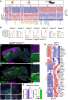
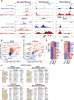
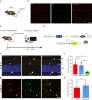
Radial glia integrin avb8 regulates cell autonomous microglial TGFβ1 signaling that is necessary for microglial identity
McKinsey, Santander, Zhang et al
Nat Commun (2025) 16 (1), 2840 DOI: 10.1038/s41467-025-57684-y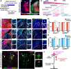
McKinsey, Santander, Zhang et al
Nat Commun (2025) 16 (1), 2840 DOI: 10.1038/s41467-025-57684-y
Cited by 26
AI Summary: OriGene’s EGFP antibody (Goat polyclonal, Cat#: R1091P) was used in this study to detect EGFP expression, aiding in the identification and analysis of genetically modified cells involved in microglial development.
Snippet: oLegend Rat monoclonal antibody. Cat#: 144302: RRID:AB_2561519. Used at 1:300. Antibody FCRLS-APC Butovsky lab Rat monoclonal antibody. Clone 4G11. Used at 1:1000. Antibody EGFP Origene Goat polyclonal antibody. Cat#: R1091P. RRID:AB_1002036. Used at 1:2000. Antibody IB4-488 Thermo Fisher Cat#: I21411. RRID:AB_2314662. Used at 1:150. Antibody IBA1 Novus
Context-dependent response of endothelial cells to PIK3CA mutation
Sabata, Roca-Coll, Dengra et al
bioRxiv (2025)
Sabata, Roca-Coll, Dengra et al
bioRxiv (2025)
Cited by 7
AI Summary: OriGene's anti-GFP antibody (R1091P) was used in this study to detect GFP-labeled endothelial cells, enabling the researchers to assess the cellular response to PIK3CA mutation in different vascular contexts.
Snippet: ubated overnight at 4°C with specific primary antibodies diluted in blocking buffer. Primary antibodies used included: anti-GFP (Abcam, ab13970;1:100), anti-GFP (Origene, R1091P;1:500), anti-GFP AlexaFluor 488 (ThermoFisher Scientific, A-21311;1:200), anti-RFP CF594 (BIOTIUM, 20422;1:200), anti-p-S6 Ser235/236 (Cell Signaling, 4857
Redefining the ontogeny of hyalocytes as yolk sac-derived tissue-resident macrophages of the vitreous body
Rosmus, Koch, Hausmann et al
J Neuroinflammation (2024) 21 (1), 168 DOI: 10.1186/s12974-024-03110-x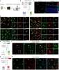
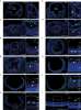
Rosmus, Koch, Hausmann et al
J Neuroinflammation (2024) 21 (1), 168 DOI: 10.1186/s12974-024-03110-x
Cited by 15
AI Summary: In this study, OriGene’s anti-P2ry12 antibody (Cat# TA326570) was used for immunofluorescence labeling to help identify and characterize the immunophenotype of murine hyalocytes, contributing to the investigation of their origin and maintenance as tissue-resident macrophages in the vitreous body.
Snippet: ght at a dilution of 1:500 for Iba-1 (234 013, Synaptic Systems, Göttingen, Germany, RRID: AB_2661873;234 004, Synaptic Systems, Göttingen, Germany, RRID: AB_2493179), 1:1000 for anti-GFP (R1091P, Acris Antibodies Inc., San Diego, USA, RRID: AB_1002036), 1:100 for F4/80 (MF48000, Invitrogen, Waltham, USA, RRID: AB_10376289), 1:500 for TMEM119 (400 002, Synaptic Systems, Göttingen, Germany, RRI
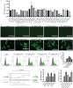
Vitamin C facilitates direct cardiac reprogramming by inhibiting reactive oxygen species
Fang, Yang, Maas et al
Stem Cell Res Ther (2024) 15 (1), 19 DOI: 10.1186/s13287-023-03615-x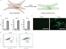
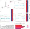
Fang, Yang, Maas et al
Stem Cell Res Ther (2024) 15 (1), 19 DOI: 10.1186/s13287-023-03615-x
Cited by 14
AI Summary: OriGene’s anti-GFP antibody (goat, R1091P) was used in this study to stain and identify newly developed cardiomyocytes during the assessment of cardiac reprogramming efficiency.
Snippet: Spheroids were incubated with antibodies anti-GFP (goat, R1091P, Origene) to stain newly developed cardiomyocytes. …Spheroids were incubated with antibodies anti-GFP (goat, R1091P, Origene) to stain newly developed cardiomyocytes. …
MYH10 activation rescues contractile defects in arrhythmogenic cardiomyopathy (ACM)
García-Quintáns, Sacristán, Márquez-López et al
Nat Commun (2023) 14 (1), 6461 DOI: 10.1038/s41467-023-41981-5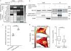
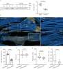
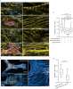
García-Quintáns, Sacristán, Márquez-López et al
Nat Commun (2023) 14 (1), 6461 DOI: 10.1038/s41467-023-41981-5
Cited by 12
AI Summary: In this study, the OriGene anti-GFP antibody (R1091P) was used for immunostaining to detect GFP-tagged proteins in tissue samples, enabling visualization and analysis of protein localization relevant to the investigation of contractile defects in arrhythmogenic cardiomyopathy.
Snippet: ere included in paraffin and cross-sections (5 μm) were then prepared. Tissues were prepared and stained following the indications of the DAB substrate kit (Vector Laboratories). Anti GFP (R1091P, ORIGENE, 1:500) was used as the primary antibody. Images were acquired using an Olympus BX51 microscope using the fitted 10x or 20x UPlanSApo objectives and Cell Sens Entry Ink acquisition software. I
Incongruence between transcriptional and vascular pathophysiological cell states
Fernández-Chacón, Mühleder, Regano et al
bioRxiv (2023)
Fernández-Chacón, Mühleder, Regano et al
bioRxiv (2023)
Cited by 3
Showing 1-6 of 27 papers.
Powered by BizGenius
*Delivery time may vary from web posted schedule. Occasional delays may occur due to unforeseen
complexities in the preparation of your product. International customers may expect an additional 1-2 weeks
in shipping.
Related Products
- Price:
- Actual Price:
Welcome to OriGene AI!
I'm your AI-powered guide to everything OriGene.
Ask me anything:
- Product specs
- Ordering help
- Technical questions
- How to find exactly what your experiment needs
Prefer a live agent? Connect with us by clicking the green chat icon, as seen in the image below or email our team at custsupport@origene.com.
