
GFP Goat Polyclonal Antibody

$1,330.00
3 Days*
Specifications
| Product Data | |
| Application | ELISA, IF, IHC, WB |
|---|---|
| Recommended Dilution | WB: 1:500 - 1:2,500 IHC: 1:200 - 1:1,000 IF: 1:200 - 1:1,000 ELISA: 1:2,000 - 1:8,000 |
| Reactivity | eGFP, GFP, rGFP |
| Antibody Host | Goat |
| Clonality | Polyclonal |
| Immunogen | Recombinant Green Fluorescent Protein (GFP) fusion protein corresponding to the full length amino acid sequence (246aa) derived from the jellyfish Aequorea victoria. |
| Specificity | Anti-GFP Antibody was prepared from monospecific antiserum by immunoaffinity chromatography using Green Fluorescent Protein (Aequorea victoria) coupled to agarose beads followed by solid phase adsorption(s) to remove any unwanted reactivities. Assay by immunoelectrophoresis resulted in a single precipitin arc against anti-Goat Serum, anti-Alkaline Phosphatase and purified and partially purified Green Fluorescent Protein (Aequorea victoria) Serum. No reaction was observed against Human, Mouse and Rat Serum Proteins. |
| Buffer | 0.05 M Tris Chloride, 0.15M Sodium Chloride, 0.001M Magnesium Chloride, 0.0001M Zinc Chloride, 50% (v/v) Glycerol; pH 8.0 Stabilizer: 10 mg/mL Bovine Serum Albumin (BSA) - Immunoglobulin and Protease free Preservative: 0.1% (w/v) Sodium Azide |
| Concentration | 1.0 mg/mL - lot specific |
| Purification | Immunoaffinity Chromatography using Green Fluorescent Protein (Aequorea victoria) coupled to agarose beads followed by solid phase adsorption(s) to remove any unwanted reactivities. |
| Conjugation | Unconjugated |
| Storage | Store vial at 4° C before opening. DO NOT FREEZE. This product is stable at 4° C as an undiluted liquid. Dilute only prior to immediate use. Freezing alkaline phosphatase conjugates will result in a substantial loss of enzymatic activity. |
| Stability | Expiration date is one (1) year from date of receipt. |
| Shipping | Blue Ice |
| Database Link | |
| Background | Conjugated GFP antibody is ideal for western blotting, ELISA and Immunohistochemistry. Green fluorescent protein is a 27 kDa protein produced from the jellyfish Aequorea victoria, which emits green light (emission peak at a wavelength of 509nm) when excited by blue light. GFP is an important tool in cell biology research. GFP is widely used enabling researchers to visualize and localize GFP-tagged proteins within living cells without the need for chemical staining. |
| Synonyms | goat anti-GFP Antibody alkaline phosphatase Conjugation, alk phos conjugated goat anti-GFP antibody, Green Fluorescent Protein, GFP antibody, Green Fluorescent Protein antibody, EGFP, enhanced Green Fluorescent Protein, Aequorea victoria, Jellyfish |
| Note | Anti-GFP is designed to detect GFP and its variants. Anti-GFP Alkaline Phosphatase conjugated antibody has been tested by ELISA, dot blot, and western blot. This antibody can be used to detect GFP by ELISA (sandwich or capture) for the direct binding of antigen and recognizes wild type, recombinant and enhanced forms of GFP. Biotin conjugated polyclonal anti-GFP used in a sandwich ELISA is well suited to titrate GFP in solution using this antibody in combination with Rockland's monoclonal anti-GFP (600-301-215) using either form of the antibody as the capture or detection antibodies. However, use the monoclonal form only for the detection of wild type or recombinant GFP as this form does not sufficiently detect 'enhanced' GFP. The detection antibody is typically conjugated to biotin and subsequently reacted with streptavidin conjugated HRP (code # S000-03). Fluorochrome conjugated polyclonal anti-GFP can be used to detect GFP by immunofluorescence microscopy in prokaryotic (E.coli) and eukaryotic (CHO cells) expression systems and can detect GFP containing inserts. Significant amplification of signal is achieved using fluorochrome conjugated polyclonal anti-GFP relative to the fluorescence of GFP alone. For immunoblotting use either alkaline phosphatase or peroxidase conjugated polyclonal anti-GFP to detect GFP or GFP containing proteins on western blots |
| Reference Data | |
Reviews


Documents
| Product Manuals |
| FAQs |
| SDS |
Citations
Regulation of Myc transcription by an enhancer cluster dedicated to pluripotency and early embryonic expression
Li-Bao, Díaz-Díaz, Raiola et al
Nat Commun (2024) 15 (1), 3931 DOI: 10.1038/s41467-024-48258-5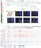
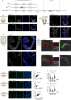
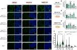
Li-Bao, Díaz-Díaz, Raiola et al
Nat Commun (2024) 15 (1), 3931 DOI: 10.1038/s41467-024-48258-5
Cited by 12
AI Summary: OriGene’s GFP Goat Polyclonal Antibody (#R1091AP) was used as a primary antibody for immunostaining experiments to detect GFP expression in the study.
Snippet: on, embryos were washed with 0.1% Triton X-100 at least 5 times. Primary and secondary antibodies were diluted in the blocking solution. Primary antibodies: GFP Goat Polyclonal Antibody (Goat, 1:200, OriGene, #R1091AP), Human/Mouse Brachyury Antibody (mouse, 1:250, R&D systems, #AF2085-SP), and Anti-SOX2 antibody (rabbit, 1:200, Abcam, #ab97959). Secondary antibodies: 648 donkey anti-goat (1:500,
Generation of a human CDX2 knock-in reporter iPSC line (MHHi007-A-1) to model human trophoblast differentiation
Malysheva, Wunderlich, Haase et al
Stem Cell Res (2018) 30, 117-121 DOI: 10.1016/j.scr.2018.05.015
Malysheva, Wunderlich, Haase et al
Stem Cell Res (2018) 30, 117-121 DOI: 10.1016/j.scr.2018.05.015
Cited by 3
Fluorescent protein-expressing neural progenitor cells as a tool for transplantation studies
Skardelly, Hempel, Hirrlinger et al
PLoS One (2014) 9 (6), e99819 DOI: 10.1371/journal.pone.0099819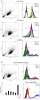
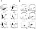
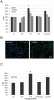
Skardelly, Hempel, Hirrlinger et al
PLoS One (2014) 9 (6), e99819 DOI: 10.1371/journal.pone.0099819
Cited by 2
AI Summary: OriGene products contributed to this study by providing the anti-GFAP antibody (Cat# TA309153) used for immunocytochemistry and immunoblotting to characterize neural progenitor cell differentiation.
DMAP1 is an essential regulator of ATM activity and function
Penicud, Behrens
Oncogene (2013)
Penicud, Behrens
Oncogene (2013)
Cited by 30
Aberrant neurogenesis after stroke: a retroviral cell labeling study
Niv, Keiner, Krishna et al
Stroke (2012) 43 (9), 2468-75 DOI: 10.1161/STROKEAHA.112.660977
Niv, Keiner, Krishna et al
Stroke (2012) 43 (9), 2468-75 DOI: 10.1161/STROKEAHA.112.660977
Panglial gap junctional communication is essential for maintenance of myelin in the CNS
Tress, Maglione, May et al
J Neurosci (2012) 32 (22), 7499-518 DOI: 10.1523/JNEUROSCI.0392-12.2012
Tress, Maglione, May et al
J Neurosci (2012) 32 (22), 7499-518 DOI: 10.1523/JNEUROSCI.0392-12.2012
AI Summary: OriGene products contributed to this study by providing the anti-Connexin 47 (Cx47) antibody (Cat# TA326043), which was used to analyze connexin expression and validate the disruption of gap junctional communication in the mouse models.
Showing 1-6 of 7 papers.
Powered by BizGenius
*Delivery time may vary from web posted schedule. Occasional delays may occur due to unforeseen
complexities in the preparation of your product. International customers may expect an additional 1-2 weeks
in shipping.
Related Products
- Price:
- Actual Price:
Welcome to OriGene AI!
I'm your AI-powered guide to everything OriGene.
Ask me anything:
- Product specs
- Ordering help
- Technical questions
- How to find exactly what your experiment needs
Prefer a live agent? Connect with us by clicking the green chat icon, as seen in the image below or email our team at custsupport@origene.com.
