
Procollagen Type III Rabbit Polyclonal Antibody
$845.00
2 Weeks*
Specifications
| Product Data | |
| Application | ELISA, IF, IHC, R |
|---|---|
| Recommended Dilution | RIA: > 1/200. ELISA: 1/100-1/200 (OD ≥ 500). Immunofluorescence (indirect, 1/40 on Frozen tissues). Immunohistochemistry on Frozen and Paraffin Embedded Tissue Sections: 1/500. Pretreatment: After removing Paraffin pre-treat with 0.2% Hyaluronidase (approx. 300 U/mg) in TBS, 15 min at 37°C, then block unspecific binding with blocking serum or 3% BSA in TBS and perform blocking of endogen Peroxidase with 1% H2O2 in TBS, if necessary. Incubation Time: 60 min at room temperature or overnight at 2-8°C. Positive Control: Human or Bovine Skin and Liver. |
| Reactivity | Bovine, Human, Porcine |
| Antibody Host | Rabbit |
| Clonality | Polyclonal |
| Immunogen | Purified Human Procollagen type III C-terminal and N-terminal PIIICP separated. |
| Specificity | Human and Bovine Pro-Collagen Type III (100%). Human and Bovine Pro-Collagen Typ I and Collagen Typ I <0.1% (RIA at 1/200 dilution). |
| Buffer | PBS without BSA or preservatives State: Purified State: Lyophilized purified Ig fraction |
| Reconstitution Method | Restore with 0.1 ml sterile distilled water. |
| Concentration | ~1.0 mg/ml (after reconstitution) |
| Purification | Affinity Chromatography |
| Conjugation | Unconjugated |
| Storage | Store lyophilized at 2-8°C for 6 months or at -20°C long term. After reconstitution store the antibody undiluted at 2-8°C for one month or (in aliquots) at -20°C long term. Avoid repeated freezing and thawing. |
| Stability | Shelf life: one year from despatch. |
| Shipping | Ambient |
| Background | Collagens consist in a family of highly specialized glycoproteins of which at least 16 genetically distinct types are known to date. The basal unit of a collagen molecule cosists in a tripel-helical structure formed by 3 alpha-chains. Predominant amino acids are glycine, proline and hydroxproline. Regularly also lysines and hydroxylysines occur, which are responsible for cross-linkage and glycosylation of the protein chains. Different composition of alpha-chains and different glycosylation contribute to the high variability of collagens in different tissues and organs. Type III collagen is an alpha1(III)-trimer, MW 95 kDa, which forms 67 nm cross-banded fibrils. Typically it can be observed in skin, cartilage and vitreous body. |
| Synonyms | Collagen, Pro-Collagen |
| Reference Data | |
Reviews


Documents
| Product Manuals |
| FAQs |
| SDS |
Citations
Tendon Regeneration After Partial-Thickness Peroneus Longus Tendon Harvesting: Magnetic Resonance Imaging Evaluation and In Vivo Animal Study
Lee, Wang, Bae et al
Am J Sports Med (2020) 48 (10), 2499-2509 DOI: 10.1177/0363546520933628
Lee, Wang, Bae et al
Am J Sports Med (2020) 48 (10), 2499-2509 DOI: 10.1177/0363546520933628
Cited by 10
AI Summary: In this study, OriGene's Mouse Anti-Tenomodulin Monoclonal Antibody (product code: TA323617) was used for immunohistochemical analysis to assess tendon regeneration quality in both patient and animal samples.
Snippet: nd degree of fibroplasia. Collagen synthesis and organization were evaluated by immunohistochemical staining for collagen type I (Abcam) and collagen type III (Acris antibodies). For detection, the polymer (Vector) method was coupled with an enzyme staining system. We used H&E-stained slides for histologic examination and evaluated 6 parameters (fiber arrang
Erythropoietin Alleviates Burn-induced Muscle Wasting
Wu, Lu, Tai et al
Int J Med Sci (2020) 17 (1), 33-44 DOI: 10.7150/ijms.38590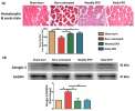
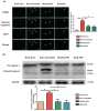
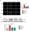
Wu, Lu, Tai et al
Int J Med Sci (2020) 17 (1), 33-44 DOI: 10.7150/ijms.38590
Cited by 12
AI Summary: In this study, OriGene's type III collagen antibody (1:200) was used for immunofluorescence analysis to assess muscular fibrotic changes following burn injury and EPO treatment.
Snippet: Muscular fibrotic changes were analyzed by immunofluorescence with fibronectin (1: 200, Novus Biologicals, Littleton, CO, USA), type I collagen (1: 200, Novus Biologicals, Littleton, CO, USA), type III collagen (1:200, Origene, Rockville, Maryland, USA) and phosphorylated;;- Muscular fibrotic changes were analyzed by immunofluorescence with fibronectin (1: 200, Novus Biologicals, Littleton, CO,...
Analysis of morphological characteristics and expression levels of extracellular matrix proteins in skin wounds to determine wound age in living subjects in forensic medicine
Fronczek, Lulf, Korkmaz et al
Forensic Sci Int (2015) 246, 86-91 DOI: 10.1016/j.forsciint.2014.11.013
Fronczek, Lulf, Korkmaz et al
Forensic Sci Int (2015) 246, 86-91 DOI: 10.1016/j.forsciint.2014.11.013
Cited by 15
AI Summary: In this study, OriGene products—specifically, the anti-collagen III antibody (TA506084), anti-collagen IV antibody (TA506082), and anti-α-SMA antibody (TA802527)—were used for immunohistochemical analysis to measure extracellular matrix protein expression in skin wound biopsies, contributing to the assessment of wound age in forensic medicine.
Laser-mediated fixation of collagen-based scaffolds to dermal wounds
Steinstraesser, Wehner, Trust et al
Lasers Surg Med (2010) 42 (2), 141-9 DOI: 10.1002/lsm.20901
Steinstraesser, Wehner, Trust et al
Lasers Surg Med (2010) 42 (2), 141-9 DOI: 10.1002/lsm.20901
Cited by 24
AI Summary: In this study, OriGene’s Mouse anti-Collagen I antibody (Cat# TA506771) was used for immunohistochemical analysis to assess extracellular matrix formation and evaluate the effectiveness of different scaffold fixation methods during wound healing.
Showing 1-4 of 4 papers.
Powered by BizGenius
*Delivery time may vary from web posted schedule. Occasional delays may occur due to unforeseen
complexities in the preparation of your product. International customers may expect an additional 1-2 weeks
in shipping.
Related Products
- Price:
- Actual Price:
Welcome to OriGene AI!
I'm your AI-powered guide to everything OriGene.
Ask me anything:
- Product specs
- Ordering help
- Technical questions
- How to find exactly what your experiment needs
Prefer a live agent? Connect with us by clicking the green chat icon, as seen in the image below or email our team at custsupport@origene.com.
