
Desmoglein 3 (DSG3) Mouse Monoclonal Antibody [Clone ID: Dsg-G194]
$515.00
2 Weeks*
Specifications
| Product Data | |
| Clone Name | Dsg-G194 |
|---|---|
| Application | IHC, WB |
| Recommended Dilution | Immunoblotting (Western Blot). Immunohistochemistry on Frozen Sections: Ready-to-use. Incubation Time: 1 h at RT. Recommendations: For better resolution, preincubation (directly after fixation) with 0.05-0.2% Triton X-100 for 5-10 min, depending on tissue type, is recommended (See Ref.2). |
| Reactivity | Human |
| Antibody Host | Mouse |
| Isotype | IgG1 |
| Clonality | Monoclonal |
| Immunogen | Recombinant Human polypeptide Desmoglein 3 (C-terminal amino acids 920-940) |
| Specificity | In Immunoblots of cytoskeletal preparations from Human epidermis the antibody reacts specifically with a 140 kD polypeptide typical for Desmoglein 3. In Immunolocalization studies, the mab shows typical staining of basal and lower suprabasal layers of the epidermis and special regions of fetal and anagen hair follicles. The staining pattern observed corresponds to those obtained with pemphigus vulgaris patient sera. Cross-reaction with Desmogleins 1 and 2 was not detected. |
| Buffer | State: Supernatant State: Liquid Culture Supernatant Preservative: 0.09% Sodium Azide |
| Conjugation | Unconjugated |
| Storage | Store the antibody undiluted at 2-8°C. |
| Stability | Shelf life: One year from despatch. |
| Shipping | Blue Ice |
| Gene Name | desmoglein 3 |
| Database Link | |
| Background | Desmosomes are cell-cell junctions between epithelial, myocardial, and certain other cell types. Desmoglein 3 is a calcium-binding transmembrane glycoprotein component of desmosomes in vertebrate epithelial cells. Currently, three desmoglein subfamily members have been identified and all are members of the cadherin cell adhesion molecule superfamily. These desmoglein gene family members are located in a cluster on chromosome 18. This protein has been identified as the autoantigen of the autoimmune skin blistering disease pemphigus vulgaris. |
| Synonyms | DSG3, CDHF6, PVA, 130 kDa pemphigus vulgaris antigen |
| Reference Data | |
| Protein Categories | Membrane Proteins |
Reviews


Documents
| Product Manuals |
| FAQs |
| SDS |
Citations
Identification of novel therapeutic targets for blocking acantholysis in pemphigus
Burmester, Flaswinkel, Thies et al
Br. J. Pharmacol. (2020)
Burmester, Flaswinkel, Thies et al
Br. J. Pharmacol. (2020)
Cited by 29
AI Summary: Based on the provided information, no OriGene products were mentioned or used in this paper; therefore, OriGene products did not contribute to this study.
Snippet: and the keratinocytes were prepared for staining with an anti-Dsg3 antibody (mouse, monoclonal (6G2C11), IgG1, Acris Antibodies, Herford, Germany) diluted 1:100 in DPBS for 1 h. The washing step with DPBS was repeated 3 times to stain the keratinocytes with a;- and the keratinocytes were prepared for staining with an anti-Dsg3 antibody (mouse, monoclonal (6G2C11), IgG1, Acris Antibodies, Herford, Germany) diluted 1:100 in DPBS for 1 h. The washing step with DPBS was repeated 3 times to stain the keratinocytes with a
Non-pathogenic pemphigus foliaceus (PF) IgG acts synergistically with a directly pathogenic PF IgG to increase blistering by p38MAPK-dependent desmoglein 1 clustering
Yoshida, Ishii, Shimizu et al
J Dermatol Sci (2017) 85 (3), 197-207 DOI: 10.1016/j.jdermsci.2016.12.010

Yoshida, Ishii, Shimizu et al
J Dermatol Sci (2017) 85 (3), 197-207 DOI: 10.1016/j.jdermsci.2016.12.010
Cited by 36
AI Summary: In this study, OriGene’s p38MAPK inhibitor (catalog # TL0016) was used to demonstrate that blocking p38MAPK prevented desmoglein 1 clustering, helping to show the pathway’s role in antibody-induced blister formation in pemphigus foliaceus.
Prevalence of pemphigus and pemphigoid autoantibodies in the general population
Prüßmann, Prüßmann, Koga et al
Orphanet J Rare Dis (2015) 10, 63 DOI: 10.1186/s13023-015-0278-x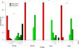
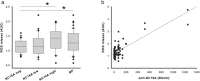
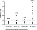
Prüßmann, Prüßmann, Koga et al
Orphanet J Rare Dis (2015) 10, 63 DOI: 10.1186/s13023-015-0278-x
Cited by 60
AI Summary: In this study, OriGene’s recombinant BP180-NC16A and BP230 proteins were used for ELISA validation and functional assays to detect and characterize autoantibodies in blood donor samples, helping to assess their prevalence and functional properties in the general population.
Showing 1-3 of 3 papers.
Powered by BizGenius
*Delivery time may vary from web posted schedule. Occasional delays may occur due to unforeseen
complexities in the preparation of your product. International customers may expect an additional 1-2 weeks
in shipping.
Related Products
- Price:
- Actual Price:
Welcome to OriGene AI!
I'm your AI-powered guide to everything OriGene.
Ask me anything:
- Product specs
- Ordering help
- Technical questions
- How to find exactly what your experiment needs
Prefer a live agent? Connect with us by clicking the green chat icon, as seen in the image below or email our team at custsupport@origene.com.
