
Synaptopodin (SYNPO) Mouse Monoclonal Antibody [Clone ID: G1D4]

$515.00
2 Weeks*
Specifications
| Product Data | |
| Clone Name | G1D4 |
|---|---|
| Application | IF, IHC, WB |
| Recommended Dilution | Immunofluorescence. Immunohistochemistry on Frozen Sections. Immunohistochemistry on Paraffin Sections (after microwave treatment). Incubation Time: 1 h at RT for Immunohistochemistry . Working Dilution: Ready-to-use for Immunohistochemistry. Immunoblotting (Western Blot). The antibody also reacts with a 44 kD degradation fragment of Synaptopodin. |
| Reactivity | Bovine, Guinea Pig, Human, Mouse, Rat |
| Antibody Host | Mouse |
| Isotype | IgG1 |
| Clonality | Monoclonal |
| Immunogen | Isolated Rat kidney glomeruli. |
| Specificity | The antibody reacts specifically with Synaptopodin. In Human tissue Synaptopodin has a Molecular Weight of 73.7 kD and pI of 9.38 (calculated from sequence data); in Mouse the corresponding data are 74 kD, pI 9.27. In SDS-PAGE the antigen appears as 100 kD polypeptide in brain and 110 kD polypeptide in kidney (the difference might be attributed to posttranslational modifications). 1. The antibody recognizes differentiated podocytes (glomerular visceral epithelial cells) in vivo and in vitro (weaker additional reaction with arterial endothelial cells), co-localization with alpha-actinin. Does not react with parietal cells. 2. Reacts with a subset of exclusively telencephalic synapses. Differentiationdependent expression during postnatal maturation of Rat brain. Differentiationdependent expression in cultured hippocampal neurons. |
| Buffer | State: Supernatant State: Liquid Culture Supernatant Preservative: 0.01% Sodium Azide |
| Conjugation | Unconjugated |
| Storage | Store the antibody undiluted at 2-8°C. |
| Stability | Shelf life: one year from despatch. |
| Shipping | Blue Ice |
| Gene Name | synaptopodin |
| Database Link | |
| Background | Synaptopodin, a prolin-rich actin-binding protein with 2 binding sites for actin, represents a new class of actin-binding proteins which has first been localized in podocytes and a subset of telencephalic postsynaptic densities. |
| Synonyms | KIAA1029 |
| Reference Data | |
| Protein Categories | Intracellular Proteins |
Reviews


Documents
| Product Manuals |
| FAQs |
| SDS |
Citations
Fetal growth restriction leads to prolonged nephrogenesis and gene dysregulation in the adult kidney
Timberline, Dailey, Johnson et al
Life Sci Alliance (2026) 9 (11) DOI: 10.26508/lsa.202603713
Timberline, Dailey, Johnson et al
Life Sci Alliance (2026) 9 (11) DOI: 10.26508/lsa.202603713
AI Summary: In this study, OriGene products, specifically the WT1 antibody (Cat# TA336404), were used for immunostaining to evaluate nephron progenitor cells and glomerular structures, contributing to the assessment of kidney development and pathology in the mouse model.
Snippet: rted (45).Podocyte density and glomerular areaDual immunofluorescent immunostaining was performed with synaptopodin (Acris Antibodies Mouse Anti-Synaptopodin, Clone G1D4, BM5086, 1:10 dilution), WT-1 (Cat# MA5-32215;Thermo Fisher Scientific, 1:50 dilution), and appropriate secondary antibodies (Jackson Immunoresear
Preterm Birth Increases Susceptibility to Hyperglycemia-Induced Kidney Injury With Sex-Specific Differences in Structural and Molecular Responses
Dailey, Cwiek, Hamil et al
Endocrinol Diabetes Metab (2026) 9 (3), e70223 DOI: 10.1002/edm2.70223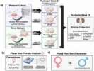
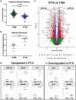
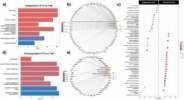
Dailey, Cwiek, Hamil et al
Endocrinol Diabetes Metab (2026) 9 (3), e70223 DOI: 10.1002/edm2.70223
AI Summary: OriGene products contributed to this study through the use of the Acris Antibodies Mouse Anti‐Synaptopodin antibody (Clone G1D4, BM5086) for dual immunofluorescence staining to assess podocyte density and glomerular area in mouse kidney tissue.
Snippet: p>2.5.5Podocyte DensityDual immunofluorescence staining for WT‐1 (Invitrogen, Rabbit Anti‐WT1, 1:50 dilution) and synaptopodin (Acris Antibodies Mouse Anti‐Synaptopodin, Clone G1D4, BM5086, 1:20 dilution) was performed. Nuclei were stained with Hoescht. Podocyte density and glomerular area were assessed using a protocol adap
Preterm birth increases susceptibility to hyperglycemia induced glomerular alterations in male mice
Cwiek, Dailey, Kameshwar et al
Sci Rep (2025) 15 (1), 18934 DOI: 10.1038/s41598-025-00103-5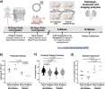
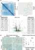
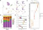
Cwiek, Dailey, Kameshwar et al
Sci Rep (2025) 15 (1), 18934 DOI: 10.1038/s41598-025-00103-5
Cited by 4
AI Summary: In this study, OriGene’s Mouse Synaptopodin antibody (BM5086) was used for dual immunofluorescence staining to quantify podocyte density and assess glomerular alterations in mouse kidney tissue.
Snippet: ensity and glomerular area were quantified using a protocol modified from Venkatareddy et al.47 Dual IF Immunostaining for WT-1 (Santa Cruz, Rabbit, Anti-WT1, sc-192, 1:50 dilution) and synaptopodin (Acris Antibodies Mouse Anti-Synaptopodin, Clone G1D4, BM5086, 1:10 dilution) was performed using 5 μm sections with sodium citrate retrieval and appropriate secondary antibodies (Jackson Immunoresea
Multiomics of synaptic junctions reveals altered lipid metabolism and signaling following environmental enrichment
Borgmeyer, Coman, Has et al
Cell Rep (2021) 37 (1), 109797 DOI: 10.1016/j.celrep.2021.109797
Borgmeyer, Coman, Has et al
Cell Rep (2021) 37 (1), 109797 DOI: 10.1016/j.celrep.2021.109797
Cited by 26
Increased dishevelled associated activator of morphogenesis 2, a new podocyte-associated protein, in diabetic nephropathy
Qi, Alsomali, Zhong et al
Nephrol Dial Transplant (2021) 36 (6), 1006-1016 DOI: 10.1093/ndt/gfab014
Qi, Alsomali, Zhong et al
Nephrol Dial Transplant (2021) 36 (6), 1006-1016 DOI: 10.1093/ndt/gfab014
Cited by 5
AI Summary: In this study, OriGene products including the DAAM2 antibody (TA503367) and DAAM2 siRNA (SR313426) were used for immunostaining and gene knockdown experiments, respectively, to investigate the expression and functional role of DAAM2 in podocytes in diabetic nephropathy.
Mutations in the gene that encodes the F-actin binding protein anillin cause FSGS
Gbadegesin, Hall, Adeyemo et al
J Am Soc Nephrol (2014) 25 (9), 1991-2002 DOI: 10.1681/ASN.2013090976
Gbadegesin, Hall, Adeyemo et al
J Am Soc Nephrol (2014) 25 (9), 1991-2002 DOI: 10.1681/ASN.2013090976
AI Summary: In this study, OriGene’s ANLN cDNA clone (RC206307) was used to investigate the functional effects of ANLN mutations associated with FSGS, enabling the overexpression and analysis of mutant and wild-type anillin in human podocytes.
Showing 1-6 of 12 papers.
Powered by BizGenius
*Delivery time may vary from web posted schedule. Occasional delays may occur due to unforeseen
complexities in the preparation of your product. International customers may expect an additional 1-2 weeks
in shipping.
Related Products
- Price:
- Actual Price:
Welcome to OriGene AI!
I'm your AI-powered guide to everything OriGene.
Ask me anything:
- Product specs
- Ordering help
- Technical questions
- How to find exactly what your experiment needs
Prefer a live agent? Connect with us by clicking the green chat icon, as seen in the image below or email our team at custsupport@origene.com.
