
Pecam1 Rat Monoclonal Antibody [Clone ID: ER-MP12]

Specifications
| Product Data | |
| Clone Name | ER-MP12 |
|---|---|
| Application | FC, IHC |
| Recommended Dilution | Immunohistochemistry on Frozen Sections: 0.25-0.5 µg/ml (1/400-1/800). Immunohistochemistry on Paraffin Sections: 20 µg/ml (1/10). Proteinase K pre-treatment for antigen retrieval is recommended. Do not use standard 4% Formalin fixation, but max. 0.05% glutaraldehyde or 1% paraformaldehyde! Suggested Positive Control: Mouse spleen. Has been described to work in FACS. |
| Reactivity | Mouse |
| Antibody Host | Rat |
| Isotype | IgG2a |
| Clonality | Monoclonal |
| Immunogen | Mouse macrophage precursor cells. The antigen is a glutaraldehyde (0.05%) and paraformaldehye (1%) resistant 140 kD surface glycoprotein (reducing and non reducing conditions) identified as CD31. The antigen expression closely follows that of CD34. |
| Specificity | Monoclonal CD31 antibody (clone ER-MP12) recognizes PECAM-1, platelet/endothelial cell adhesion molecule 1. It is a general marker for the detection of a major population of haematopoietic stem cells and is useful for the accumulation of early murine macrophage precursors from bone marrow cell suspensions by FACS. An even more precise characterization of such isolated and concentrated precursor cells is achieved when ER-MP12 is combined with other markers like ER-MP20 (anti Ly-6C, Cat.-No BM4019) or ER-MP58 (Cat.-No BM4089). The ER-MP12 antigen is also expressed by endothelial cells. Antigen Distribution Isolated Cells: The antigen is found on the surface of approximately 40% of freshly isolated bone marrow cells of adult mice, on the majority of immortalized macrophage precursor cell lines (M1, RMB-1, RMB-3) corresponding to CFU-GM cells. The antibody detects pluri- and multipotent stem cells and prothymocytes as well as particular lymphoid cells in bone marrow and peripheral lymphoid organs. Tissue Sections: The antigen is detected within the lymphopoietic islands in the spleen of newborn (day 12) and in the bone marrow of adult mice. Capillary endothelial cells of adult mice also express the antigen. Detects a major population of Mouse colony forming unit macrophage (CFU-M) precursor cells, subpopulation of pre CFU-M and monoblasts, precursor cells of granulocytes. |
| Buffer | PBS, pH 7.2 State: Purified State: Lyophilized purified IgG fraction Stabilizer: 6 mg/ml BSA Preservative: 0.05% ProClin150 |
| Reconstitution Method | Restore by adding 0.5 ml distilled water (= 0.2 mg/ml Stock Solution). |
| Concentration | 0.2 mg/ml (after reconstitution) |
| Purification | Affinity Chromatography |
| Conjugation | Unconjugated |
| Storage | Store lyophilized at 2-8°C for 6 months or at -20°C long term. After reconstitution store the antibody undiluted at 2-8°C for one month or (in aliquots) at -20°C long term. Avoid repeated freezing and thawing. |
| Stability | Shelf life: one year from despatch. |
| Shipping | Ambient |
| Gene Name | platelet/endothelial cell adhesion molecule 1 |
| Database Link | |
| Background | CD31, also known as platelet endothelial cell adhesion molecule 1 (PECAM1), is a type I integral membrane glycoprotein and a member of the immunoglobulin superfamily of cell surface receptors. It is constitutively expressed on the surface of endothelial cells, and concentrated at the junction between them. It is also weakly expressed on many peripheral lymphoid cells and platelets. CD31 has been used to measure angiogenesis in association with tumor recurrence. Other studies have also indicated that CD31 and CD34 can be used as markers for myeloid progenitor cells and recognize different subsets of myeloid leukemia infiltrates (granular sarcomas). |
| Synonyms | PECAM-1, EndoCAM, GPIIA' |
| Note | This antibody was produced serum-free, without fetal calf serum. Protocol: Protocol with Frozen, ice-cold Acetone-Fixed Sections: (The whole procedure is performed at room temperature) 1. Wash in PBS. 2. Block endogenous peroxidase. 3. Wash in PBS. 4. Block with 10% normal goat serum in PBS for 30min. in a humid chamber. 5. Incubate with primary antibody (dilution see datasheet) for 1h in a humid chamber. 6. Wash in PBS. 7. Incubate with secondary antibody (peroxidase-conjugated goat anti rat IgG (H+L) minimal-cross reaction to mouse) for 1h in a humid chamber. 8. Wash in PBS. 9. Incubate with AEC substrate (3-amino-9-ethylcarbazol) for 12min. 10. Wash in PBS. 11. Counterstain with Mayer's hemalum. Protocol with Formalin-Fixed, Paraffin-Embedded Sections: (The whole procedure is performed at room temperature) 1. Deparaffinize and rehydrate tissue section. 2. Incubate the tissue section with proteinase K for 7min. 3. Wash in distilled water. 4. Block endogenous peroxidase. 5. Wash in PBS. 6. Block with 10% normal goat serum in PBS for 30min. in a humid chamber. 7. Incubate with primary antibody (dilution see datasheet) for 1h in a humid chamber. 8. Wash in PBS. 9. Incubate with secondary antibody (peroxidase-conjugated goat anti rat IgG (H+L) minimal-cross reaction to mouse) for 1h in a humid chamber. 10. Wash in PBS. 11. Incubate with AEC substrate (3-amino-9-ethylcarbazol) for 12min. 12. Wash in PBS. 13. Counterstain with Mayer's hemalum. |
| Reference Data | |
| Protein Categories | ADC Targets, CD Markers, Intracellular Proteins, Membrane Proteins |
Reviews


Documents
| Product Manuals |
| FAQs |
| SDS |
Citations
DNA-sensing inflammasomes cause recurrent atherosclerotic stroke
Cao, Roth, Zhang et al
Nature (2024)
Cao, Roth, Zhang et al
Nature (2024)
Cited by 115
AI Summary: OriGene’s rat anti-CD31 antibody (BM4086) was used in this study to identify endothelial cells in tissue sections, contributing to the analysis of vascular changes and plaque destabilization following stroke.
Snippet: Cell Signaling), mouse anti-mouse caspase-1 (1:200;AG-20B-0042-C100, Adipogen), sheep anti-von Willebrand factor (1:100;ab11713, Abcam), rat anti-CD31 (1:200;BM4086, OriGene) and anti-collagen I (1:250;ab279711, Abcam). …
Mesenchymal loss of p53 alters stem cell capacity and models human soft tissue sarcoma traits
Sorimachi, Kobayashi, Shiozawa et al
Stem Cell Reports (2023) 18 (5), 1211-1226 DOI: 10.1016/j.stemcr.2023.03.009
Sorimachi, Kobayashi, Shiozawa et al
Stem Cell Reports (2023) 18 (5), 1211-1226 DOI: 10.1016/j.stemcr.2023.03.009
Cited by 11
AI Summary: In this study, OriGene's p53 antibody (TA500132) was used for immunohistochemistry to detect p53 expression, contributing to the analysis of p53 loss in mesenchymal stem cells and its role in soft tissue sarcoma development.
Snippet: D45 (30-F11;BD Biosciences), TER-119 (TER119;TOMBO), CD31 (MEC13.3;Bolegend), CD140a (APA5;Invitrogen), and LepR (BAF497;R&D Systems). For immunohistochemistry, a rat anti-CD31 antibody (BM4086;Acris Antibodies GmbH), a mouse anti-vimentin antibody (ab8978;Abcam), anti-rat IgG-Dylight650 (#SA5-10021;Invitrogen), and anti-mouse IgG1-Alexa Fluor 555 (#A-21127;Invitrogen) were used. Cell pr
Endothelial Retargeting of AAV9 In Vivo
Bozoglu, Lee, Ziegler et al
Adv Sci (Weinh) (2022) 9 (7), e2103867 DOI: 10.1002/advs.202103867
Bozoglu, Lee, Ziegler et al
Adv Sci (Weinh) (2022) 9 (7), e2103867 DOI: 10.1002/advs.202103867
Cited by 40
AI Summary: OriGene products contributed to this study by providing the murine CD31 antibody (BM4086) for immunofluorescence staining of endothelial cells and the AAV9 intact capsid antibody (ADK9) for detecting viral particles.
Snippet: … The following antibodies were used for immunofluorescence staining: EGFP (CAB4211, Thermo Fisher), murine CD31 (BM4086, Origene), porcine CD31 (LCI‐4, Bio‐Rad), CRISPR‐Cas9 (7A9‐3A3, Novus Biologicals), AAV9 intact capsid (ADK9, Origene). Highly crossadsorbed Alexa Fluor conjugated secondary antibodies were use;- Murine cells were immediately stained with a specific antibody for CD31 (BM4086 , Origene) for 15 min, followed by secondary antibody staining (A48265, Thermo Fisher). …
Distinct Regulatory Programs Control the Latent Regenerative Potential of Dermal Fibroblasts during Wound Healing
Abbasi, Sinha, Labit et al
Cell Stem Cell (2020) 27 (3), 396-412.e6 DOI: 10.1016/j.stem.2020.07.008
Abbasi, Sinha, Labit et al
Cell Stem Cell (2020) 27 (3), 396-412.e6 DOI: 10.1016/j.stem.2020.07.008
Cited by 200
AI Summary: OriGene products, specifically the Mouse Runx1 (NM_001146703) qPCR Primer Pair (Cat# MP218486) and Mouse Hic1 (NM_001114972) qPCR Primer Pair (Cat# MP209377), were used in this study to quantify gene expression levels of Runx1 and Hic1, contributing to the analysis of regulatory programs controlling fibroblast-mediated wound healing.
Snippet: Skip to article;Rabbit anti-Lef1 Cell Signaling Cat# 2230, RRID: AB_823558 Goat anti-CD26 R&D Systems Cat# AF954, RRID: AB_355739 Rat anti-CD31 Acris antibodies Cat# BM4086, RRID: AB_975771 Rabbit anti-FABP4 R&D Systems Cat# AF1443, RRID: AB_2102444 Goat anti-Rabbit Alexa Fluor 488/555/647 Life Technologies N/A Goat anti-Rat Alexa Fluor 488/555/647 Life Technologies N/A Donkey anti-Goat Alex Fluor 488/555/647 Life Technologies N/A
EGFR/Ras-induced CCL20 production modulates the tumour microenvironment
Hippe, Braun, Oláh et al
Br. J. Cancer (2020)
Hippe, Braun, Oláh et al
Br. J. Cancer (2020)
Cited by 44
AI Summary: OriGene products, specifically the human CCL20 ELISA Kit (EA100533) and human CCL20 qPCR primers (HP208956), were used in this study to quantify and analyze CCL20 expression in cancer cell lines and tumour tissues, thereby helping to elucidate the role of EGFR/Ras-induced CCL20 in modulating the tumour microenvironment.
Snippet: Immunohistochemistry Frozen and paraffin-embedded tumour sections from cancer patients were stained with antibodies against human CCL20 (goat IgG: R&D Systems, Minneapolis, MN, USA;horse IgG: Vector Laboratory’s Vectastain ABC peroxidase kit) or human pERK (mouse IgG1: Cell Signaling, Danvers, MA, USA;horse IgG: Vector Laboratory’s Vectastain ABC peroxidase kit). Frozen matrigel plugs were cut int...
Active Vaccination With EMMPRIN-Derived Multiple Antigenic Peptide (161-MAP) Reduces Angiogenesis in a Dextran Sodium Sulfate (DSS)-Induced Colitis Model
Simanovich, Brod, Rahat
Front Immunol (2018) 9, 2919 DOI: 10.3389/fimmu.2018.02919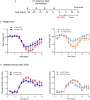
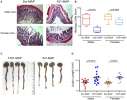
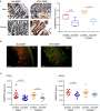
Simanovich, Brod, Rahat
Front Immunol (2018) 9, 2919 DOI: 10.3389/fimmu.2018.02919
Cited by 9
AI Summary: OriGene products, specifically the mouse CD31 (PECAM1) antibody (Cat# TA500439) and mouse EMMPRIN (BSG) ELISA kit (Cat# EKC39865), were used in this study to assess vessel density by immunohistochemistry and to measure EMMPRIN protein levels, respectively, thereby enabling evaluation of the effects of 161-MAP vaccination on angiogenesis and EMMPRIN expression in the DSS-induced colitis model.
Showing 1-6 of 12 papers.
Powered by BizGenius
*Delivery time may vary from web posted schedule. Occasional delays may occur due to unforeseen
complexities in the preparation of your product. International customers may expect an additional 1-2 weeks
in shipping.
Related Products
- Price:
- Actual Price:
Welcome to OriGene AI!
I'm your AI-powered guide to everything OriGene.
Ask me anything:
- Product specs
- Ordering help
- Technical questions
- How to find exactly what your experiment needs
Prefer a live agent? Connect with us by clicking the green chat icon, as seen in the image below or email our team at custsupport@origene.com.
