
Fibroblasts (Pan Reticular) Rat Monoclonal Antibody [Clone ID: ER-TR7]

$1,125.00
2 Weeks*
Specifications
| Product Data | |
| Clone Name | ER-TR7 |
|---|---|
| Application | FC, IHC |
| Recommended Dilution | Immunohistochemistry on Frozen Sections: 1 µg/ml (1/400) freshly prepared for Mouse tissue and 4 µg/ml (1/100) on Human/Porcine tissue. Recommended Positive Control: Mouse spleen. Does not react on routinely processed Paraffin Sections. Has been described to work in FACS. |
| Reactivity | Human, Mouse, Porcine |
| Antibody Host | Rat |
| Isotype | IgG2a |
| Clonality | Monoclonal |
| Immunogen | Murine thymic reticulum. Epitope: The antigen has not been fully characterized. The epitope may be part of reticulin. |
| Specificity | Reacts with Mouse reticular fibroblasts, reticular fibres and Human tonsil. ER-TR7 detects an antigen present in and produced by reticular fibroblasts. The recognized antigen is most likely distinct from Laminin, Fibronectin, Collagen types I-IV, Heparan Sulfate Proteoglycan, Entactin, and Nidogen. The antibody is useful to stain the microanatomy of various organs, in particular the connective tissue framework in lymphoid organs. The antibody also stains subendothelial deposits in the plaque area of atherosclerotic plaques. Antigen Distribution Isolated Cells: The antigen is found in the cytoplasm of fibroblast cell lines. Tissue Sections: The antigen is expressed in connective tissues which form a supporting network between parenchymal cells of all organs tested so far (see table below). Thus, the supportive mesenchymal structures of larger vessels can be studied. In spleen, a very clear delineation of red and white pulp is obtained. Capsule, sinuses, follicules, paracortex and medullary cords are also clearly delineated in lymph nodes. |
| Buffer | Stock solution contains PBS, pH 7.2 with 5 mg/ml BSA as a stabilizer and 0.09% Sodium Azide as preservative State: Purified State: Lyophilized purified IgG fraction |
| Reconstitution Method | Restore with 0.5 ml distilled water. |
| Concentration | 0.4 mg/ml (after reconstitution) |
| Purification | Affinity Chromatography |
| Conjugation | Unconjugated |
| Storage | Store lyophilized at 2-8°C for 6 months or at -20°C long term. After reconstitution store the antibody undiluted at 2-8°C for one month or (in aliquots) at -20°C long term. Avoid repeated freezing and thawing. |
| Stability | Shelf life: one year from despatch. |
| Shipping | Ambient |
| Background | Connective tissue consists of a ground of glycosaminoglycans, proteoglycans and glycoproteins through which various fibres run. These fibres can be collagenous, elastic or reticular. Reticular fibres are composed from the family of collagen proteins and give tensile strength. These fibres are made by reticular fibroblasts. Fibroblasts are the least specialized cells in the connective-tissue family. They are dispersed in connective tissue throughout the body, where they secrete a nonrigid extracellular matrix (ECM) that is rich in type I and/or type III collagen. Conective tissue consists of glycosaminoglycans, proteoglycans and glycoproteins through which various fibres run. These fibres can be collagenous, elastic or reticular. Reticular fibres are composed from the family of collagen proteins and give tensile strength. These fibres are made by reticular fibroblasts. The activation of fibroblasts by inflammatory stimuli results in their migration, proliferation and deposition of extracellular matrix components, important features involved in both wound healing and fibrosis. |
| Synonyms | Fibroblast Marker, Fibroblasten |
| Note | Protocol: Protocol with frozen, Ice-cold Acetone-Fixed Sections: (The whole procedure is performed at room temperature). 1. Wash in PBS. 2. Block endogenous peroxidase. 3. Wash in PBS. 4. Block with 10% normal goat serum in PBS for 30min. in a humid chamber. 5. Incubate with primary antibody (dilution see datasheet) for 1h in a humid chamber. 6. Wash in PBS. 7. Incubate with secondary antibody (peroxidase-conjugated goat anti rat IgG (H+L) minimal-cross reaction to mouse) for 1h in a humid chamber. 8. Wash in PBS. 9. Incubate with AEC substrate (3-amino-9-ethylcarbazol) for 12min. 10. Wash in PBS. 11. Counterstain with Mayers hemalum. |
| Reference Data | |
Reviews


Documents
| Product Manuals |
| FAQs |
| SDS |
Citations
Vhl safeguards thymic epithelial cell identity and thymopoietic capacity by constraining Hif1a activity during development
Grammer, Komorowska, Swann
iScience (2024) 27 (7), 110258 DOI: 10.1016/j.isci.2024.110258
Grammer, Komorowska, Swann
iScience (2024) 27 (7), 110258 DOI: 10.1016/j.isci.2024.110258
AI Summary: OriGene’s anti-ERTR7 antibody (Cat#BM4018) was used in this study to identify and characterize thymic epithelial cells, contributing to the investigation of Vhl’s role in thymic development.
Snippet: anti-EpCAM, APC BioLegend G8.8;RRID:AB_1134102 anti-ERTR7, unconjugated Origene ERTR7;Cat#BM4018 anti-Hif1a, PE Cell Signaling Technology D1S7W, RRID:AB_2799565 anti-Keratin-5, unconjugated BioLegend polyclonal;RRID:AB_2565050
Laminin regulates PDGFRβ(+) cell stemness and muscle development
Yao, Norris, Mason et al
Nat Commun (2016) 7, 11415 DOI: 10.1038/ncomms11415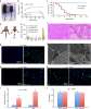
Yao, Norris, Mason et al
Nat Commun (2016) 7, 11415 DOI: 10.1038/ncomms11415
Cited by 43
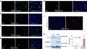
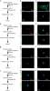
AI Summary: In this study, OriGene’s anti-PDGFRβ antibody (Cat# TA336839) and anti-Laminin antibody (Cat# TA309140) were used for immunostaining and analysis, enabling the researchers to identify and characterize PDGFRβ(+) cells and laminin expression, which were central to uncovering laminin’s regulatory role in muscle development and stem cell fate.
Proliferation of Multiple Cell Types in the Skeletal Muscle Tissue Elicited by Acute p21 Suppression
Biferi, Nicoletti, Falcone et al
Mol Ther (2015) 23 (5), 885-895 DOI: 10.1038/mt.2015.27
Biferi, Nicoletti, Falcone et al
Mol Ther (2015) 23 (5), 885-895 DOI: 10.1038/mt.2015.27
Cited by 11
AI Summary: In this study, OriGene’s p21 (Cdkn1a) shRNA lentiviral particles and p21 (Cdkn1a) qPCR primers were used to achieve and verify p21 knockdown, enabling the authors to investigate how acute suppression of p21 promotes proliferation of multiple cell types in skeletal muscle tissue.
Skin tissue generation by laser cell printing
Koch, Deiwick, Schlie et al
Biotechnol Bioeng (2012) 109 (7), 1855-63 DOI: 10.1002/bit.24455
Koch, Deiwick, Schlie et al
Biotechnol Bioeng (2012) 109 (7), 1855-63 DOI: 10.1002/bit.24455
Cited by 609
AI Summary: In the paper "Skin tissue generation by laser cell printing," OriGene products such as anti-connexin 43 antibody (OriGene, Cat# TA500372) were used to analyze gap junction formation, contributing to the assessment of cell-cell communication and tissue cohesion in the engineered 3D skin constructs.
Expansion of cortical and medullary sinuses restrains lymph node hypertrophy during prolonged inflammation
Tan, Yeo, Wong et al
J Immunol (2012) 188 (8), 4065-80 DOI: 10.4049/jimmunol.1101854
Tan, Yeo, Wong et al
J Immunol (2012) 188 (8), 4065-80 DOI: 10.4049/jimmunol.1101854
Cited by 67
AI Summary: In this study, OriGene’s Mouse Vegfa (NM_00950) qPCR primer pair was used to quantify Vegfa gene expression, helping to elucidate the spatial and temporal regulation of VEGF-A during lymph node remodeling in inflammation.
Regenerative capacity of adult cortical thymic epithelial cells
Rode, Boehm
Proc Natl Acad Sci U S A (2012) 109 (9), 3463-8 DOI: 10.1073/pnas.1118823109
Rode, Boehm
Proc Natl Acad Sci U S A (2012) 109 (9), 3463-8 DOI: 10.1073/pnas.1118823109
Cited by 90
AI Summary: In this paper, OriGene’s RNeasy Mini Kit was used for RNA isolation and the QuantiTect Reverse Transcription Kit was used for cDNA synthesis, enabling gene expression analyses crucial for investigating the regenerative capacity of adult cortical thymic epithelial cells.
Showing 1-6 of 16 papers.
Powered by BizGenius
*Delivery time may vary from web posted schedule. Occasional delays may occur due to unforeseen
complexities in the preparation of your product. International customers may expect an additional 1-2 weeks
in shipping.
Related Products
- Price:
- Actual Price:
Welcome to OriGene AI!
I'm your AI-powered guide to everything OriGene.
Ask me anything:
- Product specs
- Ordering help
- Technical questions
- How to find exactly what your experiment needs
Prefer a live agent? Connect with us by clicking the green chat icon, as seen in the image below or email our team at custsupport@origene.com.
