
Adgre1 Rat Monoclonal Antibody [Clone ID: BM8]

$765.00
2 Weeks*
Specifications
| Product Data | |
| Clone Name | BM8 |
|---|---|
| Application | FC, IF, IHC |
| Recommended Dilution | Immunohistochemistry on Frozen Sections: 0.4 µg/ml (1/500). Immunohistochemistry on Paraffin Sections: 4 µg/ml (1/50). Proteinase K pretreatment for antigen retrieval is recommended. Recommended Positive Control: Mouse spleen. Has been described to work in FACS. |
| Reactivity | Mouse |
| Antibody Host | Rat |
| Isotype | IgG2a |
| Clonality | Monoclonal |
| Immunogen | Cultured Macrophages. The antigen/epitope is a 125 kDa extracellular membrane protein sensitive to 2-Mercaptoetanol. |
| Specificity | This antibody is useful for the detection of the F4/80 antigen on major subpopulations of resident tissue macrophages. The antigen expression increases upon maturation of macrophage precursors in bone marrow and blood as well as in ontogeny. This clone is the only Macrophage marker that is able to distinguish non-destructive from destructive inflammation processes in the pancreas and has been shown to be a unique histological marker of the progression from peri-insulitis to beta-cell destruction and diabetes in a mouse diabetes model. Antigen Distribution Tissue Sections: The antigen is detected on tissue fixed macrophages in all organs tested so far (spleen, lymph nodes, thymus, liver, skin). It is also present on Langerhans cells in the skin and Kupffer cells in the liver. In complete Freund's adjuvant induced granulomas the antigen is expressed by inflammatory macrophages, but is absent from epitheloid cells. Isolated Cells: expressed in vitro on over 80% of M-CSF stimulated bone marrow derived macrophages, after a few days of culture. It is absent from granulocytes, lymphocytes and thrombocytes. |
| Buffer | Stock Solution contains PBS, pH 7.2 with 5 mg/ml BSA as a stabilizer and 0.01% Kathon as a preservative State: Aff - Purified State: Lyophilized purified IgG fraction |
| Reconstitution Method | Restore by adding 0.5 ml distilled water. |
| Concentration | 0.2 mg/ml (after reconstitution) |
| Purification | Affinity Chromatography |
| Conjugation | Unconjugated |
| Storage | Store lyophilized at 2-8°C for 6 months or at -20°C long term. After reconstitution store the antibody undiluted at 2-8°C for one month or (in aliquots) at -20°C long term. Avoid repeated freezing and thawing. |
| Stability | Shelf life: one year from despatch. |
| Shipping | Ambient |
| Gene Name | adhesion G protein-coupled receptor E1 |
| Database Link | |
| Background | F4/80 protein binds to Macrophages from different sites including the peritoneal cavity, lung, spleen and thymus, to blood monocytes and to macrophages derived from bone marrow precursors in culture. F4/80 is not expressed by lymphocytes or polymorphonuclear cells. |
| Synonyms | Emr1, Gpf480 |
| Note | Protocol: Protocol with Frozen, ice-cold Acetone-Fixed Sections: (The whole procedure is performed at room temperature) 1. Wash in PBS. 2. Block endogenous peroxidase. 3. Wash in PBS. 4. Block with 10% normal goat serum in PBS for 30min. in a humid chamber. 5. Incubate with primary antibody (dilution see datasheet) for 1h in a humid chamber. 6. Wash in PBS. 7. Incubate with secondary antibody (peroxidase-conjugated goat anti rat IgG (H+L) minimal-cross reaction to mouse) for 1h in a humid chamber. 8. Wash in PBS. 9. Incubate with AEC substrate (3-amino-9-ethylcarbazol) for 12min. 10. Wash in PBS. 11. Counterstain with Mayer's hemalum. Protocol with Formalin-Fixed, Paraffin-Embedded Sections: (The whole procedure is performed at room temperature) 1. Deparaffinize and rehydrate tissue section. 2. Incubate the tissue section with proteinase K for 7min. 3. Wash in distilled water. 4. Block endogenous peroxidase. 5. Wash in PBS. 6. Block with 10% normal goat serum in PBS for 30min. in a humid chamber. 7. Incubate with primary antibody (dilution see datasheet) for 1h in a humid chamber. 8. Wash in PBS. 9. Incubate with secondary antibody (peroxidase-conjugated goat anti rat IgG (H+L) minimal-cross reaction to mouse) for 1h in a humid chamber. 10. Wash in PBS. 11. Incubate with AEC substrate (3-amino-9-ethylcarbazol) for 12min. 12. Wash in PBS. 13. Counterstain with Mayer's hemalum. |
| Reference Data | |
| Protein Categories | GPCR, Intracellular Proteins, Membrane Proteins |
Reviews


Documents
| Product Manuals |
| FAQs |
| SDS |
Citations
Characterizing responsive and refractory orthotopic mouse models of hepatocellular carcinoma in cancer immunotherapy
Hage, Hoves, Ashoff et al
PLoS ONE (2019) 14 (7), e0219517 DOI: 10.1371/journal.pone.0219517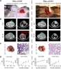
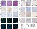
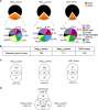
Hage, Hoves, Ashoff et al
PLoS ONE (2019) 14 (7), e0219517 DOI: 10.1371/journal.pone.0219517
AI Summary: OriGene products contributed to this study through the use of the CD8α (rat monoclonal, clone GHH8) antibody, which was employed to characterize immune cell infiltrates in the orthotopic mouse models of hepatocellular carcinoma.
Snippet: VSS software v12.3) automated slide stainer. The following anti-murine primary antibodies were used: Ki67 (rabbit monoclonal, clone 30–9, Ventana Medical Systems), F4/80 (rat monoclonal, clone BM8, Acris Antibodies GmbH), Clec4f (goat polyclonal, R&D Systems), CD4 (rabbit monoclonal, clone #1, Sino Biological Inc), CD8α (rat monoclonal, clone GHH8, Dianova), PD-L1 (rabbit monoclonal, clone E1L3
Deletion of ocular transforming growth factor β signaling mimics essential characteristics of diabetic retinopathy
Braunger, Leimbeck, Schlecht et al
Am J Pathol (2015) 185 (6), 1749-68 DOI: 10.1016/j.ajpath.2015.02.007
Braunger, Leimbeck, Schlecht et al
Am J Pathol (2015) 185 (6), 1749-68 DOI: 10.1016/j.ajpath.2015.02.007
Cited by 74
AI Summary: In this study, OriGene products, specifically the anti-HIF1A antibody (TA500494) and anti-TGFBR2 antibody (TA811496), were used to detect hypoxia-inducible factor 1α and TGF-β receptor II by immunohistochemistry, helping to elucidate the molecular changes in the retina that mimic diabetic retinopathy.
Non-redundant and redundant roles of cytomegalovirus gH/gL complexes in host organ entry and intra-tissue spread
Lemmermann, Krmpotic, Podlech et al
PLoS Pathog (2015) 11 (2), e1004640 DOI: 10.1371/journal.ppat.1004640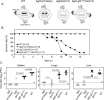
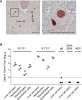
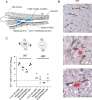
Lemmermann, Krmpotic, Podlech et al
PLoS Pathog (2015) 11 (2), e1004640 DOI: 10.1371/journal.ppat.1004640
Cited by 65
AI Summary: In this study, OriGene’s anti-IE1 antibody (Cat# AM20212PU-N) was used to detect viral infection in tissue sections, enabling the authors to assess the roles of cytomegalovirus gH/gL complexes in organ entry and intra-tissue spread.
Topically applied ZnO nanoparticles suppress allergen induced skin inflammation but induce vigorous IgE production in the atopic dermatitis mouse model
Ilves, Palomäki, Vippola et al
Part Fibre Toxicol (2014) 11, 38 DOI: 10.1186/s12989-014-0038-4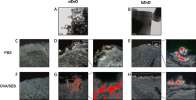
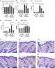
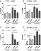
Ilves, Palomäki, Vippola et al
Part Fibre Toxicol (2014) 11, 38 DOI: 10.1186/s12989-014-0038-4
Cited by 158
AI Summary: In this study, OriGene’s Mouse IL-4 ELISA Kit (Cat# EMIL4) was used to measure cytokine levels, contributing to the assessment of immune responses following ZnO nanoparticle application in the atopic dermatitis mouse model.
Targeting membrane heat-shock protein 70 (Hsp70) on tumors by cmHsp70.1 antibody
Stangl, Gehrmann, Riegger et al
Proc Natl Acad Sci U S A (2011) 108 (2), 733-8 DOI: 10.1073/pnas.1016065108
Stangl, Gehrmann, Riegger et al
Proc Natl Acad Sci U S A (2011) 108 (2), 733-8 DOI: 10.1073/pnas.1016065108
Cited by 205
AI Summary: In this study, OriGene's recombinant mouse Hsp70 protein (Cat# TP301482) was used to confirm the specificity of the cmHsp70.1 antibody for its target epitope on tumor cells.
A murine model of epicutaneous protein sensitization is useful to study efficacies of topical drugs in atopic dermatitis
Lehto, Savinko, Wolff et al
Int Immunopharmacol (2010) 10 (4), 377-84 DOI: 10.1016/j.intimp.2010.01.001
Lehto, Savinko, Wolff et al
Int Immunopharmacol (2010) 10 (4), 377-84 DOI: 10.1016/j.intimp.2010.01.001
Cited by 17
AI Summary: In this study, OriGene’s anti-mouse CD4 (Cat# TA320097) and anti-mouse CD8 (Cat# TA320102) antibodies were used for immunohistochemical analysis to assess T cell infiltration in skin samples, contributing to the evaluation of topical drug efficacy in a murine model of atopic dermatitis.
Showing 1-6 of 9 papers.
Powered by BizGenius
*Delivery time may vary from web posted schedule. Occasional delays may occur due to unforeseen
complexities in the preparation of your product. International customers may expect an additional 1-2 weeks
in shipping.
Related Products
- Price:
- Actual Price:
Welcome to OriGene AI!
I'm your AI-powered guide to everything OriGene.
Ask me anything:
- Product specs
- Ordering help
- Technical questions
- How to find exactly what your experiment needs
Prefer a live agent? Connect with us by clicking the green chat icon, as seen in the image below or email our team at custsupport@origene.com.
