
Epcam Rat Monoclonal Antibody [Clone ID: G8.8]

Specifications
| Product Data | |
| Clone Name | G8.8 |
|---|---|
| Application | FC, IF, IHC, IP, WB |
| Recommended Dilution | Western blotting: 1/100-1/1000. Flow Cytometry: 1/25–1/200. Immunocytochemistry. Immunoprecipitation. Immunohistochemistry on Frozen Sections: 1/25–1/200 with avidin-biotinylated horseradish peroxidase complex (ABC) as detection reagent. |
| Reactivity | Mouse |
| Antibody Host | Rat |
| Isotype | IgG2a |
| Clonality | Monoclonal |
| Immunogen | This antibody is derived by fusion of X63-Ag8.653 mouse myeloma cells with spleen lymphocytes from rats repeatedly immunized with glycoconjugates from BALB/c Mouse derived medullary thymic epithelial cells. |
| Specificity | The G8.8 antibody reacts with CD326/Ep-CAM (Epithelial Cell Adhesion Molecule), also known as gp-40 in the Mouse. Ep-CAM is a 40-42-kDa cell-surface glycoprotein expressed on thymic epithelial cells, thymic dendritic cells, immature thymocytes, a small subset of peripheral T lymphocytes, intestinal epithelium, kidney-collecting tubule epithelium, keratinocytes, Langerhans cells, as well as lymph node and splenic dendrititc cells. |
| Buffer | PBS State: Purified State: Liquid purified IgG fraction Preservative: 0.09% Sodium Azide |
| Concentration | lot specific |
| Conjugation | Unconjugated |
| Storage | Store undiluted at 2-8°C for one month or (in aliquots) at -20°C for longer. Avoid repeated freezing and thawing. |
| Stability | Shelf life: one year from despatch. |
| Shipping | Blue Ice |
| Gene Name | epithelial cell adhesion molecule |
| Database Link | |
| Background | Ep-CAM is a 40 kD glycoprotein and can be detected at the basolateral membrane of the majority of epithelial tissues, where it is intricately linked with the cadherin-catenin complex and hence the fundamental WNT pathway responsible for intracellular signalling and polarity. This antigen functions as a homotypic calcium-independent cell adhesion molecule. Of particular interest, Ep-CAM appears to be overexpressed by the majority of human epithelial carcinomas, including colorectal, breast, prostate, head and neck, and hepatic carcinomas. The antigen is being used as a target for immunotherapy of human carcinomas. Formation of Ep-CAM-mediated adhesions has a negative regulatory effect on adhesions mediated by classic cadherins, which may have strong effects on the differentiation and growth of epithelial cells. Ep-CAM overexpression was suggested to be associated with enhanced epithelial proliferation. |
| Synonyms | Ep-CAM, Epithelial cell adhesion molecule, GA733-2, EGP314, KSA, TROP1, Trop-1, M1S2, M4S1, MIC18 |
| Reference Data | |
| Protein Categories | ADC Targets, Cancer: Gastrointestinal, CAR-T Targets, CD Markers, Intracellular Proteins, Membrane Proteins |
Reviews


Documents
| Product Manuals |
| FAQs |
| SDS |
Citations
The Consequences of Soluble Epoxide Hydrolase Deletion on Tumorigenesis and Metastasis in a Mouse Model of Breast Cancer
Kesavan, Frömel, Zukunft et al
Int J Mol Sci (2021) 22 (13) DOI: 10.3390/ijms22137120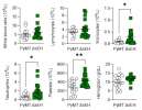
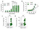
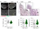
Kesavan, Frömel, Zukunft et al
Int J Mol Sci (2021) 22 (13) DOI: 10.3390/ijms22137120
Cited by 9
AI Summary: OriGene products, specifically the mouse Ephx2 (NM_007940) qPCR primer pair and mouse Ephx2 shRNA (TL502431), were used in this study to assess and manipulate the expression of the soluble epoxide hydrolase gene, thereby enabling the investigation of its role in tumor growth and metastasis in a mouse model of breast cancer.
Membrane Hsp70-A Novel Target for the Isolation of Circulating Tumor Cells After Epithelial-to-Mesenchymal Transition
Breuninger, Stangl, Werner et al
Front Oncol (2018) 8, 497 DOI: 10.3389/fonc.2018.00497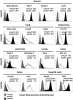
Breuninger, Stangl, Werner et al
Front Oncol (2018) 8, 497 DOI: 10.3389/fonc.2018.00497
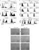
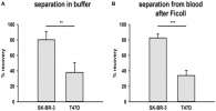
AI Summary: OriGene contributed PE-conjugated EpCAM (CD326, clone G8.8) and FITC-conjugated EpCAM (CD326, clone HEA125) antibodies, which were used for flow cytometric analysis to assess EpCAM expression on tumor cell lines in this study.
Snippet: At the Technische Universität München, flow cytometric analysis for the expression of mHsp70 and EpCAM on tumor cell lines was undertaken using either an FITC-conjugated cmHsp70.1 mAb (multimmune GmbH, Munich, Germany) and a FITC-conjugated EpCAM (CD326, clone HEA125) and PE-conjugated EpCAM (CD326, clone G8.8) mAb (Acris GmbH, an OriGene Company, Herford, Germany)
Showing 1-2 of 2 papers.
Powered by BizGenius
*Delivery time may vary from web posted schedule. Occasional delays may occur due to unforeseen
complexities in the preparation of your product. International customers may expect an additional 1-2 weeks
in shipping.
Related Products
- Price:
- Actual Price:
Welcome to OriGene AI!
I'm your AI-powered guide to everything OriGene.
Ask me anything:
- Product specs
- Ordering help
- Technical questions
- How to find exactly what your experiment needs
Prefer a live agent? Connect with us by clicking the green chat icon, as seen in the image below or email our team at custsupport@origene.com.
