
CD8A Mouse Monoclonal Antibody [Clone ID: CC63]

Specifications
| Product Data | |
| Clone Name | CC63 |
|---|---|
| Application | FC, IF, IHC, IP |
| Recommended Dilution | Immunohistochemistry on Frozen Sections. Immunohistochemistry on Paraffin Sections. Immunoprecipitation. Immunofluorescence/Immunocytochemistry. Flow Cytometry: Use 10 µl of 1/50 diluted CD8 antibody to label 106 cells in 100 µl. |
| Reactivity | Bovine, Goat, Sheep |
| Antibody Host | Mouse |
| Isotype | IgG2a |
| Clonality | Monoclonal |
| Specificity | This antibody reacts with the Bovine CD8 antigen expressed by a subset of T lymphocytes. The antibody precipitates molecules of 34kD and 38kD under reducing conditions. Clone CC63 has been reported as being suitable for use on Formalin Dichromate (FD5) Fixed Paraffin Embedded tissue with amplification and antigen retrieval techniques (See Gutierrez, M. et al. for details). |
| Buffer | PBS State: Purified State: Liquid purified IgG fraction from Tissue Culture Supernatant Preservative: 0.09% Sodium Azide |
| Concentration | lot specific |
| Purification | Affinity Chromatography on Protein G |
| Conjugation | Unconjugated |
| Storage | Store undiluted at 2-8°C for one month or (in aliquots) at -20°C for longer. |
| Stability | Shelf life: one year from despatch. |
| Shipping | Blue Ice |
| Database Link | |
| Background | The CD8 antigen is a cell surface glycoprotein found on most cytotoxic T lymphocytes that mediates efficient cell to cell interactions within the immune system. The CD8 antigen, acting as a coreceptor, and the T cell receptor on the T lymphocyte recognize antigen displayed by an antigen presenting cell (APC) in the context of class I MHC molecules. The functional coreceptor is either a homodimer composed of two alpha chains, or a heterodimer composed of one alpha and one beta chain. Both alpha and beta chains share significant homology to immunoglobulin variable light chains. |
| Synonyms | CD8 alpha chain, CD8A, MAL |
| Reference Data | |
Reviews


Documents
| Product Manuals |
| FAQs |
| SDS |
Citations
Effect of abrupt weaning at housing on leukocyte distribution, functional activity of neutrophils, and acute phase protein response of beef calves
Lynch, Earley, McGee et al
BMC Vet Res (2010) 6, 39 DOI: 10.1186/1746-6148-6-39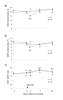
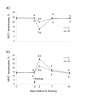
Lynch, Earley, McGee et al
BMC Vet Res (2010) 6, 39 DOI: 10.1186/1746-6148-6-39
Cited by 122
AI Summary: OriGene products, specifically mouse anti-bovine CD4 (MCA1653GA), CD8 (MCA837GA), WC1 (MCA837GA), and MHC Class II (MCA2226GA) antibodies, were used in this study to characterize lymphocyte immunophenotypes by flow cytometry, enabling assessment of immune changes in beef calves following abrupt weaning.
Repeat tuberculin skin testing leads to desensitisation in naturally infected tuberculous cattle which is associated with elevated interleukin-10 and decreased interleukin-1 beta responses
Coad, Clifford, Rhodes et al
Vet Res (2010) 41 (2), 14 DOI: 10.1051/vetres/2009062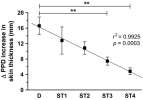
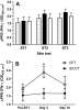
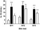
Coad, Clifford, Rhodes et al
Vet Res (2010) 41 (2), 14 DOI: 10.1051/vetres/2009062
Cited by 84
AI Summary: OriGene products, specifically the Bovine IL-10 ELISA Kit (Cat# MBS564069) and Bovine IL-1 beta ELISA Kit (Cat# MBS564043), were used in this study to quantitatively measure interleukin-10 and interleukin-1 beta levels in cattle blood samples, enabling the authors to link repeated tuberculin skin testing with elevated IL-10 and decreased IL-1 beta responses.
Screening of highly expressed mycobacterial genes identifies Rv3615c as a useful differential diagnostic antigen for the Mycobacterium tuberculosis complex
Sidders, Pirson, Hogarth et al
Infect Immun (2008) 76 (9), 3932-9 DOI: 10.1128/IAI.00150-08


Sidders, Pirson, Hogarth et al
Infect Immun (2008) 76 (9), 3932-9 DOI: 10.1128/IAI.00150-08
AI Summary: OriGene products, specifically the pCMV6-Entry expression vector, were used to clone and express candidate mycobacterial antigens for screening their immunogenicity in cattle, facilitating the identification of Rv3615c as a promising diagnostic antigen for distinguishing Mycobacterium tuberculosis complex infection from BCG vaccination.
Dissemination of bovine leukemia virus-infected cells from a newly infected sheep lymph node
Fulton, Portella, Radke
J Virol (2006) 80 (16), 7873-84 DOI: 10.1128/JVI.00529-06
Fulton, Portella, Radke
J Virol (2006) 80 (16), 7873-84 DOI: 10.1128/JVI.00529-06
AI Summary: In this study, OriGene’s anti-CD8 (clone 38.1; Cat# AM08007PU-N) and anti-IgM (clone MHM-88; Cat# AM01118PU-N) antibodies were used for immunophenotyping lymph and blood cells, enabling the researchers to track changes in CD8+ and IgM+ cell populations during early bovine leukemia virus infection in sheep.
Expression of caveolin by bovine lymphocytes and antigen-presenting cells
Harris, Werling, Koss et al
Immunology (2002) 105 (2), 190-5 DOI: 10.1046/j.1365-2567.2002.01362.x
Harris, Werling, Koss et al
Immunology (2002) 105 (2), 190-5 DOI: 10.1046/j.1365-2567.2002.01362.x
Cited by 57
AI Summary: OriGene products, specifically the anti-caveolin-1 antibody (Cat# TA325234) and anti-caveolin-3 antibody (Cat# TA326633), were used in this study to detect and analyze the expression of caveolin proteins in bovine lymphocytes and antigen-presenting cells via confocal microscopy and Western blotting.
Discordance between bovine leukemia virus tax immortalization in vitro and oncogenicity in vivo
Twizere, Kerkhofs, Burny et al
J Virol (2000) 74 (21), 9895-902 DOI: 10.1128/jvi.74.21.9895-9902.2000
Twizere, Kerkhofs, Burny et al
J Virol (2000) 74 (21), 9895-902 DOI: 10.1128/jvi.74.21.9895-9902.2000
Cited by 24
AI Summary: OriGene products contributed to this study by providing the anti-CD5 antibody (catalog #AM01105PU-N), which was used for immunophenotyping B lymphocytes infected with BLV mutants.
Showing 1-6 of 8 papers.
Powered by BizGenius
*Delivery time may vary from web posted schedule. Occasional delays may occur due to unforeseen
complexities in the preparation of your product. International customers may expect an additional 1-2 weeks
in shipping.
Related Products
- Price:
- Actual Price:
Welcome to OriGene AI!
I'm your AI-powered guide to everything OriGene.
Ask me anything:
- Product specs
- Ordering help
- Technical questions
- How to find exactly what your experiment needs
Prefer a live agent? Connect with us by clicking the green chat icon, as seen in the image below or email our team at custsupport@origene.com.
