
LYVE1 Rabbit Polyclonal Antibody

Specifications
| Product Data | |
| Application | ELISA, FC, IF, IHC, WB |
|---|---|
| Recommended Dilution | ELISA (1-15 µg/ml). Western blot (1-2 µg/ml). FACS analysis (3-20 µg/ml). Immunohistochemistry on Frozen Sections (6-30 µg/ml). Please Note: For use on Paraffin Embedded Sections the Affinity Purified antibody (Cat.-No DP3500PS) is recommended. |
| Reactivity | Human |
| Antibody Host | Rabbit |
| Clonality | Polyclonal |
| Immunogen | Recombinant Human soluble LYVE-1 (24-232) produced in insect cells. |
| Specificity | The antibody recognizes LYVE-1. |
| Buffer | 5mM PBS, pH 7.2 without preservatives or stabilizers State: Purified State: Lyophilized purified IgG fraction |
| Reconstitution Method | Restore in sterile water to a concentration of 0.1-1.0 mg/ml. |
| Purification | Protein A Chromatography (+ his tag depleted) |
| Conjugation | Unconjugated |
| Storage | The lyophilized IgG is stable at 2-8°C for one month or at -20°C for one year from despatch. When reconstituted the antibody is stable for six weeks at 2-8°C and at -20°C for longer. Aliquot to avoid repeated freezing and thawing. |
| Shipping | Ambient |
| Gene Name | lymphatic vessel endothelial hyaluronan receptor 1 |
| Database Link | |
| Background | LYVE-1 has been identified as a major receptor for HA (extracellular matrix glycosamino-glycan hyaluronan) on the lymph vessel wall. The deduced amino acid sequence of LYVE-1 predicts a 322-residue type I integral membrane polypeptide 41% similar to the CD44 HA receptor with a 212-residue extracellular domain containing a single Link module the prototypic HA binding domain of the Link protein superfamily. Like CD44, the LYVE-1 molecule binds both soluble and immobilized HA. However, unlike CD44, the LYVE-1 molecule colocalizes with HA on the luminal face of the lymph vessel wall and is completely absent from blood vessels. Hence, LYVE-1 is the first lymph-specific HA receptor to be characterized and is a uniquely powerful marker for lymph vessels themselves. |
| Synonyms | LYVE1, CRSBP-1, CRSBP1, HAR, XLKD1 |
| Note | Protocol: Double staining LYVE-1/CD31 on Humanen Cryo-Sections - dry Cryo sections for 1h at RT - fix sections for 10 min at –20°C in MeOH - wash 3x3 min in PBS at RT - block 20 min in 10% goat serum in PBS - 1. AB over night at 4°C in 10% goat serum in PBS anti h -LYVE-1 10μg/ml and anti h-CD31 1:50 (Klon JC70A) - wash 3x3 min in PBS at RT - 2. AB 30 min at RT in PBS goat anti rabbit IgG (H+L) CY3 1:500 and goat anti mouse biotin 1:500 - wash 3x3 min in PBS at RT - staining of nucleus 10 min at RT in Hoechst Dye 1:5000 in PBS - wash 3x3 min in PBS at RT - cover with DAKO Fluorescent Mountingmedium Staining protocol for Rabbit polyclonal anti-LYVE-1 on Paraffin Sections (Human Spleen) The specimens (maximum edge length 0.5cm) are fixed for 24 hours (or longer) in 3.7% formalin in tap water (low pH during fixation is advantageous). Embedding is done in paraffin carefully avoiding temperatures exceeding 60°C. The sections are cut and deposited on silanized slides and dried at 60°C overnight. They are then de-paraffinized and pretreated with protease XIV (Sigma No. P-5147) at 0.5mg/ml in TBS pH7.6 for 15min at room temperature. The primary polyclonal antibodies are applied at 1:100 to 1:150 in PBS/1%BSA/0.1%NaN3 overnight. Subsequently, the Vector anti-rabbit ABC kit for peroxidase is used according to the manufacturer´s instructions and the presence of peroxidase revealed by diaminobenzidine reaction. Note: The antibodies have to be used at a tenfold higher concentration on paraffin sections in comparison to cryo sections. High temperature antigen retrieval does not work, but protease pretreatment is mandatory. The protocol was established at the laboratory of: Prof. Dr. Birte Steiniger Institute of Anatomy and Cell Biology Robert-Koch-Str.8 D-35037 Marburg Germany E-mail: steinigb@staff.uni-marburg.de |
| Reference Data | |
| Protein Categories | Membrane Proteins |
Reviews


Documents
| Product Manuals |
| FAQs |
| SDS |
Citations
Increased CSF drainage by non-invasive manipulation of cervical lymphatics
Jin, Yoon, Hong et al
Nature (2025)
Jin, Yoon, Hong et al
Nature (2025)
Cited by 67
AI Summary: OriGene's anti-monkey LYVE1 antibody (DP3500) was used in this study to identify and visualize lymphatic vessels, aiding in the mapping of cerebrospinal fluid (CSF) drainage pathways to cervical lymph nodes.
Snippet: AChT, also known as solute carrier family 18 (vesicular acetylcholine), member 3, Slc18a3, goat polyclonal;ABN100, Merck);anti-monkey LYVE1 (rabbit polyclonal;DP3500, OriGene);anti-mouse eNOS antibody (rabbit polyclonal;ab5589, Abcam);anti-mouse phospho-eNOS antibody (rabbit polyclonal;9571, Cell Signaling);anti-mouse FOXP2 antibody (goat polyclon
A liver immune rheostat regulates CD8 T cell immunity in chronic HBV infection
Bosch, Kallin, Donakonda et al
Nature (2024) 631 (8022), 867-875 DOI: 10.1038/s41586-024-07630-7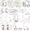
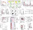
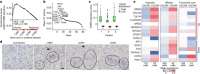
Bosch, Kallin, Donakonda et al
Nature (2024) 631 (8022), 867-875 DOI: 10.1038/s41586-024-07630-7
Cited by 95
AI Summary: OriGene's polyclonal anti-HBcAg antibody was used in this study to stain tissue sections for detecting hepatitis B core antigen, aiding the analysis of HBV-infected liver samples.
Snippet: Chronic hepatitis B virus (HBV) infection affects 300 million patients worldwide 1 , 2 , in whom virus-specific CD8 T cells by still ill-defined mechanisms lose their function;µm sections were made. Haematoxylin/eosin staining was performed according to standard protocols. Tissue sections were stained with anti-HBc and anti-GFP (1.5 µg ml−1 polyclonal anti-HBcAg (Origene);0.4 ng µl−1 of polyclona...
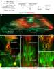
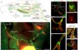
Nasopharyngeal lymphatic plexus is a hub for cerebrospinal fluid drainage
Yoon, Jin, Kim et al
Nature (2024) 625 (7996), 768-777 DOI: 10.1038/s41586-023-06899-4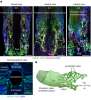
Yoon, Jin, Kim et al
Nature (2024) 625 (7996), 768-777 DOI: 10.1038/s41586-023-06899-4
Cited by 203
AI Summary: OriGene’s anti-LYVE1 (rabbit polyclonal, DP3500) antibody was used in this study to identify and visualize lymphatic endothelial cells, helping to characterize the nasopharyngeal lymphatic plexus as a key site for cerebrospinal fluid drainage.
Snippet: αSMA-Cy3 (mouse monoclonal, 1A4, C6198, Sigma-Aldrich), anti-β3 tubulin (mouse monoclonal, 2G10, ab78078, Abcam), anti-FOXC2 (sheep polyclonal, AF6989, R&D), anti-LYVE1 (rabbit polyclonal, DP3500, OriGene), anti-collagen type IV (goat polyclonal, AB769, Merck), anti-laminin α5 (rabbit polyclonal, EWL004, Kerafast), anti-tyrosine hydroxylase (rabbit polyclonal, AB152, Merck), anti-vesicular ace
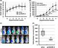
Soluble Vegfr3 gene therapy suppresses multi-organ metastasis in a mouse mammary cancer model
Shibata, Shibata, Tanaka et al
Cancer Sci (2020) 111 (8), 2837-2849 DOI: 10.1111/cas.14531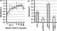
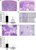
Shibata, Shibata, Tanaka et al
Cancer Sci (2020) 111 (8), 2837-2849 DOI: 10.1111/cas.14531
Cited by 12
AI Summary: In this study, OriGene’s pCMV6-Entry vector and mouse Vegfr3 cDNA (OriGene, MR207345) were used to construct the soluble VEGFR-3 expression vector (psVEGFR-3), enabling researchers to investigate and demonstrate the therapeutic effect of blocking VEGF-C/VEGFR-3 signaling on suppressing tumor growth and metastasis in a mouse mammary cancer model.
Lymphatic Endothelial Cells Are Essential Components of the Subcapsular Sinus Macrophage Niche
Mondor, Baratin, Lagueyrie et al
Immunity (2019) 50 (6), 1453-1466.e4 DOI: 10.1016/j.immuni.2019.04.002
Mondor, Baratin, Lagueyrie et al
Immunity (2019) 50 (6), 1453-1466.e4 DOI: 10.1016/j.immuni.2019.04.002
AI Summary: OriGene products are not mentioned as being used in this paper, so there is no contribution from OriGene products to summarize.
Snippet: …LYVE1 (Polyclonal Rabbit IgG) was purchased from Acris Antibodies. …LYVE1 (Polyclonal Rabbit IgG) was purchased from Acris Antibodies. … RFP and IBA1 polyclonal antibodies were purchased from Antibodies-onl;Anti-mouse LYVE-1 (Polyclonal Rabbit IgG) Acris Antibodies DP3513P Anti-mouse CSF-1 (Polyclonal Goat IgG) R&D Systems AF416
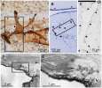
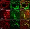
Visualization of intrapulmonary lymph vessels in healthy and inflamed murine lung using CD90/Thy-1 as a marker
Kretschmer, Dethlefsen, Hagner-Benes et al
PLoS One (2013) 8 (2), e55201 DOI: 10.1371/journal.pone.0055201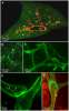
Kretschmer, Dethlefsen, Hagner-Benes et al
PLoS One (2013) 8 (2), e55201 DOI: 10.1371/journal.pone.0055201
Cited by 55
AI Summary: In this study, OriGene’s anti-CD90.2/Thy-1.2 monoclonal antibody was used to specifically label and visualize lymphatic endothelial cells in murine lung tissue, enabling the identification of lymph vessels under both healthy and inflamed conditions.
Showing 1-6 of 10 papers.
Powered by BizGenius
*Delivery time may vary from web posted schedule. Occasional delays may occur due to unforeseen
complexities in the preparation of your product. International customers may expect an additional 1-2 weeks
in shipping.
Related Products
- Price:
- Actual Price:
Welcome to OriGene AI!
I'm your AI-powered guide to everything OriGene.
Ask me anything:
- Product specs
- Ordering help
- Technical questions
- How to find exactly what your experiment needs
Prefer a live agent? Connect with us by clicking the green chat icon, as seen in the image below or email our team at custsupport@origene.com.
