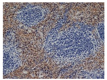
Adgre1 Rat Monoclonal Antibody [Clone ID: Cl:A3-1]
CAT#: BM4008
Adgre1 rat monoclonal antibody, clone Cl:A3-1, Purified
Other products for "Adgre1"
Specifications
| Product Data | |
| Clone Name | Cl:A3-1 |
| Applications | EM, FC, IF, IHC, IP, R, WB |
| Recommended Dilution | RIA. Western Blot. Immunoprecipitation. Immunofluorescence. Immunoelectron Microscopy. Flow Cytometry: Use 10 µl of 1/50-1/100 diluted antibody to label 106 cells in 100 µl. Immunohistochemistry on Frozen and Paraffin Embedded and Resin Sections. This product requires pre-treatment of paraffin sections (Proteinase K is recommended for tissues fixed for less than 24 hours. Citrate buffer pH 6.0 is recommended for tissues fixed for more than 24 hours). |
| Reactivities | Mouse |
| Host | Rat |
| Isotype | IgG2b |
| Clonality | Monoclonal |
| Immunogen | Thioglycollate stimulated peritoneal macrophages from C57/BL mice. Spleen cells from immunised HOB2 rats were fused with cells of the mouse NS1 myeloma cell line. |
| Specificity | This antibody recognizes the F4/80 antigen, a member of the EGF-TM7 family of proteins which shares 68% overall amino acid identity with Human EMR1. Clone CI:A31 has been reported to modulate cytokine levels released in response to Listeria monocytogenes (Ref.5). We recommend the use of BM4008LE for this purpose. |
| Formulation | PBS, pH 7.4 State: Purified State: Liquid purified IgG fraction Preservative: 0.09% Sodium Azide |
| Concentration | lot specific |
| Purification | Affinity Chromatography on Protein G |
| Conjugation | Unconjugated |
| Storage | Store undiluted at 2-8°C for one month or (in aliquots) at -20°C for longer. Avoid repeated freezing and thawing. |
| Stability | Shelf life: one year from despatch. |
| Gene Name | adhesion G protein-coupled receptor E1 |
| Database Link | |
| Background | F4/80 antigen is a 160 kD glycoprotein expressed by most murine macrophages. Expression of F4/80 is heterogeneous and is reported to vary during macrophage maturation and activation. The F4/80 antigen is expressed on a wide range of mature tissue macrophages including Kupffer cells, Langerhans, microglia, macrophages located in the gut lamina propria, peritoneal cavity, lung, thymus, bone marrow stroma and macrophages in the red pulp of the spleen. F4/80 expression has also been reported on a subpopulation of dendritic cells but is absent from macrophages located in T cell areas of the spleen and lymphnode. The ligands and biological functions of the F4/80 antigen have not yet been determined but recent studies suggest a role for F4/80 in the generation of efferent CD8+ve regulatory T cells. |
| Synonyms | Emr1, Gpf480 |
| Note | Protocol: 1. Enzyme pre-treatment using Proteinase K (Recommended for tissues fixed for 24 hours in neutral buffered formalin, NBF): Reagents A. TE buffer (50 mM Tris base, 1 mM EDTA, pH 8.0) Tris Base, 6.10 g EDTA, 0.37 g Distilled water, 1000 ml Mix to dissolve. Adjust pH to 8.0 using concentrated HCl (10 M HCl). Store at room temperature. B. Proteinase K stock solution (20x, 400 μg/ml in TE buffer, pH 8.0) Proteinase K, 4 mg TE buffer, pH 8.0, (Reagent A) 10 ml Mix well. Store in aliquots at -20°C. C. Proteinase K working solution (1x, 20 μg/ml in TE buffer, pH 8.0) Proteinase K stock solution (20x), (Reagent B) 1 ml TE Buffer, pH 8.0, (Reagent A) 19 ml Mix well. Discard working solution after use. Method 1. Dewax paraffin sections and rehydrate using preferred procedure. 2. Cover sections completely with Proteinase K working solution and incubate for 3 minutes at RT. 3. Rinse sections with Phosphate Buffered Saline (PBS). 4. Proceed with serum blocking and preferred staining protocol. 2. Heat-mediated antigen retrieval using citrate buffer, pH 6.0 (Recommended for tissues fixed for 7 days or more in neutral buffered formalin, NBF): Reagent Citrate buffer (10 mM citric acid, pH 6.0) Citric acid (anhydrous), 1.92 g Distilled water, 1000 ml Mix to dissolve. Adjust pH to 6.0 with 1 M NaOH (be sure to mix well). Store this solution at RT for 3 months, or at 4°C for longer usage. Method 1. Dewax paraffin sections and rehydrate using preferred protocol. 2. Pre-heat sodium citrate buffer in a staining vessel to 95-100°C. 3. Immerse slides in the citrate buffer and incubate for 10 minutes at 95-100°C. Check the citrate buffer level, add more if necessary, and then incubate for a further 10 minutes at 95-100°C. 4. Allow sections to cool for 20 minutes. 5. Rinse sections with PBS. 6. Proceed with serum blocking and preferred staining protocol. |
| Reference Data | |
Documents
| Product Manuals |
| FAQs |
| SDS |
{0} Product Review(s)
0 Product Review(s)
Submit review
Be the first one to submit a review
Product Citations
*Delivery time may vary from web posted schedule. Occasional delays may occur due to unforeseen
complexities in the preparation of your product. International customers may expect an additional 1-2 weeks
in shipping.


 Germany
 Japan
Japan
 United Kingdom
 China
China


