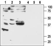
F2rl1 Rabbit Polyclonal Antibody
Frequently bought together (1)
beta Actin Mouse Monoclonal Antibody, Clone OTI1, Loading Control
USD 200.00
Other products for "F2rl1"
Specifications
| Product Data | |
| Applications | IF, WB |
| Recommended Dilution | WB: 1:200-1:2000; FC: 1:50-1:600 |
| Reactivities | Human, Mouse, Rat |
| Host | Rabbit |
| Clonality | Polyclonal |
| Immunogen | Peptide (C)KRMQISLTSNKFSRK, corresponding to amino acid residues 368-382 of rat PAR-2. Intracelluar, C- terminus. |
| Formulation | Lyophilized. Concentration before lyophilization ~0.8mg/ml (lot dependent, please refer to CoA along with shipment for actual concentration). Buffer before lyophilization: Phosphate buffered saline (PBS), pH 7.4, 1% BSA, 0.025% NaN3. |
| Reconstitution Method | Add 50 ul double distilled water (DDW) to the lyophilized powder. |
| Purification | Affinity purified on immobilized antigen. |
| Conjugation | Unconjugated |
| Storage | Store at -20°C as received. |
| Stability | Stable for 12 months from date of receipt. |
| Gene Name | F2R like trypsin receptor 1 |
| Database Link | |
| Background | Protease-activated receptor 2 (PAR-2) belongs to a family of four G protein-coupled receptors (PAR-1 - 4) that are activated as a result of proteolytic cleavage by certain serine proteases, hence their name. In this novel modality of activation, a specific protease cleaves the PAR receptor within a defined sequence in its extracellular N-terminal domain. This results in the creation of a new N-terminal tethered ligand, which subsequently binds to a site in the second extracellular loop of the same receptor. This binding results in the coupling of the receptor to G proteins and in the activation of several signal transduction pathways. Different PARs are activated by different proteases. Hence, PAR-1 is activated by thrombin, as are PAR-3 and PAR-4. PAR-2 is the only PAR family receptor that is activated by trypsin and not by thrombin. PAR-2 can be also cleaved and activated by other proteases such as tryptase, membrane-type serine protease 1, protease 3, and others. The intramolecular nature of PAR activation and the continuous presence of the tethered ligand that cannot diffuse away imply the existence of several mechanisms for the rapid termination of PAR signaling. Indeed, following receptor activation, there is rapid phosphorylation of the C-terminal end of the receptor, followed by receptor internalization and degradation. In addition, several proteases can cleave away the tethered ligand, thereby “disarming” the PAR. PAR-2-mediated intracellular signaling has not been clearly elucidated, but activators of PAR-2 induce generation of IP3 and mobilization of intracellular Ca2+. Activation of the ERK1/2 and NF-kB intracellular pathways following PAR-2 activation has been also described. Tissue distribution of PAR-2 is wide with the highest expression levels found in pancreas, small intestine, liver and kidney. In addition, PAR-2 expression was observed in dorsal root ganglion neurons, smooth muscle cells and endothelial cells. The physiological role of PAR-2 is not clearly understood, however studies using PAR-2 knockout mice have suggested roles in the cardiovascular, pulmonary and gastrointestinal systems. Several studies suggest that PAR-2 plays a central role in inflammatory diseases and nociceptive pain transduction; hence PAR-2 blockers have been proposed to be useful in the therapeutic control of inflammation and pain. |
| Synonyms | GPR11; PAR-2; PAR2 |
| Reference Data | |
Documents
| Product Manuals |
| FAQs |
| SDS |
{0} Product Review(s)
0 Product Review(s)
Submit review
Be the first one to submit a review
Product Citations
*Delivery time may vary from web posted schedule. Occasional delays may occur due to unforeseen
complexities in the preparation of your product. International customers may expect an additional 1-2 weeks
in shipping.


 Germany
 Japan
Japan
 United Kingdom
 China
China


