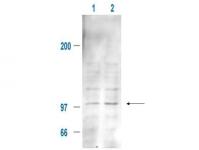
Mdm2 Rabbit Polyclonal Antibody
Frequently bought together (1)
beta Actin Mouse Monoclonal Antibody, Clone OTI1, Loading Control
USD 200.00
Other products for "Mdm2"
Specifications
| Product Data | |
| Applications | WB |
| Recommended Dilution | ELISA: 1:3,000 - 1:12,000, WB: 1:500 - 1:2,000, IP: 1:100 |
| Reactivities | Human, Mouse, Rat |
| Modifications | Phospho-specific |
| Host | Rabbit |
| Isotype | IgG |
| Clonality | Polyclonal |
| Immunogen | This affinity purified antibody was prepared from whole rabbit serum produced by repeated immunizations with a synthetic peptide corresponding to aa 177-198 of mouse MDM2. |
| Formulation | 0.02 M Potassium Phosphate, 0.15 M Sodium Chloride, pH 7.2 |
| Concentration | lot specific |
| Conjugation | Unconjugated |
| Storage | Store at -20°C as received. |
| Stability | Stable for 12 months from date of receipt. |
| Gene Name | transformed mouse 3T3 cell double minute 2 |
| Database Link | |
| Synonyms | Hdm2; HDMX; MGC5370; MGC71221; OTTHUMP00000183495 |
| Note | MDM2 is a nuclear phosphoprotein with an apparent molecular mass of 90 kD that forms a complex with the p53 tumor suppressor protein. Human MDM2 was identified as a homologous product of the 'murine double minute 2' gene (mdm2). The MDM2 gene enhances the tumorigenic potential of cells when it is overexpressed and encodes a putative transcription factor. Forming a tight complex with the p53 gene, the MDM2 oncogene can inhibit p53-mediated transactivation. MDM2 binds to p53 and amplification of MDM2 in sarcomas leads to escape from p53-regulated growth control. This mechanism of tumorigenesis parallels that for virus-induced tumors in which viral oncogene products bind to and functionally inactivate p53. Overexpression of the MDM2 oncogene was found in leukemias. Inactivation of tumor suppressor genes leads to deregulated cell proliferation and is a key factor in human tumorigenesis. MDM2 interacts physically and functionally with the retinoblastoma (RB) protein and can inhibit its growth regulatory capacity. Both RB and p53 can be subjected to negative regulation by the product of a single cellular protooncogene. The interference of binding to p53 prevents the interaction of MDM2 and its regulation of the transcriptional activity of p53 in vivo. Direct association of p53 with the cellular protein MDM2 results in ubiquitination and subsequent degradation of p53. MDM2-p53 complexes were preferentially found in S/G2M phases of the cell cycle. The MDM2 gene is alternatively spliced, producing 5 additional splice variant transcripts from the full length MDM2 gene. Four out of five of these alternatively spliced forms (MDM2a-MDMd) are missing substantial portions of the p53 binding domain and retain the acidic domain and the zinc-finger domains. The fifth and smallest transcript (MDM2e) retains the largest spliced region encoding the p53 binding domain; however, it lacks the nuclear localization signal, the acidic domain and zinc-finger domains. The alternatively spliced transcripts tend to be expressed in tumorigenic tissue, whereas the full-length MDM2 transcript is expressed in normal tissue. MDM2 is found in the nucleus and cytoplasm, however, it is expressed predominantly in the nucleoplasm. Interaction with ARF (P14) results in the localization of both proteins to the nucleus. The nucleolar localization signals in both ARF and MDM2 may be necessary to allow efficient nucleolar localization of both proteins. |
| Reference Data | |
Documents
| Product Manuals |
| FAQs |
| SDS |
{0} Product Review(s)
0 Product Review(s)
Submit review
Be the first one to submit a review
Product Citations
*Delivery time may vary from web posted schedule. Occasional delays may occur due to unforeseen
complexities in the preparation of your product. International customers may expect an additional 1-2 weeks
in shipping.


 Germany
 Japan
Japan
 United Kingdom
 China
China


