


 Islands")


")


")


")


")
")


 Germany
 Japan
Japan
 United Kingdom
 China
China
MHC Class I H2 Kd/Dd Mouse Monoclonal Antibody [Clone ID: 34-1-2S]
CAT#: AM08081FC-S
MHC Class I H2 Kd/Dd mouse monoclonal antibody, clone 34-1-2S, FITC
Conjugation: Unconjugated Biotin PE
Product Images

Specifications
| Product Data | |
| Clone Name | 34-1-2S |
| Applications | FC |
| Recommended Dilution | Flow Cytometry. |
| Reactivities | Mouse |
| Host | Mouse |
| Isotype | IgG2a |
| Clonality | Monoclonal |
| Immunogen | BDF splenocytes Donor: C3H spleen Fusion Partner: Sp2/0-Ag14 |
| Specificity | Anti H-2KdDd mAb reacts with both H-2Kd and H-2Dd products. The antibody also cross reacts with Kbsrpq. This K-D cross reaction indicates the presence of shared specificities between the two separate H-2 regions. |
| Formulation | PBS containing 0.02% sodium azide (NaN3) as preservative and EIA grade BSA as a stabilizing protein Label: FITC State: Liquid purified Ig fraction Label: Fluorescein isothiocyanate isomer 1 |
| Concentration | lot specific |
| Purification | Affinity chromatography on Protein G |
| Conjugation | FITC |
| Storage | Store the antibody undiluted at 2-8°C for one month or (in aliquots) at -20°C for longer. Avoid repeated freezing and thawing. |
| Stability | Shelf life: one year from despatch. |
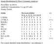
| Note | Protocol: FLOW CYTOMETRY ANALYSIS: Method: 1. Prepare a cell suspension in media A. For cell preparations, deplete the red blood cell population with Lympholyte®-M; cell separation medium. 2. Wash 2 times. 3. Resuspend the cells to a concentration of 2x10e7 cells/ml in media A. Add 50 μl of this suspension to each tube (each tube will then contain 1 x 10e6 cells, representing 1 test). 4. To each tube, add 0.5 - 0.2 μg of this antibody per 10e6 cells. 5. Vortex the tubes to ensure thorough mixing of antibody and cells. 6. Incubate the tubes for 30 minutes at 4°C. (It is recommended that the tubes are protected from light, since most fluorochromes are light sensitive.) 7. Wash 2 times at 4°C. 8. Resuspend the cell pellet in 50 μl ice cold media B. 9. Transfer to suitable tubes for flow cytometric analysis containing 15 μl of propidium iodide at 0.5 mg/ml in PBS. This stains dead cells by intercalating in DNA. Media: A. Phosphate buffered saline (pH 7.2) + 5% normal serum of host species + sodium azide (100 μl of 2M sodium azide in 100 mls). B. Phosphate buffered saline (pH 7.2) + 0.5% Bovine serum albumin + sodium azide (100 μl of 2M sodium azide in 100 mls). Results: Tissue Distribution by Flow Cytometry Analysis: Mouse Strain: BALB/c Cell Concentration: 1x10e6 cells per test Antibody Concentration Used: 0.5 μg/10e6 cells Isotypic Control: FITC Mouse IgG2a Percentage of cells stained above control: Thymus 91.9% Spleen 95.1% Lymph Node 100% |
| Reference Data | |
Documents
| Product Manuals |
| FAQs |
| SDS |
{0} Product Review(s)
Be the first one to submit a review


