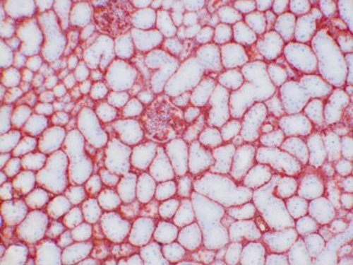
A2M Mouse Monoclonal Antibody [Clone ID: PAP-F101]
Frequently bought together (1)
beta Actin Mouse Monoclonal Antibody, Clone OTI1, Loading Control
USD 200.00
Other products for "A2M"
Specifications
| Product Data | |
| Clone Name | PAP-F101 |
| Applications | IHC |
| Recommended Dilution | IHC (f), WB (native). Each lot has been tested and validated for immunohistochemistry (IHC). Approximate working dilution for IHC: Frozen sections: 0.2-0.4µg/ml (1:1000-1:2000) Paraffin sections: does not react on routinely processed paraffin sections. Optimal dilutions should be determined by the end user. Suggested positive control: swine kidney. |
| Reactivities | Porcine |
| Host | Mouse |
| Isotype | IgG1 |
| Clonality | Monoclonal |
| Immunogen | Porcine lung extract. |
| Specificity | Porcine: positive.
Other: not tested Epitope: The antigen is alpha-2-Macroglobulin, according to the results obtained in IHC and in Western Blot with purified α-2-macro- globulin. The epitope has not been further characterized. Distribution: Tissue sections: The antibody reacts with tissue sections of kidney, pancreas, brain, stomach, adrenal, lung, intestine, skin, testis, liver, lymph node and uterus. Staining is prominent with endothelial structures in all organs, and with serum. |
| Formulation | Affinity purified from cell culture supernatant, lyophilized. Reconstitute by adding 0.5ml distilled water. This stock solution contains 0.4mg/ml IgG, phosphate buffered saline pH 7.2 (PBS), 5mg/ml bovine serum albumin (BSA) as a stabilizer and 0.05% (v/v) Kathon CG as a preservative. |
| Concentration | N/A |
| Conjugation | Unconjugated |
| Storage | Original vial: 1 year at 4° - 8°C. Minimize repeated thawing and freezing of the stock solution. |
| Background | Clone PAM-F101 is a unique monoclonal antibody developed against porcine α-2-Macro- globulin. α-2-Macroglobulin (α2M) is a broad spectrum proteinase inhibitor synthesized mainly by hepatocytes, and locally by macrophages. The inhibitory mode of action uses a capturing mechanism through a peptide sequence (“bait region”) which contains specific cleavage sites for different proteinases. When a proteinase cleaves the bait region, a conformational change and concomitant thioester bond hydrolysis is induced, leading to covalent binding of α2M to the proteinase. The bound enzyme remains active against low molecular weight substrates while the activity against high molecular weight substrates is reduced. The proteinase-α2M complex is recognized by macrophage receptors and cleared from the system. α2M is composed of four identical subunits arranged as a pair of disulfide-linked dimers, altogether with 720kDa molecular weight. α2M is sensitive to hypochlorite which induces dissociation of native α2M tetramers into stable dimers that are no longer able to trap proteases. Electrophoresis typically yields a 360kDa band in the native state, 180kDa with SDS-PAGE under non-reducing conditions. Reducing conditions (e.g. dithiothreitol DTT) will generate two different fragments with molecular mass of 93 and 87 kDa, respectively. This antibody was produced serum-free, without fetal calf serum. |
| Reference Data | |
Documents
| Product Manuals |
| FAQs |
| SDS |
{0} Product Review(s)
0 Product Review(s)
Submit review
Be the first one to submit a review
Product Citations
*Delivery time may vary from web posted schedule. Occasional delays may occur due to unforeseen
complexities in the preparation of your product. International customers may expect an additional 1-2 weeks
in shipping.


 Germany
 Japan
Japan
 United Kingdom
 China
China


