


 Islands")


")


")


")


")
")


 Germany
 Japan
Japan
 United Kingdom
 China
China
Itgal Mouse Monoclonal Antibody [Clone ID: WT.1]
Product Images
Other products for "Itgal"
Specifications
| Product Data | |
| Clone Name | WT.1 |
| Applications | FC, IHC |
| Recommended Dilution | Immunoprecipitation. Flow cytometry. Immunohistochemistry (cryostat sections). Functional studies: in vivo/in vitro |
| Reactivities | Rat |
| Host | Mouse |
| Isotype | IgG2a |
| Clonality | Monoclonal |
| Immunogen | Rat Splenic PHA blasts Donor: BALB/c spleen Fusion Partner: Mouse myeloma cell line PAI |
| Specificity | This Antibody is specific for the a subunit of LFA-1. It inhibits homeotypic aggregation of PHA blasts and blocks the binding of rat lymphocytes to purified rat ICAM-1. |
| Formulation | PBS, 0.09% NaN3 and EIA grade BSA as a stabilizing protein to bring total protein concentration to 4-5 mg/ml. Label: Biotin State: Liquid, purified. |
| Concentration | lot specific |
| Conjugation | Biotin |
| Storage | Store the antibody undiluted at 2-8°C for one month or (in aliquots) at -20°C for longer. Avoid repeated freezing and thawing. |
| Stability | Shelf life: one year from despatch. |
| Gene Name | integrin subunit alpha L |
| Database Link | |
| Background | LFA-1 (lymphocyte function associated molecule-1) is one of the leukocyte integrins. It is a heterodimer consisting of a and b subunits of 160-170 kDa and 95-100 kDa respectively. LFA-1 promotes non-antigen dependent adhesion of T-cells to a variety of lymphoid cells that bear its complementary receptor I-CAM-1 (1). It has a broad distribution and is found on most common lymphocytes. |
| Synonyms | Integrin alpha-L, LFA1, LFA-1 |
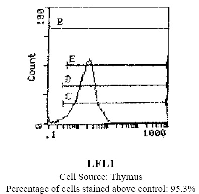
| Note | Protocol: FLOW CYTOMETRY ANALYSIS: Method: 1. Prepare cell suspension in Media A. For cell preparations, deplete the red blood cell population with Lympholyte®-Rat cell separation medium. 2. Wash 2 times. 3. Resuspend cells to a concentration of 2x10e7 cells/ml in media A. Add 50 µl of this suspension to each tube (each tube will then contains 1x10e6 cells representing 1 test). 4. To each tube add 1.0 µg of this Ab. 5. Vortex the tubes to ensure thorough mixing of antibody and cells. 6. Incubate the tubes for 30 minutes at 4°C. 7. Wash 2 times at 4°C. 8. Add 100 µl of secondary antibody (Streptavidin-FITC) at a 1/700 dilution. 9. Incubate tubes at 4°C for 30-60 minutes (It is recommended that the tubes are protected from light since most fluorochromes are light sensitive). 10. Wash 2 times at 4°C in Media B. 11. Resuspend the cell pellet in 50 µl ice cold Media B. 12. Transfer to suitable tubes for flow cytometric analysis containing 15 µl of propidium iodide at 0.5 mg/ml in phosphate buffered saline. (This stains dead cells by intercalating DNA). Media: A. Phosphate buffered saline (pH 7.2) + 5% normal serum of host species + sodium azide (100 µl of 2M sodium azide in 100 mls). B. Phosphate buffered saline (pH 7.2) + 0.5 % bovine serum albumin + sodium azide (100 µl of 2M sodium azide in 100 mls). Results - Tissue Distribution by Flow Cytometric Analysis: Rat Strain: Wistar Cell Concentration: 1x10e6 cells per test Antibody Concentration Used: 1.0 µg/10e6 cells Isotypic Control: Biotin Mouse IgG2a,k Cell Source Percentage of cells stained above control: Thymus: 95.3% |
| Reference Data | |
Documents
| Product Manuals |
| FAQs |
| SDS |
{0} Product Review(s)
0 Product Review(s)
Submit review
Be the first one to submit a review


Product Citations
*Delivery time may vary from web posted schedule. Occasional delays may occur due to unforeseen
complexities in the preparation of your product. International customers may expect an additional 1-2 weeks
in shipping.