


 Islands")


")


")


")


")
")


 Germany
 Japan
Japan
 United Kingdom
 China
China
Ly6g Rat Monoclonal Antibody [Clone ID: RB6-8C5]
Product Images
Other products for "Ly6g"
Specifications
| Product Data | |
| Clone Name | RB6-8C5 |
| Applications | FC, WB |
| Recommended Dilution | Flow cytometry (1,2,3). Western blot (5). |
| Reactivities | Mouse |
| Host | Rat |
| Isotype | IgG2b |
| Clonality | Monoclonal |
| Specificity | This antibody reacts with the Mouse myeloid differentiation antigen GR-1 (1,2). |
| Formulation | PBS, 0.02% NaN3 and EIA grade BSA as a stabilizing protein to bring total protein concentration to 4-5 mg/ml Label: FITC State: Liquid Ig fraction Absorption emission: 495 nm / 528 nm |
| Concentration | lot specific |
| Purification | Protein G chromatography |
| Conjugation | FITC |
| Storage | Store the antibody at 2 - 8 °C for up to one month. For long term storage, aliquot and freeze unused portion at -20 °C in volumes appropriate for single usage. Avoid repeated freezing and thawing This product is photosensitive and should be protected from light. |
| Stability | Shelf life: one year from despatch. |
| Gene Name | lymphocyte antigen 6 complex, locus G |
| Database Link | |
| Background | GR-1 is a 25-30 kDa cell surface antigen and is expressed on myeloid cells but not lymphoid or erythroid cells. The expression of the Gr-1 antigen increases with granulocyte maturation (3) as shown by the distinct populations of bone-marrow cells this monoclonal antibody labels: negative, low positive and high positive. Expression is transient on cells of monocytic lineage (3). |
| Synonyms | Gr-1 Granulocyte marker |
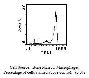
| Note | Protocol: FLOW CYTOMETRY ANALYSIS: Method: 1. Prepare a cell suspension in media A. For cell preparations, deplete the red blood cell population. 2. Wash 2 times. 3. Resuspend the cells to a concentration of 2x10e7 cells/ml in media A. Add 50 µl of this suspension to each tube (each tube will then contain 1 x 10e6 cells, representing 1 test). 4. To each tube, add 0.1 - 0.5 µg of antibody per 10e6 cells. 5. Vortex the tubes to ensure thorough mixing of antibody and cells. 6. Incubate the tubes for 30 minutes at 4°C. (It is recommended that the tubes are protected from light, since most fluorochromes are light sensitive.) 7. Wash 2 times at 4°C. 8. Resuspend the cell pellet in 50 µl ice cold media B. 9. Transfer to suitable tubes for flow cytometric analysis containing 15 µl of propidium iodide at 0.5 mg/ml in PBS. This stains dead cells by intercalating in DNA. Media: A. Phosphate buffered saline (pH 7.2) + 5% normal serum of host species + sodium azide (100 µl of 2M sodium azide in 100 mls). B. Phosphate buffered saline (pH 7.2) + 0.5% Bovine serum albumin + sodium azide (100 µl of 2M sodium azide in 100 mls). Results: Tissue Distribution by Flow Cytometry Analysis: Mouse Strain: CBA/J Cell Concentration : 1x10e6 cells per test Antibody Concentration Used: 0.1 µg/10e6 cells Isotypic Control: FITC Rat IgG2b Cell Source Percentage of cells stained above control: Thymus 1.5% Whole Blood Monocytes 87.2% Bone Marrow Macrophages 90.0% (see picture below) Strain Distribution by Flow Cytometry Analysis: Procedure: see above Cell Concentration: 1x10e6 cells per test Antibody Concentration Used: 0.1 µg/10e6 cells Strains Tested: BALB/c, C57BL/6, CBA, C3H/he, AKR Positive: BALB/c, C57BL/6, CBA, C3H/he, AKR Negative: none |
| Reference Data | |
Documents
| Product Manuals |
| FAQs |
| SDS |
{0} Product Review(s)
0 Product Review(s)
Submit review
Be the first one to submit a review


Product Citations
*Delivery time may vary from web posted schedule. Occasional delays may occur due to unforeseen
complexities in the preparation of your product. International customers may expect an additional 1-2 weeks
in shipping.