


 Islands")


")


")


")


")
")


 Germany
 Japan
Japan
 United Kingdom
 China
China
Cd8a Mouse Monoclonal Antibody [Clone ID: AD4(15)]
Product Images

Other products for "Cd8a"
Specifications
| Product Data | |
| Clone Name | AD4(15) |
| Applications | CT, FC |
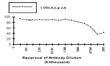
| Recommended Dilution | Cytotoxicity assays. Results: Antibody Titration by Cytotoxicity Analysis: Cell Source: Thymus Donor: C57BL/6 (Ly-2.2) Cell Concentration: 1.1x10⁶ cells/ml Complement: Low-Tox®-M Rabbit Complement. Complement Concentration: 1/18 Flow Cytometry. Results: Tissue Distribution by Flow Cytometry Analysis: Mouse Strain: BALB/c Cell Concentration : 1x10⁶ cells per test Antibody Concentration Used: 1/2500 in 50 μl /106 cells Isotypic Control: Mouse IgM. |
| Reactivities | Mouse |
| Host | Mouse |
| Isotype | IgM |
| Clonality | Monoclonal |
| Immunogen | C57BL/6 Donor: B6-Ly-2a spleen Fusion Partner: Myeloma P3/X63-Ag8 |
| Specificity | Anti-Mouse CD8a (Ly 2.2) monoclonal antibody reacts with a sub-population of T-lymphocytes from Mouse strains expressing the Ly-2.2 phenotype but does not react with lymphocytes from strains expressing the Ly-2.1 phenotype. |
| Formulation | State: Supernatant State: Lyophilized Cell Culture Supernatant with no additional preservatives. |
| Reconstitution Method | Restore with 1.0 ml of cold distilled water |
| Conjugation | Unconjugated |
| Storage | Store lyophilized at 2-8°C for 6 months or at -20°C long term. After reconstitution store the antibody undiluted at 2-8°C for one month or (in aliquots) at -20°C long term. Avoid repeated freezing and thawing. |
| Stability | Shelf life: one year from despatch. |
| Gene Name | CD8 antigen, alpha chain |
| Database Link | |
| Synonyms | CD8 alpha chain, CD8A, MAL |
| Note | Protocol: CYTOTOXICITY ANALYSIS: Method: 1. Prepare a cell suspension from the appropriate tissue in Cytotoxicity Medium or equivalent. Remove red cells and dead cells (where necessary) by purification of viable lymphocytes on Lympholyte®-M cell separation medium. After washing, adjust the cell concentration to 1x10⁶ cells per ml in Cytotoxicity Medium. 2. Add the antibody to a final concentration of 1/1000 and mix. 3. Incubate for 60 minutes at 4°C. 4. Centrifuge to pellet the cells and discard the supernatant. 5. Resuspend to the original volume in Low-Tox®-M Rabbit Complement diluted to the recommended concentration in Cytotoxicity Medium. 6. Incubate for 60 minutes at 37°C. 7. Place on ice. 8. Add Trypan Blue, 10% by volume of 1% Trypan Blue (w/v) added 3-5 minutes before scoring works well. Score live versus dead cells in a hemacytometer. Cytotoxic Index (C. I.) can be calculated as follows: % cyt (antibody + complement) - % cyt (complement alone) = x 100 = C.I. 100% - % cyt (complement alone) CYTOTOXICITY DEPLETION ASSAY: Method: 1. Prepare a cell suspension from the appropriate tissue in Cytotoxicity Medium or equivalent. Remove red cells and dead cells (where necessary) by purification of viable lymphocytes on Lympholyte®-M density cell separation medium. After washing, adjust the cell concentration to 1x10⁶ cells per ml in Cytotoxicity Medium. 2. Add the antibody to a final concentration of 1/1000 and mix. Alternatively, pellet the cells and resuspend in antibody diluted 1/1000 in Cytotoxicity Medium. 3. Incubate for 60 minutes at 4°C. 4. Centrifuge to pellet the cells and discard the supernatant. 5. Resuspend to the original volume in Low-Tox-M® Rabbit Complement, diluted to the appropriate concentration in Cytotoxicity Medium. (Recommended concentration included with each batch of Low-Tox-M® Rabbit Complement). 6. Incubate for 60 minutes at 37°C. 7. Monitor for percent cytotoxicity at this stage, before further processing. For this purpose, remove a small sample from each tube, dilute 1/10 with medium, and add 1/10 volume of 1% Trypan Blue. After 3-5 minutes, score live versus dead cells in a hemacytometer. 8. For functional studies, remove the dead cells from the treated groups before further processing, particularly if the treated cells are to be cultured. This can be done by layering the cell suspension over a separation medium and centrifuging at room temperature as per the instructions provided. Live cells will form a layer at the interface, while the dead cells pellet. The interface can then be collected and washed in Cytotoxicity Medium before being resuspended in the appropriate medium for further processing. Alternatively, the cells can be washed and resuspended in the appropriate medium for further processing immediately after Step#6, provided that the dead cells will not interfere with subsequent assays. FLOW CYTOMETRY ANALYSIS: Method: 1. Prepare a cell suspension in media A. For cell preparations, deplete the red blood cell population with Lympholyte®-M cell separation medium. 2. Wash 2 times. 3. Resuspend the cells to a concentration of 2x10⁷ cells/ml in media A. Add 50 μl of this suspension to each tube (each tube will then contain 1x10⁶ cells, representing 1 test). 4. To each tube, add 2.0 μg of this antibody. 5. Vortex the tubes to ensure thorough mixing of antibody and cells. 6. Incubate the tubes for 30 minutes at 4°C. 7. Wash 2 times at 4°C. 8. Add 100 μl of secondary antibody (FITC Goat anti-mouse IgM-3 (H+L)) at 1/500 dilution. 9. Incubate the tubes at 4°C for 30-60 minutes. (It is recommended that the tubes are protected from light since most fluorochromes are light sensitive). 10. Wash 2 times at 4°C in media B. 11. Resuspend the cell pellet in 50 μl ice cold media B. 12. Transfer to suitable tubes for flow cytometric analysis containing 15 μl of propidium iodide at 0.5 mg/ml in PBS. This stains dead cells by intercalating in DNA. Media: A. Phosphate buffered saline (pH 7.2) + 5% normal serum of host species + sodium azide (100 μl of 2M sodium azide in 100 mls). B. Phosphate buffered saline (pH 7.2) + 0.5% Bovine serum albumin + sodium azide (100 μl of 2M sodium azide in 100 mls). |
| Reference Data | |
Documents
| Product Manuals |
| FAQs |
| SDS |
{0} Product Review(s)
0 Product Review(s)
Submit review
Be the first one to submit a review


Product Citations
*Delivery time may vary from web posted schedule. Occasional delays may occur due to unforeseen
complexities in the preparation of your product. International customers may expect an additional 1-2 weeks
in shipping.