


 Islands")


")


")


")


")
")


 Germany
 Japan
Japan
 United Kingdom
 China
China
Cd8a Mouse Monoclonal Antibody [Clone ID: 49-31.1]
Product Images
Other products for "Cd8a"
Specifications
| Product Data | |
| Clone Name | 49-31.1 |
| Applications | CT, FC |
| Recommended Dilution | Flow Cytometry. |
| Reactivities | Mouse |
| Host | Mouse |
| Isotype | IgG3 |
| Clonality | Monoclonal |
| Immunogen | Recipient: 129/ReJ Donor: CBA Fusion Partner: Spleen from immunized recipient fused with Myeloma P3 NSI-Ag 4-1 |
| Specificity | Anti-mouse Ly-2.1 monoclonal antibody reacts with a sub-population of lymphocytes from mouse strains expressing the Ly 2.1 (CD8a) phenotype, but does not react with lymphocytes from mouse strains expressing the Ly 2.2 phenotype. |
| Formulation | PBS containing 0.02% sodium azide (NaN3) as preservative State: Purified State: Liquid purified Ig fraction |
| Concentration | lot specific |
| Purification | Affinity chromatography on Protein G |
| Conjugation | Unconjugated |
| Storage | Store the antibody undiluted at 2-8°C for one month or (in aliquots) at -20°C for longer. Avoid repeated freezing and thawing. |
| Stability | Shelf life: one year from despatch. |
| Gene Name | CD8 antigen, alpha chain |
| Database Link | |
| Synonyms | CD8 alpha chain, CD8A, MAL |
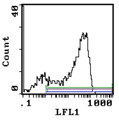
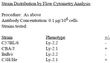
| Note | Protocol: FLOW CYTOMETRY ANALYSIS: Method: 1. Prepare a cell suspension in media A. For cell preparations, deplete the red blood cell population with Lympholyte®-M cell separation medium. 2. Wash 2 times. 3. Resuspend the cells to a concentration of 2x10e7 cells/ml in media A. Add 50 μl of this suspension to each tube (each tube will then contain 1x10e6 cells, representing 1 test). 4. To each tube, add 0.1 - 0.05μg of this antibody. 5. Vortex the tubes to ensure thorough mixing of antibody and cells. 6. Incubate the tubes for 30 minutes at 4°C. 7. Wash 2 times at 4°C. 8. Add 100 μl of secondary antibody (FITC Goat anti-mouse IgG-3 (H+L)) at 1/500 dilution. 9. Incubate the tubes at 4°C for 30-60 minutes. (It is recommended that the tubes are protected from light since most fluorochromes are light sensitive). 10. Wash 2 times at 4°C in media B. 11. Resuspend the cell pellet in 50 μl ice cold media B. 12. Transfer to suitable tubes for flow cytometric analysis containing 15 μl of propidium iodide at 0.5 mg/ml in PBS. This stains dead cells by intercalating in DNA. Media: A. Phosphate buffered saline (pH 7.2) + 5% normal serum of host species + sodium azide (100 μl of 2M sodium azide in 100 mls). B. Phosphate buffered saline (pH 7.2) + 0.5% Bovine serum albumin + sodium azide (100 μl of 2M sodium azide in 100 mls). Results: Tissue Distribution by Flow Cytometry Analysis: Mouse Strain: C3H/He Cell Concentration: 1x10e6 cells per test Antibody Concentration Used: 0.1 μg/10e6 cells Isotypic Control: Purified Mouse IgG-3 Percentage of cells stained above control: Thymus 78.9% |
| Reference Data | |
Documents
| Product Manuals |
| FAQs |
| SDS |
{0} Product Review(s)
0 Product Review(s)
Submit review
Be the first one to submit a review


Product Citations
*Delivery time may vary from web posted schedule. Occasional delays may occur due to unforeseen
complexities in the preparation of your product. International customers may expect an additional 1-2 weeks
in shipping.