


 Islands")


")


")


")


")
")


 Germany
 Japan
Japan
 United Kingdom
 China
China
Cd8a Mouse Monoclonal Antibody [Clone ID: 49-31.1]
Product Images
Other products for "Cd8a"
Specifications
| Product Data | |
| Clone Name | 49-31.1 |
| Applications | FC |
| Recommended Dilution | Flow Cytometry. |
| Reactivities | Mouse |
| Host | Mouse |
| Isotype | IgG3 |
| Clonality | Monoclonal |
| Immunogen | Immunization: Recipient: 129/ReJ Donor: CBA Fusion Partner: Spleen from immunized recipient fused with Myeloma P3 NSI-Ag 4-1 |
| Specificity | Anti-mouse Ly-2.1 monoclonal antibody reacts with a sub-population of lymphocytes from mouse strains expressing the Ly 2.1 (CD8a) phenotype, but does not react with lymphocytes from mouse strains expressing the Ly 2.2 phenotype. |
| Formulation | PBS containing 0.02% sodium azide (NaN3) as preservative and EIA grade BSA as a stabilizing protein to bring total protein concentration to 4-5 mg/ml. Label: FITC State: Liquid purified Ig fraction Label: Fluorescein isothiocyanate isomer 1 |
| Concentration | lot specific |
| Purification | Affinity chromatography on Protein G |
| Conjugation | FITC |
| Storage | Store the antibody undiluted at 2-8°C for one month or (in aliquots) at -20°C for longer. Avoid repeated freezing and thawing. |
| Stability | Shelf life: one year from despatch. |
| Gene Name | CD8 antigen, alpha chain |
| Database Link | |
| Synonyms | CD8 alpha chain, CD8A, MAL |
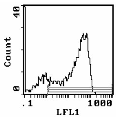
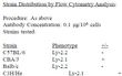
| Note | Protocol: FLOW CYTOMETRY ANALYSIS: Method: 1. Prepare a cell suspension in media A. For cell preparations, deplete the red blood cell population with Lympholyte®-M cell separation medium. 2. Wash 2 times. 3. Resuspend the cells to a concentration of 2x10e7 cells/ml in media A. Add 50 μl of this suspension to each tube (each tube will then contain 1 x 10e6 cells, representing 1 test). 4. To each tube, add 2.0-1.0 μg of this antibody per 10e6 cells. 5. Vortex the tubes to ensure thorough mixing of antibody and cells. 6. Incubate the tubes for 30 minutes at 4°C. (It is recommended that the tubes are protected from light, since most fluorochromes are light sensitive.) 7. Wash 2 times at 4°C. 8. Resuspend the cell pellet in 50 μl ice cold media B. 9. Transfer to suitable tubes for flow cytometric analysis containing 15 μl of propidium iodide at 0.5 mg/ml in PBS. This stains dead cells by intercalating in DNA. Media: A. Phosphate buffered saline (pH 7.2) + 5% normal serum of host species + sodium azide (100 μl of 2M sodium azide in 100 mls). B. Phosphate buffered saline (pH 7.2) + 0.5% Bovine serum albumin + sodium azide (100 μl of 2M sodium azide in 100 mls). Results: Tissue Distribution by Flow Cytometry Analysis: Mouse Strain: BALB/c Cell Concentration: 1x10e6 cells per test Antibody Concentration Used: 2,0 μg/10e6 cells Isotypic Control: FITC Mouse IgG3 Percentage of cells stained above control: Thymus 80.7% |
| Reference Data | |
Documents
| Product Manuals |
| FAQs |
| SDS |
{0} Product Review(s)
0 Product Review(s)
Submit review
Be the first one to submit a review


Product Citations
*Delivery time may vary from web posted schedule. Occasional delays may occur due to unforeseen
complexities in the preparation of your product. International customers may expect an additional 1-2 weeks
in shipping.