


 Islands")


")


")


")


")
")


 Germany
 Japan
Japan
 United Kingdom
 China
China
CD45 / LCA (CD45R) Rat Monoclonal Antibody [Clone ID: RA3-6B2]
Product Images
Other products for "Ptprc"
Specifications
| Product Data | |
| Clone Name | RA3-6B2 |
| Applications | FC, IHC, IP |
| Recommended Dilution | Flow Cytometry (for details please see Protocols). Immunoprecipitation. Reported to work in Immunohistochemical applications. |
| Reactivities | Mouse |
| Host | Rat |
| Isotype | IgG2a |
| Clonality | Monoclonal |
| Immunogen | Mouse pre-B tumour cells (RAW112). |
| Specificity | This antibody reacts with a form of the CD45 antigen found on B cells and lytically active subsets of NK cells and non - MHC restricted CTL's (1,2,3,4). It immunoprecipitates the high molecular weight (220,000 Da) surface molecule of the leukocyte common antigen B220 (1) on B cells. |
| Formulation | PBS, no preservative, 0.2 µm filtered State: Azide Free State: Liquid purified Ig fraction |
| Concentration | lot specific |
| Purification | Protein G Chromatography |
| Conjugation | Unconjugated |
| Storage | Store the antibody undiluted at 2-8°C for one month or (in aliquots) at -20°C for longer. Avoid repeated freezing and thawing. |
| Stability | Shelf life: one year from despatch. |
| Gene Name | protein tyrosine phosphatase, receptor type, C |
| Database Link | |
| Background | CD45R is a member of the protein tyrosine phosphatase (PTP) family and a major cell surface glycoprotein. PTPs are known to be signaling molecules that regulate a variety of cellular processes including cell growth, differentiation, mitotic cycle, and oncogenic transformation. CD45R represents a restricted form of the CD45 family which primarily recognizes only cells of B lineage from proB cell through mature B lymphocytes and, prior to the availability of anti CD19 MAbs, was commonly used as a pan B cell marker. It also reacts with certain activated T cells, as well as non MHC restricted lytically active lymphokine activated killer (LAK) cells. CD45R contains an extracellular domain, a single transmembrane segment and two tandem intracytoplasmic catalytic domains. It is specifically expressed in hematopoietic cells and has been shown to be an essential regulator of T and B cell antigen receptor signaling. It functions through either direct interaction with components of the antigen receptor complexes, or by activating various Src family kinases required for the antigen receptor signaling. CD45R also suppresses JAK kinases, and thus functions as a regulator of cytokine receptor signaling. Four alternatively spliced transcripts variants of this gene, which encode distinct isoforms, have been reported. |
| Synonyms | PTPRC, Leukocyte common antigen, L-CA, T200 |
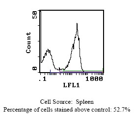
| Note | Protocol: FLOW CYTOMETRY ANALYSIS: Method: 1. Prepare a cell suspension in media A. For cell preparations, deplete the red blood cell population. 2. Wash 2 times. 3. Resuspend the cells to a concentration of 2x10e7 cells/ml in media A. Add 50 µl of this suspension to each tube (each tube will then contain 1x10⁶ cells, representing 1 test). 4. To each tube, add 0.2-0.5 µg of antibody. 5. Vortex the tubes to ensure thorough mixing of antibody and cells. 6. Incubate the tubes for 30 minutes at 4°C. 7. Wash 2 times at 4°C. 8. Add 100 µl of secondary antibody FITC Goat anti-rat IgG (H+L) at 1:500 dilution. 9. Incubate the tubes at 4°C for 30-60 minutes. (It is recommended that the tubes are protected from light since most fluorochromes are light sensitive). 10. Wash 2 times at 4°C in media B. 11. Resuspend the cell pellet in 50 µl ice cold media B. 12. Transfer to suitable tubes for flow cytometric analysis containing 15 µl of propidium iodide at 0.5 mg/ml in PBS. This stains dead cells by intercalating in DNA. Media: A. Phosphate buffered saline (pH 7.2) + 5% normal serum of host species + sodium azide (100 µl of 2M sodium azide in 100 mls). B. Phosphate buffered saline (pH 7.2) + 0.5% Bovine serum albumin + sodium azide (100 µl of 2M sodium azide in 100 mls). Results: Tissue Distribution by Flow Cytometry Analysis: Mouse Strain: CBA/J Cell Concentration : 1x10e6 cells per test Antibody Concentration Used: 0.2 µg/10e6 cells Isotypic Control: Purified Rat IgG2a Cell Source: Percentage of cells stained above control: Thymus: 0.6% Spleen: 52.7% Lymph Node: 14.3% |
| Reference Data | |
Documents
| Product Manuals |
| FAQs |
| SDS |
{0} Product Review(s)
0 Product Review(s)
Submit review
Be the first one to submit a review


Product Citations
*Delivery time may vary from web posted schedule. Occasional delays may occur due to unforeseen
complexities in the preparation of your product. International customers may expect an additional 1-2 weeks
in shipping.