Islands")


")


")


")


")
")


 Germany
 Japan
Japan
 United Kingdom
 China
China
AKT1 (NM_005163) Human Tagged Lenti ORF Clone
CAT#: RC220257L1
- LentiORF®
Lenti ORF clone of Human v-akt murine thymoma viral oncogene homolog 1 (AKT1), transcript variant 1, Myc-DDK-tagged
"NM_005163" in other vectors (7)
Product Images


Specifications
| Product Data | |
| Type | Human Tagged ORF Clone |
| Tag | Myc-DDK |
| Symbol | AKT1 |
| Synonyms | AKT; PKB; PKB-ALPHA; PRKBA; RAC; RAC-ALPHA |
| Vector | pLenti-C-Myc-DDK |
| E. coli Selection | Chloramphenicol (34 ug/mL) |
| Mammalian Cell Selection | None |
| Sequence Data |
The ORF insert of this clone is exactly the same as(RC220257).
|
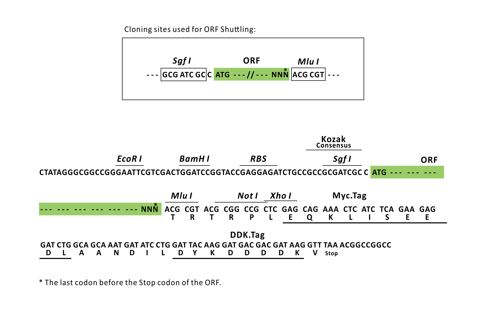
| Restriction Sites |
SgfI-MluI
Cloning Scheme for this gene
Plasmid Map

|
| ACCN | NM_005163 |
| ORF Size | 1440 bp |
| OTI Disclaimer | The molecular sequence of this clone aligns with the gene accession number as a point of reference only. However, individual transcript sequences of the same gene can differ through naturally occurring variations (e.g. polymorphisms), each with its own valid existence. This clone is substantially in agreement with the reference, but a complete review of all prevailing variants is recommended prior to use. More info |
| OTI Annotation | This clone was engineered to express the complete ORF with an expression tag. Expression varies depending on the nature of the gene. |
| Product Components | The ORF clone is ion-exchange column purified and shipped in a 2D barcoded Matrix tube containing 10ug of transfection-ready, dried plasmid DNA (reconstitute with 100 ul of water). |
| Reconstitution | 1. Centrifuge at 5,000xg for 5min. 2. Carefully open the tube and add 100ul of sterile water to dissolve the DNA. 3. Close the tube and incubate for 10 minutes at room temperature. 4. Briefly vortex the tube and then do a quick spin (less than 5000xg) to concentrate the liquid at the bottom. 5. Store the suspended plasmid at -20°C. The DNA is stable for at least one year from date of shipping when stored at -20°C. |
| Reference Data | |
| RefSeq | NM_005163.2, NP_005154.2 |
| RefSeq Size | 3008 bp |
| RefSeq ORF | 1443 bp |
| Locus ID | 207 |
| UniProt ID | P31749 |
| Cytogenetics | 14q32.33 |
| Domains | pkinase, S_TK_X, TyrKc, PH, S_TKc |
| Protein Families | Druggable Genome, ES Cell Differentiation/IPS, Protein Kinase |
| Protein Pathways | Acute myeloid leukemia, Adipocytokine signaling pathway, Apoptosis, B cell receptor signaling pathway, Chemokine signaling pathway, Chronic myeloid leukemia, Colorectal cancer, Endometrial cancer, ErbB signaling pathway, Fc epsilon RI signaling pathway, Fc gamma R-mediated phagocytosis, Focal adhesion, Glioma, Insulin signaling pathway, Jak-STAT signaling pathway, MAPK signaling pathway, Melanoma, mTOR signaling pathway, Neurotrophin signaling pathway, Non-small cell lung cancer, Pancreatic cancer, Pathways in cancer, Progesterone-mediated oocyte maturation, Prostate cancer, Renal cell carcinoma, Small cell lung cancer, T cell receptor signaling pathway, Tight junction, Toll-like receptor signaling pathway, VEGF signaling pathway |
| MW | 55.7 kDa |
| Gene Summary | This gene encodes one of the three members of the human AKT serine-threonine protein kinase family which are often referred to as protein kinase B alpha, beta, and gamma. These highly similar AKT proteins all have an N-terminal pleckstrin homology domain, a serine/threonine-specific kinase domain and a C-terminal regulatory domain. These proteins are phosphorylated by phosphoinositide 3-kinase (PI3K). AKT/PI3K forms a key component of many signalling pathways that involve the binding of membrane-bound ligands such as receptor tyrosine kinases, G-protein coupled receptors, and integrin-linked kinase. These AKT proteins therefore regulate a wide variety of cellular functions including cell proliferation, survival, metabolism, and angiogenesis in both normal and malignant cells. AKT proteins are recruited to the cell membrane by phosphatidylinositol 3,4,5-trisphosphate (PIP3) after phosphorylation of phosphatidylinositol 4,5-bisphosphate (PIP2) by PI3K. Subsequent phosphorylation of both threonine residue 308 and serine residue 473 is required for full activation of the AKT1 protein encoded by this gene. Phosphorylation of additional residues also occurs, for example, in response to insulin growth factor-1 and epidermal growth factor. Protein phosphatases act as negative regulators of AKT proteins by dephosphorylating AKT or PIP3. The PI3K/AKT signalling pathway is crucial for tumor cell survival. Survival factors can suppress apoptosis in a transcription-independent manner by activating AKT1 which then phosphorylates and inactivates components of the apoptotic machinery. AKT proteins also participate in the mammalian target of rapamycin (mTOR) signalling pathway which controls the assembly of the eukaryotic translation initiation factor 4F (eIF4E) complex and this pathway, in addition to responding to extracellular signals from growth factors and cytokines, is disregulated in many cancers. Mutations in this gene are associated with multiple types of cancer and excessive tissue growth including Proteus syndrome and Cowden syndrome 6, and breast, colorectal, and ovarian cancers. Multiple alternatively spliced transcript variants have been found for this gene. [provided by RefSeq, Jul 2020] |
Documents
| Product Manuals |
| FAQs |
| SDS |
Resources
Other Versions
| SKU | Description | Size | Price |
|---|---|---|---|
| RC220257 | AKT1 (Myc-DDK-tagged)-Human v-akt murine thymoma viral oncogene homolog 1 (AKT1), transcript variant 1 |
USD 457.00 |
|
| RC220257L2 | Lenti ORF clone of Human v-akt murine thymoma viral oncogene homolog 1 (AKT1), transcript variant 1, mGFP tagged |
USD 757.00 |
|
| RC220257L3 | Lenti ORF clone of Human v-akt murine thymoma viral oncogene homolog 1 (AKT1), transcript variant 1, Myc-DDK-tagged |
USD 757.00 |
|
| RC220257L4 | Lenti ORF clone of Human v-akt murine thymoma viral oncogene homolog 1 (AKT1), transcript variant 1, mGFP tagged |
USD 757.00 |
|
| RG220257 | AKT1 (tGFP-tagged) - Human v-akt murine thymoma viral oncogene homolog 1 (AKT1), transcript variant 1 |
USD 657.00 |
|
| SC116883 | AKT1 (untagged)-Human v-akt murine thymoma viral oncogene homolog 1 (AKT1), transcript variant 1 |
USD 457.00 |
|
| SC323430 | AKT1 (untagged)-Kinase deficient mutant (K179M) of Human v-akt murine thymoma viral oncogene homolog 1 (AKT1), transcript variant 1 |
USD 457.00 |
{0} Product Review(s)
Be the first one to submit a review