


 Islands")


")


")


")


")
")


 Germany
 Japan
Japan
 United Kingdom
 China
China
CD43 / Leukosialin Mouse Monoclonal Antibody [Clone ID: W3/13HLK]
Product Images
Other products for "Spn"
Specifications
| Product Data | |
| Clone Name | W3/13HLK |
| Applications | FC |
| Recommended Dilution | Flow cytometry (protocol see below). This clone has been reported to work in immunohistochemistry (frozen sections). |
| Reactivities | Rat |
| Host | Mouse |
| Isotype | IgG1 |
| Clonality | Monoclonal |
| Immunogen | Rat thymocyte membrane |
| Specificity | This antibody is specific for CD43. It recognizes a monomorphic determinant expressed on rat thymocytes, polymorphonuclear cells, plasma cells and stem cells, but not B lymphocytes or pre-B cells (1,3). This antibody is useful for labelling T but not B lymphocytes and in studies on stem cells since pre-B cells are not labelled while the multipotential stem cell is. It may also be used in analysis of NK cells (5) and in molecular studies in the sialoglycoprotein which it recognizes. |
| Formulation | PBS, without preservatives State: Azide Free State: Liquid Ig fraction |
| Concentration | lot specific |
| Purification | Protein G chromatography |
| Conjugation | Unconjugated |
| Storage | Store the antibody at 2 - 8 °C up to one month or (in aliquots) at -20 °C for longer. Avoid repeated freezing and thawing. |
| Stability | Shelf life: one year from despatch. |
| Database Link | |
| Background | The antigen is a heavily glycosylated glycoprotein of apparent molecular weight 95,000 and has a high content of O-linked carbohydrate structures (3). The carbohydrate structures of leukosialin account for approximately 60% of its weight (2). On thymocytes, this glycoprotein is the main target for binding of peanut lectin (4). |
| Synonyms | Leukocyte sialoglycoprotein, Sialophorin, Galactoglycoprotein, SPN |
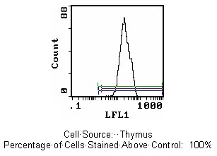
| Note | Protocol: FLOW CYTOMETRY ANALYSIS: 1. Prepare cell suspension in Media A. For cell preparations, deplete the red blood cell population. 2. Wash 2 times. 3. Resuspend cells to 1x10e6 cells in approximately 50 µl Media A in a microcentrifuge tube. (i.e. 50 µl of cells resuspended to 2x10e7 cells/ml.) (The Contents Of 1 Tube Represent 1 Test.) 4. To each tube add 1.0 – 0.5µg of antibody per 10e6 cells. 5. Vortex the tubes to ensure thorough mixing of antibody and cells. 6. Incubate the tubes for 30 minutes at 4°C. 7. Wash 2 times at 4°C. 8. Add 100 µl of secondary antibody FITC Goat anti-mouse IgG (H+L) at 1:500 dilution. 9. Incubate tubes at 4°C for 30-60 minutes. (It is recommended that the tubes are protected from light since most fluorochromes are light sensitive.) 10. Wash 2 times at 4°C in media B. 11. Resuspend the cell pellet in 50 µl ice cold media B. 12. Transfer to suitable tubes for flow cytometric analysis containing 15 µl of propidium iodide at 0.5 mg/ml in phosphate buffered saline. (This stains dead cells by intercalating DNA.) MEDIA: A. Phosphate buffered saline (pH 7.2) + 5% normal serum of host species + sodium azide (100 µl of 2 M sodium azide in 100 mls.) B. Phosphate buffered saline (pH 7.2) + 0.5 % bovine serum albumin + sodium azide (100 µl of 2 M sodium azide in 100 mls.) Rat Strain: Fisher Cell Concentration: 1x10e6 cells per test Antibody Concentration: 0.5µg / 10e6 cells Isotypic Control: Purified Mouse IgG1 CELL SOURCE PERCENT STAINING Thymus 100% Spleen 33.3% Lymph Node 58.9% (see picture below) STRAIN DISTRIBUTION: Procedure: As above Antibody Concentration: 1:200 Strains Tested: Lewis, Wistar, ACI, Brown Norway, Fischer 344, Buffalo Positive: Lewis, Wistar, ACI, BN, Fischer 344, Buffalo Negative: none |
| Reference Data | |
Documents
| Product Manuals |
| FAQs |
| SDS |
{0} Product Review(s)
0 Product Review(s)
Submit review
Be the first one to submit a review


Product Citations
*Delivery time may vary from web posted schedule. Occasional delays may occur due to unforeseen
complexities in the preparation of your product. International customers may expect an additional 1-2 weeks
in shipping.