


 Islands")


")


")


")


")
")


 Germany
 Japan
Japan
 United Kingdom
 China
China
HLA Class II DR Rat Monoclonal Antibody [Clone ID: YD1/63.4.10]
CAT#: SM2201F
HLA Class II DR rat monoclonal antibody, clone YD1/63.4.10, FITC
Conjugation: Unconjugated Biotin
Product Images
Specifications
| Product Data | |
| Clone Name | YD1/63.4.10 |
| Applications | FC |
| Recommended Dilution | Flow cytometry. Immunohistochemistry on cryostat and Paraffin-embedded sections. |
| Reactivities | Human |
| Host | Rat |
| Isotype | IgG2a |
| Clonality | Monoclonal |
| Immunogen | DAUDI cells Donor: immunized DA rat spleen cells Fusion Partner: Y3 Ag1.2.3 rat myeloma |
| Specificity | Anti-human HLA-DR monoclonal antibody recognizes the HLA-DR (MHC class II) antigen. |
| Formulation | PBS containing 0.02% sodium azide (NaN3) as preservativeand EIA grade BSA as a stabilizing protein to bring total protein concentration to 4-5 mg/ml. Label: FITC State: Liquid purified Ig fraction Label: Fluorescein isothiocyanate isomer 1 |
| Concentration | lot specific |
| Purification | Affinity chromatography on Protein G |
| Conjugation | FITC |
| Storage | Store the antibody undiluted at 2-8°C for one month or (in aliquots) at -20°C for longer. Avoid repeated freezing and thawing. |
| Stability | Shelf life: one year from despatch. |
| Synonyms | HLA-DR, HLA class II histocompatibility antigen DR, MHC class II antigen DR |
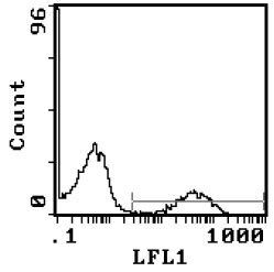
| Note | Protocol: FLOW CYTOMETRYANALYSIS: Method: 1. Prepare a cell suspension in media A. For cell preparations, deplete the red blood cell population with Lympholyte®-H cell separation medium. 2. Wash 2 times. 3. Resuspend the cells to a concentration of 2x10e7 cells/ml in media A. Add 50 μl of this suspension to each tube (each tube will then contain 1x10e6 cells, representing 1 test). 4. To each tube, add 50μl of a 0.2-0.1μg dilution of this antibody per 10e6 cells. 5. Vortex the tubes to ensure thorough mixing of antibody and cells. 6. Incubate the tubes for 30 minutes at 4°C. 7. Wash 2 times at 4°C. 8. Resuspend the cell pellet in 50 μl ice cold media B. 9. Transfer to suitable tubes for flow cytometric analysis containing 15 μl of propidium iodide at 0.5 mg/ml in PBS. This stains dead cells by intercalating in DNA. Media: A. Phosphate buffered saline (pH 7.2) + 5% normal serum of host species + sodium azide (100 μl of 2M sodium azide in 100 mls). B. Phosphate buffered saline (pH 7.2) + 0.5% Bovine serum albumin + sodium azide (100 μl of 2M sodium azide in 100 mls). Results: Tissue Distribution by Flow Cytometry Analysis: Cell Concentration: 1x10e6 cells per test Antibody Concentration Used: 0.1 μg/10e6 cells Isotypic Control: FITC Rat IgG2a |
| Reference Data | |
Documents
| Product Manuals |
| FAQs |
| SDS |
{0} Product Review(s)
0 Product Review(s)
Submit review
Be the first one to submit a review


Product Citations
*Delivery time may vary from web posted schedule. Occasional delays may occur due to unforeseen
complexities in the preparation of your product. International customers may expect an additional 1-2 weeks
in shipping.