


 Islands")


")


")


")


")
")


 Germany
 Japan
Japan
 United Kingdom
 China
China
T Cell Receptor (TCR) alpha/beta Hamster Monoclonal Antibody [Clone ID: H57-597]
CAT#: CL075F
T Cell Receptor (TCR) alpha/beta hamster monoclonal antibody, clone H57-597, FITC
Product Images
Specifications
| Product Data | |
| Clone Name | H57-597 |
| Applications | FC |
| Recommended Dilution | Flow Cytometry. Immunohistochemistry on Frozen Sections: Clone has reportedly worked with this application (See also Reference 6). |
| Reactivities | Mouse |
| Host | Hamster |
| Isotype | IgG |
| Clonality | Monoclonal |
| Immunogen | Affinity-purified DO-11.10 TCR from Armenian Hamster. Fusion Partner: Mouse myeloma variant P3X63 Ag.653. |
| Specificity | This anti-Mouse antibody T cell receptor monoclonal antibody reacts with the surface of all ab TCR bearing cells and does not react with receptors on gd TCR positive T cells. This monoclonal antibody when used in an immobilized form was able to activate all ab TCR bearing T cell hybridomas tested to produce IL-2. Use of this antibody in conjunction with an anti-CD3e monoclonal antibody allows for accurate measurements of the mutually exclusive sub-populations of ab TCR and gd TCR bearing T cells. |
| Formulation | PBS, 0.02% Sodium Azide and EIA grade BSA as a stabilizing protein to bring total protein concentration to 4-5 mg/ml. Label: FITC State: Liquid purified IgG fractiom |
| Concentration | lot specific |
| Purification | Protein G Chromatography |
| Conjugation | FITC |
| Storage | Store undiluted at 2-8°C for one month or (in aliquots) at -20°C for longer. This product is photosensitive and should protected from light. Avoid repeated freezing and thawing. |
| Stability | Shelf life: one year from despatch. |
| Synonyms | TCRA, TCRB, T-Cell Receptor alpha, T-Cell Receptor beta, T-Cell Receptor alpha beta |
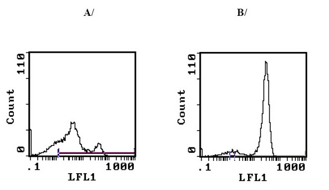
| Note | Protocol: Floy Cytometry Analysis: Method: 1. Prepare a cell suspension in media A. For cell preparations, deplete the red blood cell population with Lympholyte®-M cell separation medium. 2. Wash 2 times. 3. Resuspend the cells to a concentration of 2x10⁷ cells/ml in media A. Add 50 µl of this suspension to each tube (each tube will then contain 1 x 10⁶ cells, representing 1 test). 4. To each tube, add 0.2-0.1 µg* of this Ab per 106 cells. 5. Vortex the tubes to ensure thorough mixing of antibody and cells. 6. Incubate the tubes for 30 minutes at 4°C. (It is recommended that the tubes are protected from light, since most fluorochromes are light sensitive.) 7. Wash 2 times at 4°C. 8. Resuspend the cell pellet in 50 µl ice cold media B. 9. Transfer to suitable tubes for flow cytometric analysis containing 15 µl of propidium iodide at 0.5 mg/ml in PBS. This stains dead cells by intercalating in DNA. Media: A. Phosphate buffered saline (pH 7.2) + 5% normal serum of host species + sodium azide (100 µl of 2M sodium azide in 100 mls). B. Phosphate buffered saline (pH 7.2) + 0.5% Bovine serum albumin + sodium azide (100 µl of 2M sodium azide in 100 mls). Results - Tissue Distribution by Flow Cytometry Analysis: Mouse Strain: BALB/c Cell Concentration: 1x10⁶ cells per test Antibody Concentration Used: 0.2 µg/106 cells. Isotypic Control: FITC Hamster IgG Cell Source: Percentage of Cells Stained Above Control Thymus: 76.9% see Figure A Splenic T Cells: 90.3% see Figure B Strain Distribution by Flow Cytometry Analysis: Cell Concentration: 1x10e6 cells per test Antibody Concentration Used: 1.0 µg/106 cells Strains Tested: C57BL/6, CBA/J, AKR, BALB/c, C3H/He Positive: C57BL/6, CBA/J, AKR, BALB/c, C3H/He Negative: none |
| Reference Data | |
Documents
| Product Manuals |
| FAQs |
| SDS |
{0} Product Review(s)
0 Product Review(s)
Submit review
Be the first one to submit a review


Product Citations
*Delivery time may vary from web posted schedule. Occasional delays may occur due to unforeseen
complexities in the preparation of your product. International customers may expect an additional 1-2 weeks
in shipping.