


 Islands")


")


")


")


")
")


 Germany
 Japan
Japan
 United Kingdom
 China
China
Thy1 (Thy1.2) Mouse Monoclonal Antibody [Clone ID: 5a-8]
Product Images
Other products for "Thy1"
Specifications
| Product Data | |
| Clone Name | 5a-8 |
| Applications | FC, IHC |
| Recommended Dilution | Flow Cytometry (protocol see below). Appropriate control samples should always be included in any labelling studies. |
| Reactivities | Mouse |
| Host | Mouse |
| Isotype | IgG2b |
| Clonality | Monoclonal |
| Immunogen | CBA/J Donor: AKR/J Spleen |
| Specificity | This antibody detects CD90 (Thy 1.2). It reacts with all T lymphocytes from mouse strains expressing the Thy 1.2 phenotype (i.e. C57BL/6, C3H/He, DBA/2, CBA/J, BALB/c), but does not react with lymphocytes expressing the Thy 1.1 phenotype (i.e. AKR/J, B6.PL (74 NS). |
| Formulation | PBS containing 0.02% Sodium Azide and EIA grade BSA as a stabilizing protein to bring total protein concentration to 4-5 mg/ml. Label: Biotin State: Liquid purified Ig fraction. |
| Concentration | lot specific |
| Purification | Protein G Chromatography. |
| Conjugation | Biotin |
| Storage | Store the antibody undiluted at 2-8°C for one month or (in aliquots) at -20 °C for longer. Avoid repeated freezing and thawing. |
| Stability | Shelf life: one year from despatch. |
| Gene Name | thymus cell antigen 1, theta |
| Database Link | |
| Background | CD90 / Thy1 antigen is a GPI linked glycoprotein member of the Immunoglobulin superfamily. It is expressed on murine T cells, thymocytes, neural cells, cells of granulocytic lineage, early hematopoietic progenitors, fibroblasts, neurons and Kupffer's cells. Thy1 may play a role in cell to cell or cell to ligand interactions during synaptogenesis and other events in the brain. It is found in most mouse strains except AKR/J, A, Thy1.1 and B6.PL (74NS) expressing Thy1.1. |
| Synonyms | Thy-1, THY1, CDw90 |
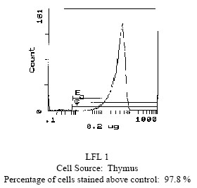
| Note | Protocol: FLOW CYTOMETRY ANALYSIS: Method: 1. Prepare a cell suspension in media A. For cell preparations, deplete the red blood cell population. 2. Wash 2 times. 3. Resuspend the cells to a concentration of 2x10e7 cells/ml in media A. Add 50 µl of this suspension to each tube (each tube will then contain 1 x 10e6 cells, representing 1 test). 4. To each tube, add 0.2-0.5 µg of this antibody per 10e6 cells. 5. Vortex the tubes to ensure thorough mixing of antibody and cells. 6. Incubate the tubes for 30 minutes at 4°C. 7. Wash 2 times at 4°C. 8. Add 100 µl of secondary antibody (Streptavidin-FITC) at a 1/500 dilution. 9. Incubate tubes at 4°C for 30-60 minutes (It is recommended that tubes are protected from light since most fluorochromes are light sensitive). 10. Wash 2 times at 4°C. 11. Resuspend the cell pellet in 50 µl ice cold media B. 12. Transfer to suitable tubes for flow cytometric analysis containing 15 µl of propidium iodide at 0.5 mg/ml in PBS. This stains dead cells by intercalating in DNA. Media: A. Phosphate buffered saline (pH 7.2) + 5% normal serum of host species + sodium azide (100 µl of 2M sodium azide in 100 mls). B. Phosphate buffered saline (pH 7.2) + 0.5% Bovine serum albumin + sodium azide (100 µl of 2M sodium azide in 100 mls). Results: Tissue Distribution by Flow Cytometry Analysis: Mouse Strain: CBA/J Cell Concentration : 1x10e6 cells per tests Antibody Concentration Used: 0.2 µg/10e6 cells Isotypic Control: Biotin Mouse IgG2b,k Cell Source: Percentage of cells stained above control: Thymus: 97.8% Spleen: 35.4% |
| Reference Data | |
Documents
| Product Manuals |
| FAQs |
| SDS |
{0} Product Review(s)
0 Product Review(s)
Submit review
Be the first one to submit a review


Product Citations
*Delivery time may vary from web posted schedule. Occasional delays may occur due to unforeseen
complexities in the preparation of your product. International customers may expect an additional 1-2 weeks
in shipping.