


 Islands")


")


")


")


")
")


 Germany
 Japan
Japan
 United Kingdom
 China
China
Glycophorin A (GYPA) Rabbit Polyclonal Antibody
Product Images
Other products for "Glycophorin A"
Specifications
| Product Data | |
| Applications | FC |
| Recommended Dilution | Flow Cytometry. |
| Reactivities | Human |
| Host | Rabbit |
| Isotype | IgG |
| Clonality | Polyclonal |
| Immunogen | Human Glycophorin A transmembrane protein Donor: Rabbit serum |
| Specificity | Anti-human Glycophorin A (GpA) Polyclonal Antibody targets the human Glycophorin A protein. This FITC conjugated polyclonal antibody recognizes various epitopes on the GpA protein. The 23 residue hydrophobic transmembrane domain has been shown to mediate non-covalent dimerization of the protein under conditions of SDS page (and in other detergents). We also sell an ascites purified version of this antibody (AP31671PU-N). |
| Formulation | PBS containing 0.02% sodium azide (NaN3) as preservative and EIA grade BSA as a stabilizing protein to bring total protein concentration to 4-5 mg/ml. Label: FITC State: Liquid purified Ig fraction Label: Fluorescein isothiocyanate isomer 1 |
| Concentration | lot specific |
| Purification | Affinity chromatography on Protein G |
| Conjugation | FITC |
| Storage | Store the antibody undiluted at 2-8°C for one month or (in aliquots) at -20°C for longer. Avoid repeated freezing and thawing. |
| Stability | Shelf life: one year from despatch. |
| Gene Name | glycophorin A (MNS blood group) |
| Database Link | |
| Background | This 131 amino acid, 36 KDa, transmembrane sialoglycoprotein spans the membrane of erythrocytes once. GpA presents its N terminus at the extracellular surface of the human red blood cell, has a single transmembrane domain and an internal cytoplasmic C terminus. GpA contributes to the expression of blood group antigen M and N specificities, which work to prevent red blood cell aggregation in circulation. Studies show that GpA is expressed early on the cell surface during erythropoiesis as pluripotent stem cells are recruited to differentiate into erythrocytes. This makes GpA a useful marker for studying the developmental biology of hematopoietic progenitor cells. |
| Synonyms | Glycophorin-A, GPA, PAS-2, Sialoglycoprotein alpha, MN sialoglycoprotein |
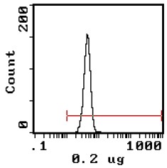
| Note | Protocol: FLOW CYTOMETRY ANALYSIS: Method: 1. Prepare a cell suspension in media A and wash twice. 2. Resuspend the cells to a concentration of 2x10e7 cells/ml in media A. Add 50μl of this suspension to each tube (each tube will then contain 1 x 10e6 cells, representing 1 test). 3. To each tube, add 0.2 μg of this antibody per 10e6 cells. 4. Vortex the tubes to ensure thorough mixing of antibody and cells. 5. Incubate the tubes for 30 minutes at 4°C. (It is recommended that the tubes are protected from light, since most fluorochromes are light sensitive.) 6. Wash 2 times at 4°C. 7. Resuspend the cell pellet in 50 μl ice cold media B. 8. Transfer to suitable tubes for flow cytometric analysis containing 15 μl of propidium iodide at 0.5 mg/ml in PBS. This stains dead cells by intercalating in DNA. Media: A. Phosphate buffered saline (pH 7.2) + 5% normal serum of host species + sodium azide (100 μl of 2M sodium azide in 100 mls). B. Phosphate buffered saline (pH 7.2) + 0.5% Bovine serum albumin + sodium azide (100 μl of 2M sodium azide in 100 mls). Results: Tissue Distribution by Flow Cytometry Analysis: Cell Concentration: 1x10e6 cells per test Antibody Concentration Used: 0.2 μg/10e6 cells Isotypic Control: FITC Rabbit IgG Percentage of cells stained above control: Human RBC’s 99.2% |
| Reference Data | |
| Protein Families | ES Cell Differentiation/IPS, Transmembrane |
| Protein Pathways | Hematopoietic cell lineage |
Documents
| Product Manuals |
| FAQs |
| SDS |
{0} Product Review(s)
0 Product Review(s)
Submit review
Be the first one to submit a review


Product Citations
*Delivery time may vary from web posted schedule. Occasional delays may occur due to unforeseen
complexities in the preparation of your product. International customers may expect an additional 1-2 weeks
in shipping.