CD95 (FAS) Mouse Monoclonal Antibody [Clone ID: UB2]
Specifications
| Product Data | |
| Clone Name | UB2 |
| Applications | IF, IHC |
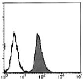
| Recommended Dilution | Immunohistochemistry on frozen and paraffin sections: 10-20 µg. Flow cytometry: 10 μg/mL (final concentration), details see protocol. |
| Reactivities | Human |
| Host | Mouse |
| Isotype | IgG1 |
| Clonality | Monoclonal |
| Immunogen | Recombinant human Fas |
| Specificity | This antibody recognizes the human Fas antigen specifically. |
| Formulation | PBS (pH 7.2) State: Aff - Purified State: Liquid purified Ig fraction containing 50% Glycerol without preservatives |
| Purification | Ammonium sulfate precipitation and affinity chromatography on protein A agarose |
| Conjugation | Unconjugated |
| Storage | Upon receipt, store (in aliqouts) at -20 °C. Avoid repeated freezing and thawing. |
| Stability | Shelf life: one year from despatch. |
| Gene Name | Fas cell surface death receptor |
| Database Link | |
| Background | It is now widely accepted that apoptosis plays an important role in the selection of immature thymocytes and Ag-primed peripheral T cells. Fas antigen is a cell-surface protein that mediates apoptosis. It is expressed in various tissues including the thymus and has structural homology with a number of cell-surface receptors, including tumor necrosis factor receptor and nerve growth factor receptor. |
| Synonyms | FASLG receptor, Apo-1 antigen, APT1, FAS1, TNFRSF6 |
| Note | This product was originally produced by MBL International. Protocol: Flow cytometric analysis for floating cells |
| Reference Data | |
Documents
| Product Manuals |
| FAQs |
| SDS |
{0} Product Review(s)
Be the first one to submit a review


 Germany
 Japan
Japan
 United Kingdom
 China
China


