RFP-Tag Mouse Monoclonal Antibody [Clone ID: 3G5]

CAT#: AM26655AG-N

RFP-Tag mouse monoclonal antibody, clone 3G5, Agarose, Agarose


Size
    • 200 ul

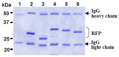
Product Images

Specifications

Product Data
Clone Name 3G5
Applications IP
Recommended Dilution Immunoprecipitation: 20 μL of gel slurry. For details see protocol below.
Host Mouse
Isotype IgG2a
Clonality Monoclonal
Immunogen RFP
Specificity

This antibody reacts with RFP fusion proteins

Formulation PBS
Label: Agarose
State: Agarose
State: 400 μg of anti-RFP monoclonal antibody covalently coupled to 200 μL of agarose gel and provided as a 50% gel slurry, total volume of 400 μL
Preservative: 0.09% sodium azide
Conjugation Agarose
Storage Store at 2-8 °C.
Stability Shelf life: one year from despatch.
Background

Expression vector containing a tag sequence is commonly used to introduce and express a specific gene into a target cell. Red Fluorescent Protein (RFP) fusion protein expression system is preferably used in various laboratories, because it’s easy monitoring of fusion proteins. This specific antibody for RFP is useful tool for monitoring of the fusion protein expression.

Synonyms Red fluorescent protein Tag, DsRed Tag
Note

This product was originally produced by MBL International.


Protocol:

Immunoprecipitation
1) Wash the transfectant cells 3 times with PBS and suspend with 10 volume of cold Lysis buffer (50 mM Tris-HCl pH 7.5, 150 mM NaCl, 0.05% NP-40) containing appropriate protease inhibitors. Incubate it at 4°C with rotating for 15 minutes, then sonicate briefly (up to 10 seconds).
2) Centrifuge the tube at 12,000 x g for 10 minutes at 4°C and transfer the supernatant to another tube.
3) Add primary antibody as suggest in the APPLICATIONS into 200 µL of cell extract. Mix well and incubate with gentle agitation for 60-120 minutes at 4°C.
4) Wash the beads 3-5 times with the cold Lysis buffer (centrifuge the tube at 2,500 x g for 10 seconds).
5) Resuspend the agarose in 20 µL of Laemmli’s sample buffer, boil for 3-5 minutes, and centrifuge for 5 minutes.
6) Load 10 µL of the sample per lane in a 1-mm-thick SDS-polyacrylamide gel for electrophoresis.
7) Blot the protein to a polyvinylidene difluoride (PVDF) membrane at 1 mA/cm2 for 1 hour in a semi-dry transfer system (Transfer Buffer: 25 mM Tris, 190 mM glycine, 20% MeOH). See the manufacturer's manual for precise transfer procedure.
8) To reduce nonspecific binding, soak the membrane in 10% skimmed milk (in PBS, pH 7.2) for 1 hour at room temperature, or overnight at 4°C.
9) Wash the membrane with PBS-T [0.05% Tween-20 in PBS] (5 minutes x 3 times).
10) Incubate the membrane with 1 µg/mL of anti-RFP monoclonal antibody diluted with PBS, pH 7.2 containing 1% skimmed milk for 1 hour at room temperature. (The concentration of antibody will depend on condition.)
11) Wash the membrane with PBS-T [0.05% Tween-20 in PBS] (5 minutes x 3 times).
12) Incubate the membrane with the 1:10,000 HRP-conjugated anti-mouse IgG diluted with 1% skimmed milk (in PBS, pH 7.2) for 1 hour at room temperature.
13) Wash the membrane with PBS-T (5 minutes x 3 times).
14) Wipe excess buffer on the membrane, then incubate it with appropriate chemiluminescence reagent for 1 minute. Remove extra reagent from the membrane by dabbing with paper towel, and seal it in plastic wrap.
15) Expose to an X-ray film in a dark room for 3 minutes. Develop the film as usual. The condition for exposure and development may vary.

Reference Data

{0} Product Review(s)

0 Product Review(s) Submit review

Be the first one to submit a review

Product Citations

*Delivery time may vary from web posted schedule. Occasional delays may occur due to unforeseen complexities in the preparation of your product. International customers may expect an additional 1-2 weeks in shipping.