PML Protein (PML) Mouse Monoclonal Antibody [Clone ID: 1B9]

CAT#: AM26594AF-N

PML Protein (PML) mouse monoclonal antibody, clone 1B9, Azide Free


Size
    • 100 ug

Product Images

Specifications

Product Data
Clone Name 1B9
Applications FC, IF, WB
Recommended Dilution Western blot: 1 µg/ml (for a chemiluminescence detection system).
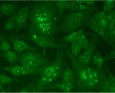
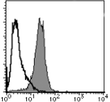
Immunocytochemistry: 1 µg/ml. Immunostaining features in the APL cell line NB-4 showed the microgranular pattern. Exposure to 0.1 μM of ATRA for 48 hours restored the normal immunostaining pattern. Flow cytometry: 10 µg/ml. For details see protocols below. It is reported that this clone 1B9 can be used in Immunoprecipitation in the reference number 1).
Reactivities Human
Host Mouse
Isotype IgG1
Clonality Monoclonal
Immunogen Recombinant human PML
Specificity This antibody reacts with PML.
Formulation PBS containing 50% glycerol, pH 7.2. Contains no preservative.
State: Azide Free
State: Liquid Ig fraction
Concentration lot specific
Purification Protein A agarose
Conjugation Unconjugated
Storage Upon receipt, store undiluted (in aliquots) at -20°C. Avoid repeated freezing and thawing.
Stability Shelf life: One year from despatch.
Gene Name promyelocytic leukemia
Background Acute promyelocytic leukemia (APL) is associated with a t(15;17) translocation that creates the promyelocyte-retinoic acid receptor α (PML-RAR α ) fusion protein and successfully differentiated by all- trans -retinoic acid (ATRA). PML-RAR α consists of all amino acid of RAR α except the first 59 amino acids and includes its DNA-binding and ligand-binding domains. PML-RAR α contains the functional domains of PML which includes the DNA binding and dimerization property. Thus, the functions of PML and/or retinoid X receptor are sequestrated by PML-RAR α in a dominant negative manner. In APL cells, the PML-RAR α and PML are immunologically localized as microgranules in the nuclei and cytoplasm, whereas in normal cells, PML is immunologically found as a discrete speckled pattern in nuclei. Th e ATRA treatment of the APL cells triggers a reorganization of PML to generate normal localization. Anti-PML antibody is a strong tool for the detection of the chromosomal translocation t(15;17) on the APL cells and/or determination of the sensitivity of the APL cells to the ATRA differentiation of hematopoietic cells and apoptosis.
Synonyms RING finger protein 71, MYL, TRIM19
Note

This product was originally produced by MBL International.


Protocol:

Immunocytochemistry
1) Culture the cells in the appropriate condition on a glass slide. (for example, spread 10 4 of cells per one well, then incubate in a CO 2 incubator for one night.)
2) Fix the cells by immersing the slide in Acetone for 10 minutes on ice.
3) Air dry the slides.
4) Add 30 μ L of normal goat serum containing 1 mg/mL normal human IgG and 0.1% NaN 3 on to the cells. Incubate for 10 minutes at room temperature.
5) Add the primary antibody diluted with PBS as suggested in the APPLICATIONS onto the cells and incubate for 1 hour at room temperature. (Optimization of antibody concentration or incubation condition are recommended if necessary.)
6) Prepare a wash container such as a 500 mL beaker with a magnetic stirrer. Then wash the cultured cells on the glass slide by soaking the slide with a plenty of PBS in the wash container for 5 minutes. Take care not to touch the cells. Repeat another washes once more.
7) Add 30 μ L of 1:100 FITC conjugated anti-mouse IgG diluted with PBS onto the cells. Incubate for 30 minutes at room temperature. Keep out light by aluminum foil.
8) Wash the slide in a plenty of PBS as in the step 6).
9) Wipe excess liquid from slide but take care not to touch the cells. Never leave the cells to dry.
10) Promptly add mounting medium onto the slide, then put a cover slip on it.
(Positive controls for Immunocytochemistry; NB-4, HEp-II)

SDS-PAGE & Western Blotting
1) Wash the 1 x 10e7 cells 3 times with PBS and resuspend in 1 mL of Laemmli’s sample buffer.
2) Boil the samples for 2 minutes and centrifuge. Load 5 μ L of sample per lane on a 1-mm-thick SDS-polyacrylamide gel and carry out electrophoresis.
3) Blot the protein to a polyvinylidene difluoride (PVDF) membrane at 1 mA/cm 2 for 1 hour in a semi-dry transfer system (Transfer Buffer: 25 mM Tris, 190 mM glycine, 20% MeOH). See the manufacture's manual for precise transfer procedure.
4) To reduce nonspecific binding, soak the membrane in 10% skimmed milk (in PBS, pH 7.2) for 1 hour at room temperature, or overnight at 4 o C. 5) Incubate the membrane for 1 hour at room temperature with primary antibody dilu ted with PBS, pH 7.2 containing 1% skimmed milk as suggested in the APPLICATIONS. (The concentration of antibody will depend on the conditions.)
6) Wash the membrane with PBS-T [0.05% Tween-20 in PBS] (5 minutes x 3 times).
7) Incubate the membrane with 1:10,000 HRP-conjugated anti-mouse IgG diluted with 1% skimmed milk (in PBS, pH 7.2) for 1 hour at room temperature.
8) Wash the membrane with PBS-T (5 minutes x 3 times).
9) Wipe excess buffer off the membrane, and incubate membrane with an appropriate chemiluminescence reagent for 1 minute. Remove extra reagent from the membrane by dabbing with a paper towel, and seal it in plastic wrap.
10) Expose the membrane onto an X-ray film in a dark room for 3 minutes. Develop the film under usual settings. The conditions for exposure and development may vary.
(Positive control for Western blotting; HeLa)

Flow cytometric analysis for floating cells We usually use Fisher tubes or equivalents as reaction tubes for all steps described below.
1) Wash the cells 3 times with washing buffer [PBS containing 2% fetal calf serum (FCS) and 0.1% NaN3]
2) Add 200 μ L of PBS containing 4% paraformaldehyde (PFA) to the cell pellet after tapping. Mix well, then fix the cells for 15 minutes at 4 o C.
3) Wash the cells 3 times with washing buffer.
4) Add 200 μ L of 0.1% Triton X-100 to the cell pellet after tapping. Mix well, then permeabilize the cells for 15 minutes at room temperature.
5) Wash the cells 3 times with washing buffer.
6) Add 20 μ L of human Fc receptor blocking reagent to the cell pellet after tapping. Mix well and incubate for 5 minutes at room temperature.
7) Add 40 μ L of the primary antibody at the concentration as suggested in the APPLICATIONS diluted in the washing buffer. Mix well and incubate for 30 minutes at room temperature.
8) Add 1 mL of the washing buffer followed by centrifugation at 500 x g for 1 minute at room temperature. Remove supernatant by careful aspiration.
9) Add 40 μ L of 1:100 FITC conjugated anti-mouse IgG diluted with the washing buffer. Mix well and incubate for 30 minutes at room temperature.
10) Add 1 mL of the washing buffer followed by centrifugation at 500 x g for 1 minute at room temperature. Remove supernatant by careful aspiration.
11) Resuspend the cells with 500 μ L of the washing buffer and analyze by a flow cytometer.
(Positive control for Flow cytometry; Jurkat)

Reference Data

{0} Product Review(s)

0 Product Review(s) Submit review

Be the first one to submit a review

Product Citations

*Delivery time may vary from web posted schedule. Occasional delays may occur due to unforeseen complexities in the preparation of your product. International customers may expect an additional 1-2 weeks in shipping.