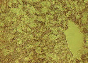
Dlk1 Rat Monoclonal Antibody [Clone ID: 24-11]

CAT#: AM26511AF-N

Dlk1 rat monoclonal antibody, clone 24-11, Azide Free


Size
    • 100 ug

Product Images

Other products for "Dlk1"

Specifications

Product Data
Clone Name 24-11
Applications FC, IHC
Recommended Dilution

Immunohistochemistry on Frozen Sections: 1 μg/ml.
Immunohistochemistry Paraffin Sections: 10 μg/ml.
Flow Cytometry: 5-10 μg/ml.
For details See Protocol below.

Reactivities Mouse
Host Rat
Isotype IgG1
Clonality Monoclonal
Immunogen Pref-1-Fc protein
Specificity This antibody reacts with Dlk.
Formulation PBS containing 50% Glycerol, pH 7.2
State: Azide Free
State: Liquid purified Ig fraction
Preservative: None
Concentration lot specific
Purification Protein G Agarose
Conjugation Unconjugated
Storage Upon receipt, store (in aliqouts) at -20°C. 
Avoid repeated freezing and thawing.
Stability Shelf life: One year from despatch.
Gene Name delta-like 1 homolog (Drosophila)
Background Delta like protein (Dlk), also known as Preadipocyte factor-1 (Pref-1) or zona glomerulosa-specific factor (ZOG), is an EGF-like transmembrane protein expressed preadipocytes but not in mature adipocytes. It is highly expressed in fetal liver, the adrenal gland, and placenta, as well as some neuroendocrine tumors and small cell lung carcinomas, where it plays a role in differentiation and proliferation. Dlk positively and negatively regulates adipocyte differentiation via at least four major variants (45-60 kDa) of Dlk generated by alternatively splicing. Constitutive expression of Dlk inhibits adipogenesis, but insulin or insulin like growth factor-1 (IGF-1) can circumvent this inhibition. Regulated processing of Dlk releases a 50 kDa soluble form that was previously characterized as Fetal Antigen-1, a protein involved in pancreatic island cell differentiation.
Synonyms DLK-1, DLK, Protein delta homolog 1, pG2, PREF1, Preadipocyte factor 1
Note This product was originally produced by MBL International.

Protocol:

Flow Cytometric analysis for floating cells
We usually use Fisher tubes or equivalents as reaction tubes for all steps described below.
1) Wash the cells 3 times with washing buffer [PBS containing 2% fetal calf serum (FCS) and 0.09% Sodium Azide].
2) Resuspend the cells with washing buffer (5x10cells/ml).
3) Add 50 µl of the cell suspension into each tube, and centrifuge at 500 x g for 1 minute at room temperature (20~25°C). Remove supernatant by careful aspiration.
4) Add 10 µl of normal goat serum containing 1 mg/mL normal human IgG and 0.09% Sodium Azide to the cell pellet after tapping. Mix well and incubate for 5 minutes at room temperature.
5) Add 40 µl of the primary antibody at the concentration of as suggested in the APPLICATIONS diluted in the washing buffer. Mix well and incubate for 30 minutes at room temperature.
6) Add 1 ml of the washing buffer followed by centrifugation at 500 x g for 1 minute at room temperature. Remove supernatant by careful aspiration.
7) Add 30 µl of 1/100 PE conjugated anti-Rat IgG diluted with the washing buffer. Mix well and incubate for 15 minutes at room temperature.
8) Add 1 ml of the washing buffer followed by centrifugation at 500 x g for 1 minute at room temperature. Remove supernatant by careful aspiration.
9) Resuspend the cells with 500 µL of the washing buffer and analyze by a flow cytometer.
Positive Control for Flow Cytometry: Mouse fetal hepatocytes, E14.5

Immunohistochemical staining for Frozen Sections
1) Mouse fetal liver was embedded in OCT compound.
2) Make the frozen sections using Microtome.
3) Fix the sections with 4% paraformaldehyde in PBS.
4) Cover tissues with normal goat serum to block non-specific staining. Do not wash.
5) Tip off the blocking buffer, wipe gently around each section and cover tissues with primary antibody diluted with PBS containing 3% normal goat serum as suggested in the APPLICATIONS.
6) Incubate the sections for 1 hour at room temperature.
7) Wash the slides 3 times in PBS for 5 minutes each.
8) Wipe gently around each section and cover tissues with Biotinylated secondary antibody (Vector). Incubate for 30 minutes at room temperature. Wash as in step 7).
9) Wipe gently around each section and cover tissues with Vectastain® ABC Kit (Vector). Incubate for 30 minutes at room temperature. Wash as in step 7).
10) Visualize by reacting for 10-20 minutes with substrate solution containing 7.5 mg DAB, 40 µL of 30% H2O2 in 150 mL PBS.
*DAB is a suspect carcinogen and must be handled with care. Always wear gloves.
11) Wash the slides in water for 5 minutes.
12) Now ready for mounting.
Positive Control for Immunohistochemistry: Mouse fetal liver, E14

Immunohistochemical staining for Paraffin-Embedded Sections: SAB method
1) Deparaffinize the sections with Xylene 3 times for 3-5 minutes each.
2) Wash the slides with Ethanol 3 times for 3-5 minutes each.
3) Wash the slides with PBS 3 times for 3-5 minutes each.
4) Remove the slides from the PBS and cover each section with 3% H2O2 for 10 minutes at room temperature to block endogenous peroxidase activity. Wash 3 times in PBS for 5 minutes each.
5) Remove the slides from PBS, wipe gently around each section and cover tissues with Protein Blocking Agent for 5 minutes to block non-specific antibody staining. Do not wash.
6) Tip off the blocking buffer, wipe gently around each section and cover tissues with primary antibody diluted with PBS containing 1% BSA as suggested in the APPLICATIONS.
7) Incubate the sections for 1 hour at room temperature.
8) Wash the slides 3 times in PBS for 5 minutes each.
9) Wipe gently around each section and cover tissues with Polyvalent Biotinylated Antibody. Incubate for 10 minutes at room temperature. Wash as in step 8).
10) Wipe gently around each section and cover tissues with Streptavidin-Peroxidase. Incubate for 10 minutes at room temperature. Wash as in step 8).
11) Visualize by reacting for 10-20 minutes with substrate solution containing 7.5 mg DAB, 40 µL of 30% H2O2 in 150 mL PBS.
*DAB is a suspect carcinogen and must be handled with care. Always wear gloves.
12) Wash the slides in water for 5 minutes.
13) Counter stain in hematoxylin for 1 minute, wash the slides 3 times in water for 5 minutes each, and then immerse the slides in PBS for 5 minutes. Dehydrate by immersing in Ethanol 3 times for 3 minutes each, followed by immersing in Xylene 3 times for 3 minutes each.
14) Now ready for mounting.
Positive Control for Immunohistochemistry: Mouse fetal liver, E14.5

Reference Data

{0} Product Review(s)

0 Product Review(s) Submit review

Be the first one to submit a review

Product Citations

*Delivery time may vary from web posted schedule. Occasional delays may occur due to unforeseen complexities in the preparation of your product. International customers may expect an additional 1-2 weeks in shipping.