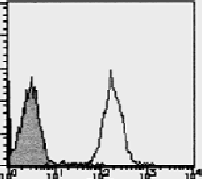
CXCR4 (Extracell. Dom)(N-term) Rat Monoclonal Antibody [Clone ID: A145]
CAT#: AM26473FC-N
CXCR4 (Extracell. Dom)(N-term) rat monoclonal antibody, clone A145, FITC
Specifications
| Product Data | |
| Clone Name | A145 |
| Applications | FC |
| Recommended Dilution | Flow Cytometry: 10-20 μg/ml (final concentration). For details see Protocol below. |
| Reactivities | Human |
| Host | Rat |
| Isotype | IgG1 |
| Clonality | Monoclonal |
| Immunogen | CXCR4 transfected Cos-1 cells |
| Specificity | This clone A145 recognizes N-terminus extracellular domain of CXCR4. |
| Formulation | PBS Label: FITC State: Liquid purified Ig fraction Stabilizer: 1% BSA Preservative: 0.09% Sodium Azide |
| Concentration | lot specific |
| Purification | Ammonium sulfate precipitation followed by gel filtration through Superdex 200 in PBS |
| Conjugation | FITC |
| Storage | Store undiluted at 2-8°C. This product is photosensitive and should be protected from light. Avoid repeated freezing and thawing. |
| Stability | Shelf life: one year from despatch. |
| Gene Name | C-X-C motif chemokine receptor 4 |
| Database Link | |
| Background | CXCR4/CD184/LESTR/fusin/NPY3R is a G protein-coupled receptor for the CXC chemokine SDF-1. Binding of SDF-1 induces CXCR4 phosphorylation by Ser/Thr kinases, leading to CXCR4 internalization via clathrin-coated pits. CXCR4 functions include co-stimulation in pre-B cell proliferation, induction of apoptosis, and HIV entry, since CXCR4 is one of the 2 major HIV/SIV co-receptors. Early infection with HIV-1 is dominated by CCR5-tropic (R5) viruses. The evolution of CXCR4-tropic (X4) viruses occurs later in the infection and is associated with rapid disease progression. CXCR4 mediates chemotaxis in mature and progenitor blood cells and is essential for B lympho- and myelopoiesis, cardiogenesis, blood vessel formation and cerebellar development. Although ubiquitously expressed in blood and tissue cells, its role in blood and tissue homeostasis is not fully understood. CXCR4 is predominantly stored intracellularly, and may contribute to the inefficiency in transmission and propagation of X4-tropic viruses. |
| Synonyms | CXC-R4, CXCR-4, Fusin, LCR1, FB22, NPYRL, HM89, SDF1 receptor, LESTR |
| Note | This product was originally produced by MBL International. Protocol: Flow cytometric analysis for floating cells Flow cytometric analysis for whole blood cells |
| Reference Data | |
Documents
| Product Manuals |
| FAQs |
| SDS |
{0} Product Review(s)
Be the first one to submit a review


 Germany
 Japan
Japan
 United Kingdom
 China
China


